|
|
 |
|
Calanoida ( Order ) |
|
|
|
Calanoidea ( Superfamily ) |
|
|
|
Megacalanidae ( Family ) |
|
|
|
Bathycalanus ( Genus ) |
|
|
| |
Bathycalanus richardi Sars, 1905 (F,M) | |
| | | | | | | Syn.: | ? Megacalanus bradyi Wolfenden, 1905 a (p.1, figs.F); Grice & Hulsemann, 1967 (p.13);
? Megacalanus princeps Wolfenden, 1905 a (p.3, figs.F: 7-9, Rem.: p.25);
? Bathycalanus maximus Wolfenden, 1906 (p.28);
? Bathycalanus bradyi : Farran, 1939 (p.360); Vervoort, 1946 (p.71); Sewell, 1947 (p.32, figs.F); Grice & Hulsemann, 1967 (p.13).
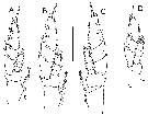
Synonymies after Bradford-Grieve & al;, 2017 (p.76) : Bathycalanus richardi Sars, 1905 (p.7, 8); Wolfenden, 1911 (p.200, pl.23, fig.8); Sars, 1924, 1925 (pp 16-19, pls 14, 15); Sewell, 1929 (p.31). | | | | Ref.: | | | Sars, 1905 b (p.8); Wolfenden, 1906 (Rem.: p.25-26); 1911 (p.200, fig.F, Rem.); Sars, 1925 (p.16, figs.F,M); Sewell, 1929 (p.31); 1932 (Pl.II: figs.F); Rose, 1933 a (p.65, figs.F,M); Jespersen, 1934 (p.45); Farran, 1939 (p.360, Rem.); Lysholm & al., 1945 (p.7); Vervoort, 1946 (p.63, Rem.); Vervoort, 1957 (p.32, Rem.); Owre & Foyo, 1967 (p.34, figs.F,M); Tanaka & Omori, 1967 (p.243, figs.M, Rem.); Brodsky & al., 1983 (p.193, figs.F,M); Michel, 1994 (p.183, Rem.); Chihara & Murano, 1997 (p.834, Pl.127: F,M); Bradford-Grieve & al., 1999 (p.877, 906, figs.F,M); Boxshall & Halsey, 2004 (p.141: figs.M); Andronov, 2014 (p.41, figs.: P3 & P5); Fornshell & Ferrari, 2014 (p.112, fig.9: Von Vaupel Klein's organ); | 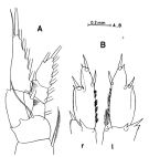 issued from : O. Tanaka & M. Omori in Information Bull on Planktology in Japan. Commemoration Number of Dr. Y. Matsue's Sixtieth Birthday, December 1967. [p.243, Fig.2]. Male (from NW Pacific): A, P1; B, 2nd and 3rd exopod segments of P5 (l = left, r = right). Nota: The 5th pair of legs in male is slightly asymmetrical: the inner distal margin of the 2nd segment of the left leg is furnished with a densely plumosed spine, while this is absent in the right leg.
|
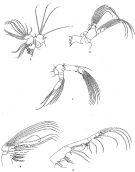
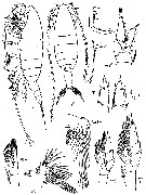
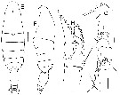
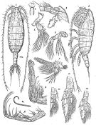 Issued from: G.O. Sars in Résult. Camp. Scient. Prince Albert I, 69, pls.1-127 (1924). [Pl.IV, figs. 1-12]. Female: 1, habitus (dorsal); 2, idem (lateral left side); 3, forehead (lateral); 4, rostrum (frontal view); 5, A2; 6, Md; 7, Mx1; 8, Mx2; 8a, distal part of a seta of Mx2 (enlarged); 9, Mxp; 10, P1; 11, P3; 12, P5.
|
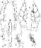
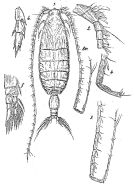 Issued from: G.O. Sars in Résult. Camp. Scient. Prince Albert I, 69, pls.1-127 (1924). [Pl.V, , figs. 1-6]. Male: 1, habitus (dorsal); 2, proximal segments of right A1; 3, distal segments of right A1; 4, grasping segments of right A1 (enlarged); 5, P1; 6, exopodal segments of P5. Nota: Right A1 geniculate.
|
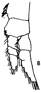
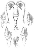 Issued from : R.B.S. Sewell in Mem. Indian Mus., 1932, X (continued). [Pl.I]. Female (from N Indian Ocean): 1, habitus (dorsal); 2,P1; 3, P2: 4, P3; 5, P4; 6, P5.
|
 Issued from : R.B.S. Sewell in Mem. Indian Mus., 1932, X (continued). [Pl.II]. Female: 1, A2; 2, Md; 3, Mx1; 4, Mx2; 5, Mxp.
|
 issued from : R.N. Wolfenden in Die Marinen Copepoden der Deutschen Südpolar-Expedition 1901-1903, 1911. [Pl.XXIII, Fig.8]. Female: 8, P1.
|
 issued from : K.A. Brodsky, N. V. Vyshkvartzeva, M.S. Kos & E.L. Markhaseva in Opred. Faune SSSR, 1983, 135 (1). [p.194, Fig.85]. Female & Male. r = right leg of P5 male; l = left leg of P5 male. P1 and P5 female from Tanaka & Omori (1967); remaining figures from Sars (1924). Nota: - Genital segment amphora-like; - Exopod of P1 2-segmented.
|
 issued from : H.B. Owre & M. Foyo in Fauna Caribaea, 1967, 1, Crustacea, 1: Copepoda. [p.35, Figs.174-175]. Female (from Florida Current): 174, Mx2; 175, terminal setae of Mx2.
|
 issued from : J.M. Bradford-Grieve, E.L. Markhaseva & C.E.F. Rocha & B. Abiahy in South Atlantic Zooplankton, Edit. D. Boltovskoy. 1999. Vol.2. Copepoda. [p.977, Fig. 7.5]. Female: Mx1. Nota: Rostrum with pair of chitinous projections. A1 extends beyond caudal rami by about 4-6 segments.
|
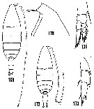 issued from : H.B. Owre & M. Foyo in Fauna Caribaea, 1, Crustacea, 1: Copepoda. Copepods of the Florida Current. 1967. [p.34, Figs.169-173. Redrawings after Sars (1925). Female (from 25°40'N, 79°43'W): 169, habitus (dorsal); 170, forehead (lateral); 171, P1. Male: 172, habitus (dorsal); 173, exopod of P5.
|
 Issued from : V.N. Andronov in Russian Acad. Sci. P.P. Shirshov Inst. Oceanol. Atlantic Branch, Kaliningrad, 2014. [p.41, Fig.11, 1, 2]. Bathycalanus richardi after Brodsky & al., 1983. 1, P3; 2, P5.
|
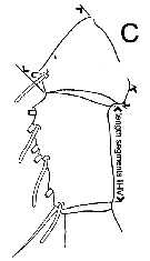
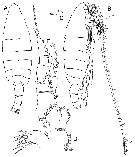 Issued from : J.M. Bradford-Grieve, L. Blanco-Bercial & G.A. Boxshall in Zootaxa, 2017, 4229 (1). [p.77, Fig.41]. Female (from 27.133°N, 138.233°E): A-B, habitus (dorsal and lateral, respectively); C, forehead (lateral); D, Mx1; E, P1. Scale bars: 1.0 mm (A, B); 0.1 mm (C, D, E). Nota: - Forehead in dorsal view with rounded prominence extending into pair of small anteriorly-directed spine-like processes. - Pedigerous somite 5 with short, symmetrical, rounded posterior lappets extending 1/4 of way along genital double-somite. - Genital double-somite in dosrsal view widest at anterior 1/3, length about 1.4 times widest width (See figure 1 below concerning measurements taken). - A1 extends about 7 segments beyond caudal rami. - Mx1 praecoxal arthrite (formerly named inner lobe 1) with 13 setae and spines, including 2 setae on posterior surface; coxal endite without setae (formerly named 2nd inner lobe); basal endite 2 (formerly named 3rd inner lobe) with 3 short setae; endopod segments with 2 (subequal), 2 (subequal), 6 setae (including 1 smaller seta arising from posterior surface), respectively. - P1 exopod with articulation between exopod segments 2 and 3 undeveloped.
|
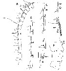 Issued from : J.M. Bradford-Grieve, L. Blanco-Bercial & G.A. Boxshall in Zootaxa, 2017, 4229 (1). [p.78, Fig.42]. Female A1 ancestral segments: A,
segments I-XV; B, segments XVI-XIX; C, segments XX-XXIII; D, segments XXIV-XXVIII; E, detail of segment XXVIII; F, dorsal surface of segments I-V (hs = hair sensillum; mc = macula crivrosa). Scale bars: 1.0 mm (A-D); 0.1 mm (E, F).
|
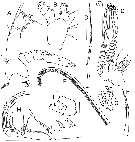 Issued from : J.M. Bradford-Grieve, L. Blanco-Bercial & G.A. Boxshall in Zootaxa, 2017, 4229 (1). [p.79, Fig.43]. Female: A, A2; B, Md palp; C, Mx2; D, seta from praecoxal endite 1 of Mx2; E, part of seta from coxal endite 2 of Mx2; F, inner seta of praecoxal endite 2 of Mx2; G, detail of endopod of Mx2 (inner surface); H, Mxp; I, terminal part of endopod of Mxp (note outer seta of endopod segment 5 knocked off). Scale bars: 1.0 mm (C, H); 0.1 mm on remaining figures. Nota: - A2 exopod ancestral segment IV with short seta, not extending beyond distal border of ancestral segment VI, lined with spinules. - Mxp syncoxal endite 4 with longest spinulose seta extending to distal border, or beyond, of endopod segment 1.
|
 Issued from : J.M. Bradford-Grieve, L. Blanco-Bercial & G.A. Boxshall in Zootaxa, 2017, 4229 (1). [p.80, Fig.44, A-D]. Female: A, P2 (anterior view); B, P3 (anterior view); C, P4 (posterior view); D, P5 (anterior view). Scale bar = 1.0 mm in all figures.
|
 Issued from : J.M. Bradford-Grieve, L. Blanco-Bercial & G.A. Boxshall in Zootaxa, 2017, 4229 (1). [p.80, Fig.44, E-J]. Male: E-F, habitus (dorsal and lateral, respectively); G, forehead (lateral); H, P1 (anterior view); I, endopod of Md palp; J, basal endite 2 and endopod of Mx1. Scale bars: 1.0 mm (E-F); 0.1 mm (G-J). A-D from 27.133°N, 138.233°E; E from 38.0.33 N, 124.183°W; F-J from 11.683°N, 20.419°W. Nota: - Forehead in dorsal view with rounded prominence extending into pair of small anteriorly-directed spine-like processes. - Pedigerous somite 5 with short, rounded posterior lappets. - Urosomal measurements made in lateral view (see below figure 1 B, p.13,, tabulated in table 9). In lateral view urosomite II of enlarged and swollen appearance, constricted anteriorly : Ur II: 2.17-2.58 (mean = 2.37, SD = 0.17, n = 4) times longer than urossomite III and constricted anteriorly such that ratio UrII ant/UrII mx = 0.64-0.68 (mean = 0.67; SD = 0.03, n = 5).
|
 Issued from : J.M. Bradford-Grieve, L. Blanco-Bercial & G.A. Boxshall in Zootaxa, 2017, 4229 (1). [p.81, Fig.45]. Male: A, right A1 ancestral segments XVIII-XX; B, right A1 ancestral segments XVIII-XIX; C, A1 right ancestral segments XX-XXIII; D, right A1 ancestral segments XXVI-XXVIII; E, P5 (posterior view); F, detail of inner distal corner of left exopod segment 2; G, detail of inner distal corner of left exopod segment 2 of another specimen; Scale bars = 0.1 mm for all figures; A, E-F from 11.683°N, 20.419°W: B-D, G from 38.033°N, 124.183°W. Nota: Right A1 ancestral segments XIX- 1 ms, 1 fused gripping element extending beyond base of aesthetasc, 1a; XX- 1 gripping element, 1ms, 1a; XXI-XXIII- 2 gripping elements extending well beyond base of following element, 1a, 1 short fused gripping element, 1ms. - P5 left exopodal segment 2 specialised seta with long lash extending almost to distal border of endopod&l segment 3, basal part rectangular in shape; inner border of right exopodal segment 3 lined with setules. Male specimens taken from the Atlantic Ocean are clearly Bathycalanus richardi Sars, 1905 in that they have a 2-segmented P1 exopod. The authors, also, note that the right A1, ancestral segment XIX, was drawn by Sars (1925, pl.5, fig.3) as having 2 fused gripping elements whereas, in the specimens examined by the authors, the proximal-most gripping element is not predent although there is a short, low ridge on the anterior border that is additional to the notmal complement of setae. A number of setae were apparently missed by Sars (1924, pl.5, figs 6, 7, 9) from female Md endopod segment 1, Mx1 endopod segment 2,Mxp endopod segments 4 to 6.
|
 Issued from : J.M. Bradford-Grieve, L. Blanco-Bercial & G.A. Boxshall in Zootaxa, 2017, 4229 (1). [p.91, Table 10]. Morphological characters after identification key of Bathycalanus females and males. Compare to other species of genus. Main characters identification after species key : 1 - Mx1 praecoxal endite without setae. 2 - Anterior margin of head with 2 very small spine-like processes. 3 - P1 exopod segments 2 and 3 fused.
|
 Issued from : J.M. Bradford-Grieve, L. Blanco-Bercial & G.A. Boxshall in Zootaxa, 2017, 4229 (1). [p.82, Table 9]. Proportions of male urosomites. See below diagram illustrating how measurements were made (Fig.1, D). Stations: Antipode IV Stn 53D = 26.441°N, 139.037E; MV66-11 = 38.033°N, 124.183°W; ANTXIV/1 CoO224 = 11.683°N, 20.419°W; Antipode IV Stn 55D = 20.933°N, 138.533°E. Main characters identification after species key : 1 - Mx1 praecoxal endite without setae. 2 - Anterior margin of head with 2 very small spine-like processes. 3 - P1 exopod segments 2 and 3 fused; Urosomite segment II in lateral view enlarged and swollen: UrII 2.17-2.58 times longer than UrIII
|
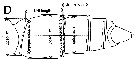 Issued from : J.M. Bradford-Grieve, L. Blanco-Bercial & G.A. Boxshall in Zootaxa, 2017, 4229 (1). [p.13, Table 1, D]. Diagram illustrating measurements. Example taken for Bathycalanus bradyi in lateral view.. male. Ur = urosomite
|
 Issued from : J.M. Bradford-Grieve, L. Blanco-Bercial & G.A. Boxshall in Zootaxa, 2017, 4229 (1). [p.13, Table 1, B]. Diagram illustrating measurements (width and length). Example taken for Bathycalanus tumidus. Gns = genital double-somite.
|
 Issued from : J.M. Bradford-Grieve, L. Blanco-Bercial & G.A. Boxshall in Zootaxa, 2017, 4229 (1). [p.13, Table 1, BC. Diagram illustrating measurements on ancestral segments I-IV along posterior border of A1. Example taken for Megacalanus ohmani female.
|
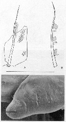 Issued from : J.A. Fornshell & F.D. Ferrari in Crustaceana, 2014, 87 (1). [p.112, Fig.9, A-C]. Von Vaupel Klein's organ on right endopodal segment 1 and right basal seta of P1. A, anterior, distal up §ventral seta not drawn; B, right basal seta; C, distodorsal tip of endopodal proximal segment anterioventral, distodorsal tip from this view appears rounded rather than cleft, only a few denticles dorsal to be pores remain, most marked by scars (scale line: 50 µm) Scale line; 300 µm. The Von Vaupel Klein organ is an association (srtucture) of the distal seta from basis and the proximal endopodal segment of P1. This structure shows significant variability among many gymnoplean copepods, in the shape of the distodorsal corner of the proximal endopodal segment, presence and location of denticles on the anterior face of the segment, presence and size of denticles along the distal margin of the segment , number of pores on the segment, shape of the seta that originates on the basis, and the morphology of the basis at the origin of the seta. The function of this complex structure is not known.
| | | | | Compl. Ref.: | | | C.B. Wilson, 1950 (p.171); Wickstead, 1962 (p.551, food & feeding); Grice & Hulsemann, 1968 (tab.2); Deevey & Brooks, 1977 (p.256, Table 2, Station "S"); Razouls & al., 2000 (p.343, tab. 5, Appendix); Holmes, 2001 (p.35); Hsiao & al., 2004 (p.326, tab.1) | | | | NZ: | 13 | | |
|
Distribution map of Bathycalanus richardi by geographical zones
|
| | | | | | | | | | | | 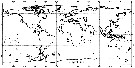 Issued from : J.M. Bradford-Grieve, L. Blanco-Bercial & G.A. Boxshall in Zootaxa, 2017, 4229 (1). [p.84, Fig.46]. Issued from : J.M. Bradford-Grieve, L. Blanco-Bercial & G.A. Boxshall in Zootaxa, 2017, 4229 (1). [p.84, Fig.46].
Distribution of Bathycalanus species;
Bathycalanus richardi (filled squareç; Bathycalanus bradyi (filled triangle); Bathycalanus milleri (open triangle); Bathycalanus dentatus (open square); Bathycalanus tumidus (open circle); Bathycalanus adornatus (open diamond); Bathycalanus pustulosus (filled star); Bathycalanus unicornis ( (filled diamond); Bathycalanus bucklinae (filled circle); Bathycalanus eximius (cross). |
| | | | Loc: | | | Antarct. (Drake Passage, SW Atlant., S & SE Pacif.), sub-Antarct. (SE Pacif.), Cape Verde Is., off Azores, Bay of Biscay, Barbada Is., Florida, Sargasso Sea, Station "S" (32°10'N, 64°30'W), Strait of Davis, SW Iceland, G. of Oman, Arabian Sea, SW Indian, G. of Bengal, Indonesia-Malaysia, Philippines, China Seas (South China Sea), Taiwan E, SW Japan, Japan Sea, Aleutian Is., SW Galapagos, N Marquesas Is., off Juan Fernandez Is.
Type locality: W and S Azores Islands to out of Bay of Biscay. | | | | N: | 19 | | | | Lg.: | | | (1) F: 10,2; (11) F: 11,93; (28) F: 11,15-8,25; M: 8,15-7,8; (49) F: 10,8-9,9; (70) F: 9,7-9,4; M: 9,6; (131) F: 12-9,5; M: 10-8; (140) F: 9,5; (866) M: 6-10; (1316) F: 8,6-10,6; M: 8,8-9,7; {F: 8,25-12,0; M: 6,0-10,0}
(140) Prosome length : 7.1 mm; urosome length: 2.4 mm. | | | | Rem.: | Meso-bathypelagic.
Sampling depth (Antarct., sub-Antarct.) :1000-2000 m. In Sargasso Sea: 500-1500 m (Deevey & Brooks, 1977, Station "S");
A boubt subsists concerning the synonymy between B. bradyi and B. richardi.
H.B. Michel (1994, p.185) regards Bathycalanus bradyi as a synonym of Bathycalanus richardi, certain populations of which incompletely develop the disyal septum which defines the middle segment of the exopod on P1.
For Bradford-Grieve & al. (2017, p.83) there is some uncertainty over the previous identifications of Bathycalanus that have a pair of small spine-like processes on the rostral base.
Concerning morphological variations, P1 usually does not have a functional articulation expressed between exopod segments 1 and 2 although some specimens have these segments more or less expressed. Male P5 left exopod segment 2 inner distal border specialised setulose seta is of variable length ranging from extending short of the distal border of endopod segment 2 to beyond it; sometimes, but not always, there is a small outer border spinule on the basal part of the setulose seta. There were slight differences in the male right A1 between the specimens from Atlantic and Pacific oceans: ancestral segments XVIII and XIX had a proximal ridge in the Atlantic specimen but were without such a ridge in Pacific specimens. | | | Last update : 14/09/2018 | |
|
|
 Any use of this site for a publication will be mentioned with the following reference : Any use of this site for a publication will be mentioned with the following reference :
Razouls C., Desreumaux N., Kouwenberg J. and de Bovée F., 2005-2026. - Biodiversity of Marine Planktonic Copepods (morphology, geographical distribution and biological data). Sorbonne University, CNRS. Available at http://copepodes.obs-banyuls.fr/en [Accessed January 06, 2026] © copyright 2005-2026 Sorbonne University, CNRS
|
|
|
|


;)
;)
;)
;)
;)
;)
;)
;)
;)
;)
;)
;)
;)
;)
;)
;)
;)
;)
;)
;)

;)
