|
|
 |
|
Calanoida ( Order ) |
|
|
|
Metridinidae ( Family ) |
|
|
|
Metridia ( Genus ) |
|
|
| |
Metridia princeps Giesbrecht, 1889 (F,M) | |
| | | | | | | Syn.: | no Metridia princeps : Esterly, 1906 a (p.69, figs.F,M); ? Lee & al., 1971 (p.1151);
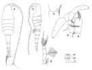
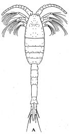
? Metridia bicornuta Davis, 1949 (p.47, Descr.F, figs.F) | | | | Ref.: | | | Giesbrecht, 1892 (p.340, 346, 773, Descr.F, figs.F); Giesbrecht & Schmeil, 1898 (p.107, Rem. F); Canu, 1896 (p.433, Rem.); I. C. Thompson, 1903 a (p.24, Descr.M, figs.M); Farran, 1908 b (p.61); Wolfenden, 1908 (p.15, figs.F); A. Scott, 1909 (p.121, figs.F,M); Wolfenden, 1911 (p.287, figs.F,M); Lysholm & Nordgaard, 1921 (p.24); Sars, 1925 (p.194, figs.F,M); Farran, 1926 (p.271); Rose, 1929 (p.29); Farran, 1929 (p.209, 258); Sewell, 1932 (p.252, Rem. F,M, figs. juv.); Wilson, 1932 a (p.122, figs.F,M); Rose, 1933 a (p.176, figs.F,M); Jespersen, 1934 (p.92); Jespersen, 1940 (p.43); Wilson, 1942 a (p.194, fig.F); Lysholm & al., 1945 (p.29); Sewell, 1947 (p.167, fig.44, Rem.); Farran, 1948 c (n°14, p.3, figs.F); Brodsky, 1950 (1967) (p.301, figs.F,M); Vervoort, 1957 (p.118, fig., Rem.); Tanaka, 1963 (p.17, figs.F,M); Vervoort, 1965 (p.104, Rem.); Owre & Foyo, 1967 (p.70, figs.F,M); Arashkevich, 1969 (p.703, figs.F); Vaupel-Klein, 1970 (p.4, 35, figs.F, Rem., Tabl.III); Wheeler, 1970 (p.12, fig.juv.M, Rem.); Björnberg & al., 1981 (p.640, figs.F,M); Gardner & Szabo,1982 (p.330, figs.F,M); Szabo & Gardner, 1986 (p.1560: Appendix, Rem.); Hulsemann, 1988 (p.188, Rem.); Chihara & Murano, 1997 (p.837, tab.6); Cuoc & al., 1997 (p.652, figs.F: genital complex); Bradford-Grieve & al., 1999 (p.884, 948, figs.F,M); Bradford-Grieve,1999 b (p.116, figs.F,M, Rem., figs.178, 192); Markhaseva, 2001 (p.47, figs.F,M, Rem.); Boxshall & Halsey, 2004 (p.147: figs.F,M); Vives & Shmeleva, 2007 (p.367, figs.F,M, Rem.) | 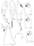 issued from : E.L. Markhaseva in Zoosyst. Rossica, 2001, 9 (1). [p.66, Figs. 148-156]. Female: 148, habitus (right lateral view); 149, idem (dorsal view); 150, cephalosome (right lateral view); 151, anterior part of cephalosome; 152, rostrum; 153, urosome (right lateral view); 154-156, genital somite (ventral view). Nota : Prosome 1.33-1.47 times as long as urosome. Cephalosome without collar on both left and right sides. Rostrum of 2 filaments at subdivided base. Genital somite 1.8-2.0 times as long as wide, with elongate spermathecae, right one usually darker. Caudal rami about 6 times as long as wide. A1 24-segmented, exceeding the body length by 4-5 distal segments. Oral parts and Mxp as in M. ferrarii.
|
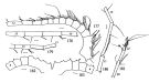
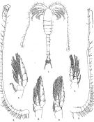
 issued from : E.L. Markhaseva in Zoosyst. Rossica, 2001, 9 (1). [p.67, Figs. 157-167]. Female: 157, left A1 (segments 1-6); 158, idem (segments 7-11); 159, idem (segments12-15); 160, idem (segments 16-18); 161, idem (segments19-24); 162, first leg ( (endopod); 163, second leg (endopod); 164, idem (endopod.1); 165, third leg (exopod); 166, fourth leg (exopod); 167, fifth legs.
|
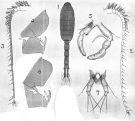
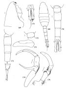 issued from : E.L. Markhaseva in Zoosyst. Rossica, 2001, 9 (1). [p.68, Figs.168-176]. Male: 168, habitus (dorsal view); 169, cephalosome (right lateral view); 170, posterior prosomal somite and urosome; 171, idem (right lateral view); 172, posterior prosomal somite and genital somite (dorsal view); 173, anal somite and caudal rami (dorsal view); 174, left fifth leg; 175, idem (exopod.3); 176, coxopodof fifth legs and right fifth leg. Nota : Cephalosome collar absent. Genital somite with small projection on the right lacking spinules ; spinules present at the posterior prosomal somite on the left. Urosomal somites lacking spinules. Caudal rami about 6 times as long as wide. Right A1 geniculated.
|
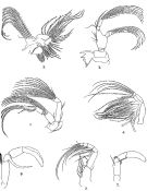
 issued from : E.L. Markhaseva in Zoosyst. Rossica, 2001, 9 (1). [p.69, Figs.177-183]. Male: 177, right A1 (segments 1-12); 178, idem (segments 13-16); 179, idem (segments 15-17); 180, idem (segments 17-19); 181, idem (segments 20-22); 182, left A1 (segments 1-4); 183, right A1 (segments 1-4).
|
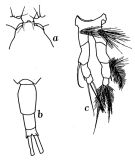
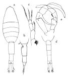 issued from : O. Tanaka in Publs Seto Mar. Biol. Lab., 1963, XI (1). [p.18, Fig.156]. Female: a, habitus (dorsal); b, last thoracic segment and urosome (left lateral side); c, P5. Nota: The urosome segments and furca are in the proportional lengths as 42:22:12:24 = 100. Male: d, last thoracic segment and urosome (dorsal); e, P5. Nota: The urosome segments and furca are in the proportional lengths as 11:19:17:19:9:25 = 100. The right A1 exceeds the end of the furca by last 2 segments; the left A1 forms a grasping organ.
|
 isued from : J.M. Bradford-Grieve in The Marine Fauna of New Zealand: Pelagic Calanoid Copepoda. National Institute of Water and Atmospheric Research (NIWA). NIWA Biodiversity Memoir, 111, 1999. [p.117, Fig.78]; Female: A, habitus (dorsal); B, P5. Male: C, habitus (left lateral side); D, P5; E, detail of joint between left exopod segment 1 and exopod segments 2+3.
Female characteristics: Metasome oval. - Rostrum in the form of a bifid lamella with 2 slender filaments. - Posterior metasomal corners short and rounded. - Urosome large, almost equal in length to metasome. - Genital segment exceeding length of remaining urosome. - Anal segment widened posteriorly, half as long as preceding segment. - Caudal rami long, narrow, more than 5 times as long as wide, twice the length of anal segment. - A1 longer than body, proximal segments toothed anteriorly. - P5 3-segmented with long hairs on basal segment, distal segment short and terminated by 3 subequal plumose setae. Male characteistics: Left A1 shorter than right and prehensile. - P5 asymmetrical stronger on right, 4-segmented, segment 2 with a long gently curving inner process, distal segment with a long parallel outer plate separated from the segment, segment terminated by 2 spinules; left leg shorter, 2nd segment withouy spines on inner side, 2 distal joints are fused. On Southwest Pacific, specimens in P5 right exopod segment 2 relatively short; exopod segment 3 does not appear to have an outer edge lobe but is constructed as in other Metridia<=em>; left basipod 1 has a tuft of long hairs on the posterior surface; left exopod segment 2+3 has large, hairy spine near its base, directed proximally, in a hairy depression.
|
 Issued from : J.C. von Vaupel-Klein in Zool. Verh. Leiden, 1970, 110. [p.31, Fig.132, a-c]. Female: a, forehead (dorsal); b, urosome (dorsal); c, P5.
|
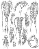 Issued from : G.O. Sars in Résult. Camp. Scient. Prince Albert I, 69, pls.1-127 (1924). [Pl.LIII, figs.1-12]. Female: 1, habitus (dorsal); 2, idem (lateral left side); 3, forehead (lateral); 4, rostrum (frontal view); 5, P1; 6, P2; 7, 1st endopodal segment of P2 (with adjacent segments); 8, P3; 9, P5. Male: 10, habitus (dorsal); 11, P5; 12, anal segment and caudal rami (dorsal).
|
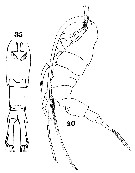
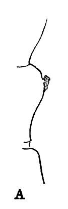 issued from : R.B.S. Sewell in The John Murray Expedition, 1933-34, Scientific Reports, VIII (1), 1947. [p.166, Fig.44, A]. Process on the posterior aspect of the 2nd basal segment of P1. Remarks: The shape of the hook-like or spine-like process shows some variation in different genera, but it is undoubtedly homologous throughout the whole series. The function of this organ is unknown.
|
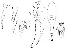
 Issued from : R.B.S. Sewell in Mem. Indian Mus., 1932, X (continued). [P;.III. Male (from N Indian Ocean): 1, habitus (dorsal); 2, left A1; 3, right A1; 4, P1; 5, P2; 6, P3; 7, P4.
|
 Issued from : R.B.S. Sewell in Mem. Indian Mus., 1932, X (continued). [P;.IV]. Male: 1, A2; 2, Md; 3, Mx1; 4, Mx2; 5, Mxp; 6, left P5; 7, right P5.
|
 Issued from : R.B.S. Sewell in Mem. Indian Mus., 1932, X (continued). [p.248, Fig.87, A]. male: habitus, dorsal (arrangement pf cutaneous pores).
|
 issued from : A. Scott in Siboga-Expedition, 1909, XIX a. [Plate XXXVIII, Figs.1-7]. Male (from Indonesia-Malaysia): 1, habitus (dorsal); 2, right A1; 3, left A1; 4, P2 (hooks on basal segment of endopodite); 5, P5. Female: 6, P2 (hooks on basal segment of endopodite); 7, P5.
|
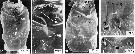 issued from : C. Cuoc, D. Defaye, M. Brunet, R. Notonier & J. Mazza in Mar. Biol., 1997, 129. [p.655, Figs.14-18]. Scanning electron micrographs of genital area of females. 14, 16, ventral face of genital double-somite of two specimens; 15, detail of genital area; 17, 18, copulatory pores ( cp) of females with single insemination on right side (17) and with insemination on both sides (18) [note spermatophoral plugs (black arrowheads), closed copulatory pore (arrow), and sensory pore (?) (white arrowhead)]
|
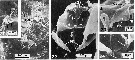 issued from : C. Cuoc, D. Defaye, M. Brunet, R. Notonier & J. Mazza in Mar. Biol., 1997, 129. [p.655, Figs.20-24]. Scanning electron micrographs of genital area of females. 20, fixation sites of 3 spermatophores, note fixation disk level with right copulatory pore (large arrow), 2 posterior disks (triangles), and superficial furrows (small arrow); 21, posterior part of genital double-somite, with spermatophoral remnants and some plandular pores (arrows); 22-24, inner dorsal view of genital area (22), with details of right egg-laying duct ( d3) (23) and copulatory ducts (arrow) (24). cg: closed gonopores; cp: copulatory pores; d1: seminal duct; d2: shell duct; gp: gonoporal plates; gs: gonoporal slits; f: folds of superficial furrows; : muscle of egg-laying duct; sgsuperficial furrows between copulatory pores and gonopores; sr: seminal receptacles.
|
 issued from : R.N. Wolfenden in Die Marinen Copepoden der Deutschen Südpolar-Expedition 1901-1903, 1911. [Pl.XL, Figs.8-13]. Female: 8, habitus (lateral); 9, idem (dorsal); 10, P5. Male: 11, last thoracic segment and urosome (lateral, right side); 12, P2; 13, P5.
|
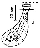 issued from : J. Mauchline in J. mar. biol. Ass, U.K., 1977, 57 (4). [p.980, Fig.4, J]. Integumental organs: J, opening of luminescence gland.
|
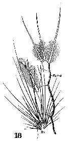 Issued from : W. Giesbrecht in Systematik und Faunistik der Pelagischen Copepoden des Golfes von Neapel und der angrenzenden Meeres-Abschnitte. Fauna Flora Golf. Neapel, 1892, 19 , Atlas von 54 Tafeln. [Taf.33, Fig.18]. Female: 18, P5.
|
 Issued from : W. Giesbrecht in Systematik und Faunistik der Pelagischen Copepoden des Golfes von Neapel und der angrenzenden Meeres-Abschnitte. Fauna Flora Golf. Neapel, 1892, 19 , Atlas von 54 Tafeln. [Taf.33, Figs.3, 35]. Female: 3, A1 (proximal segments); 35, urosome (ventral).
|
 issued from : H.B. Owre & M. Foyo in Fauna Caribaea, 1, Crustacea, 1: Copepoda. Copepods of the Florida Current. 1967. [p.15, Fig.33]. Female: 33, P2.
| | | | | Compl. Ref.: | | | Cleve, 1904 a (p.192); Pearson, 1906 (p.24); Hardy & Gunther, 1935 (1936) (p.179, Rem.); Sewell, 1948 (p.329, 503, 514, 519, 521, 525, 530, 539, 547, 549, 567); C.B. Wilson, 1950 (p.265); Fagetti, 1962 (p.27); Grice, 1963 a (p.496); Grice & Hulsemann, 1965 (p.224); 1968 (tab.2); Park, 1970 (p.477); Gueredrat & Friess, 1971 (p.187, fig.2); Roe, 1972 (p.277, tabl.1, tabl.2); Harding, 1974 (p.141, tab.2, gut contents); Vives & al., 1975 (tab.II, XII); Björnberg, 1973 (p.337, 387); Deevey & Brooks, 1977 (p.256, tab.2, Station "S"); Vaissière & Séguin, 1980 (p.23, tab.1); Vives, 1982 (p.293); Kovalev & Shmeleva, 1982 (p.84); Roe, 1984 (p.358); Guangshan & Honglin, 1984 (p.118, tab.); Brenning, 1985 a (p.28, Table 2); Lozano Soldevilla & al., 1988 (p.59); Hopkins & Torres, 1988 (tab.1); Cervantes-Duarte & Hernandez-Trujillo, 1989 (tab.3); Heinrich, 1990 (p.18); Madhupratap & Haridas, 1990 (p.305, fig.3: vertical distribution night/day; fig.7: cluster); Hernandez-Trujillo, 1991 (1993) (tab.I); Shih Young, 1995 (p.70); Suarez-Morales & Gasca, 1998 a (p.110); Lapernat, 2000 (tabl.3, 4); Razouls & al., 2000 (p.343, tab. 5, Appendix); Madhupratap & al., 2001 (p. 1345, vertical distribution vs. O2, figs.4, 5: clusters, p.1353); Holmes, 2001 (p.19); Yamaguchi & al., 2002 (p.1007, tab.1); Hsiao & al., 2004 (p.326, tab.1); Neumann-Leitao & al., 2008 (p.799: Tab.II, fig.6); Fernandes, 2008 (p.465, Tabl.2); Gaard & al., 2008 (p.59, Table 1, N Mid-Atlantic Ridge); Galbraith, 2009 (pers. comm.); Park & Ferrari, 2009 (p.143, Table 4, Appendix 1); Mazzocchi & Di Capua, 2010 (p.426); Palomares-Garcia & al., 2013 (p.1009, Table I, abundance vs environmental factors, very rare); in CalCOFI regional list (MDO, Nov. 2013; M. Ohman, comm. pers.); Hirai & al., 2013 (p.1, Table I, molecular marker); Bonecker & a., 2014 (p.445, Table II: frequency, horizontal & vertical distributions); Bode & al., 2015 (p.268, Table 1, chemical components, trophic level, geographic zone); El Arraj & al., 2017 (p.272, table 2); Belmonte, 2018 (p.273, Table I: Italian zones); Bode & al., 2018 (p.840, Table 1, respiration & ingestion rates, depth) | | | | NZ: | 20 | | |
|
Distribution map of Metridia princeps by geographical zones
|
| | | | | | | | | | | | | | | | | | 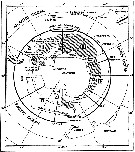 issued from : W. Vervoort in B.A.N.Z. Antarctic Reseach Expedition, Reports - Ser. B, Vol. III, 1957 [Fig.108] issued from : W. Vervoort in B.A.N.Z. Antarctic Reseach Expedition, Reports - Ser. B, Vol. III, 1957 [Fig.108]
Chart showing the geographical distribution (white triangle) in the seas surrounding the Antarctic continent.
Nota: In this chart the area frequented by whaling vessels has been hatched. The Antarctic circle (66°.5 S) has been drawn as a broken line. The numbers I to VI refer to the sectors into which the Antarctic seas are divided according to Mackintosh (1942) (after Vervoort, 1951). |
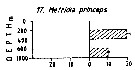 Issued from : M. Madhupratap & P. Haridas in J. Plankton Res., 12 (2). [p.310, Fig.3]. Issued from : M. Madhupratap & P. Haridas in J. Plankton Res., 12 (2). [p.310, Fig.3].
Vertical distribution of calanoid copepod (mean +1 SE), abundance No/100 m3. 17- Metridia princeps.
Night: shaded, day: unshaded.
Samples collected from 6 stations located off Cochin (India), SE Arabian Sea, November 1983, with a Multiple Closing Plankton Net (mesh aperture 300 µm), in vertical hauls at 4 depth intervalls (0-200, 200-400, 400-600, 600-1000 m). |
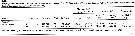 Issued from : M. Bode, R. Koppelmann, L. Teuber, W. Hagen & H. Auel inGlobal Biogeochemical Cycles, 2018, 32. [p.844, Table 1). Issued from : M. Bode, R. Koppelmann, L. Teuber, W. Hagen & H. Auel inGlobal Biogeochemical Cycles, 2018, 32. [p.844, Table 1).
Cf. explanations of these measures in Calanoides natalis from the same authors. |
| | | | Loc: | | | Cosmopolite : Antarct. (SW Atlant., Weddel Sea, Indian, SW & SE Pacif., Ross Sea, Mc Murdo), South Georgia, Atlant., off Bermuda: Station S (32°10N, 64°30W), off E Cape Cod, S Iceland, North Sea, Moroccan coast, Ibero-moroccan Bay, Medit. (Ligurian Sea, Tyrrhenian Sea, Adriatic Sea), Indian, Indonesia-Malaysia, New Zealand, China Seas (South China Sea), E Taiwan, SE Japan, Pacif. (W equatorial), Gulf of California (rare), Station Knot, Kuril-Kamchatka Trench, off British Columbia, Arctic | | | | N: | 81 | | | | Lg.: | | | (1) F: 8,1; M: 8; (5) F: 8,8-7,9; M: 8,3; (16) F: 8,5-7,13; M: 8,15-6,75; (26) F: 7,34; M: 7,31; (32) F: 9-8; M: 6-5,8; (35) M: 8,5; (38) F: 7,9-7,1; (44) F: 7,75; M: 7,4; (45) F: 8,5-8; M: 7-6; (46) F: 8,5; (199) F: 7,52-6,69; M: 7,68-6,7; (205) F: 7,5; (411) F: 7,42; M: 7,65; (823) F: 7,8-7,4; M: 7,4; (909) F: 6,9-7,8; M: 6,75-7,6; {F: 6,69-9,00; M: 5,80-8,50}
The mean female size is 7.798 mm (n = 22; SD = 0.6082), and the mean male size is 7.235 mm (n = 17; SD = 0.8156). The size ratio (male : female) is 0.94 (n = 11; SD = 0.1040). | | | | Rem.: | bathy-abyssopelagic.
Sampling depth (Antarct.) : 0-1000 m. Sargasso Sea: 500-2000 m (Deevey & Brooks, 1977, Station "S"); 2000-1000 m (Harding, 1974).
For Vaupel-Klein (1970, p.36) the specimen from the Station "P" belongs to a geographical vatiety of M. princeps and that M. ornata as described by Brodsky (1950, p.303, fig.210) must also be regarded as a variety or a subspecies of M. princeps already noticed the close relationship between M. princeps, M; ornata, M. macrura, and M. ignota.
According to Markhaseva (2001, p.48) : probably the distributional range for this species is not so wide (see geographical chart in Razouls & al. 2008), as it could have been mixed with other large-sized Metridia, the taxonomic status of which are not clear.
R. Stephen, 2007 : Data sheets of NIO, Kochi, India (on line). | | | Last update : 25/10/2022 | |
|
|
 Any use of this site for a publication will be mentioned with the following reference : Any use of this site for a publication will be mentioned with the following reference :
Razouls C., Desreumaux N., Kouwenberg J. and de Bovée F., 2005-2025. - Biodiversity of Marine Planktonic Copepods (morphology, geographical distribution and biological data). Sorbonne University, CNRS. Available at http://copepodes.obs-banyuls.fr/en [Accessed November 17, 2025] © copyright 2005-2025 Sorbonne University, CNRS
|
|
|
|


;)
;)
;)
;)
;)
;)
;)
;)
;)
;)
;)
;)
;)
;)
;)
;)
;)
;)

;)
;)
