|
|
 |
Fiche d'espèce de Copépode |
|
|
Calanoida ( Ordre ) |
|
|
|
Diaptomoidea ( Superfamille ) |
|
|
|
Pontellidae ( Famille ) |
|
|
|
Labidocera ( Genre ) |
|
|
| |
Labidocera japonica Mori, 1935 (F,M) | |
| | | | | | | Ref.: | | | Mori, 1937 (1964) (p.94, figs.F,M); Sewell, 1948 (p.408); Brodsky, 1950 (1967) (p.411, figs.F,M); Tanaka, 1964 c (p.258, figs.F,M); Greenwood, 1978 c (p.538: Rem.); Fleminger & al., 1982 (p.261, Rem., figs.F,M); Ohtsuka & Onbé, 1991 (p.213, figs.F); Chihara & Murano, 1997 (p.867, Pl.149,151: F,M); Ohtsuka & al., 2015 (p.123, Table 2); Sanu & al., 2016 (p.99, Table 1, 2, 3, molecular database) | 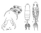 issued from : Mori T. in The Pelagic Copepoda from the neighbouring waters of Japan. S. Shirai ed., Tokyo, 1964. [Pl.43, Figs.9-12]. Male: 9, P5; 10, dorsal view. Female (from 38°17'0''N, 141°42'0''E): 11, dorsal view; 12, P5. Nota female: - Head with lateral hooks. - Rostrum symmetrical. - Last thoracic segment symmetrical; posterior angles produced into pointed projections. - Urosme 3-segmented. - Genital segment swelled at the right side. - Anal segment very short and fused with caudal rami. - Caudal rami symmetrical. - P5 nearly symmetrical; exopodite curved and furnished with 3 outer marginal soines ; apex terminates into a simple spine; apex of the endopodite furnished with many spinules. Nota male: - Lateral angles of the last thoracic segment pointed and asymmetricak; righy angle longer than the left. - Urosome 5-segmented. - 1st urosomal segment with 1 spine on the right ventral side. - Grasping A1 resembles that of L. minuta but the spine on the 22nd segment more slender than that of the latter. - P5 asymmetrical; terùinal segment of the left leg with 3 spines and 2 setae; the thumb and terminal claw of the forceps trlatively slender; 1st segment of exopodite of the right leg with a broad tooth-like process on the external margin.
|
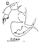
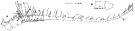 issued from : S. Ohtsuka & T. Onbé in Mar. Biol., 1991, 111. [p.217, Fig.2, A-B]. Female (from Bungo Channel: Inland Sea of Japan), A, right A1 (S = seta with setules, aesthetascs are covered with dots); B, basal two segments of left A1 (setae and aesthetascs omitted). Scale bar = 0.2 mm.
|
 issued from : S. Ohtsuka & T. Onbé in Mar. Biol., 1991, 111. [p.217, Fig.2, A]. Female (from Bungo Channel: Inland Sea of Japan): A, Md (cutting edge). Scale bar = 0.01 mm.
|
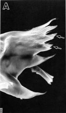
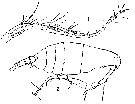
 Issued from : K.A. Brodskii in Calanoida of the Far Eastern Seas and Polar Basin of the USSR. Opred. Fauna SSSR, 1950, 35 (Israel Program for Scientific Translations, Jerusalem, 1967) [p.412, Fig.292]. Female (from Sea of Japan): habitus (dorsal); forehead (lateral); S5, P5; S5 variant, P5(variant form); posterior corners of the last thoracic segment and urosome (dorsal; another specimen). Male (from Sea of Japan): habitus (dorsal); S5, P5 (Ri = right leg). Eye pigment dark blue, the blue pigment of the 3 rd larger ventral eye is visible through the transparent head region.
|
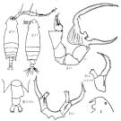
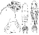 issued from : T. Mori in The pelagic Copepoda from the neighbouring waters of Japan, 1937 (2nd edit., 1964). [Pl.43, Figs.9-12]. Female: 11, habitus (dorsal); 12, P5. Male: 9, P5; 10, habitus (dorsal).
|
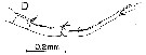
 issued from : T. Mori in The pelagic Copepoda from the neighbouring waters of Japan, 1937 (2nd edit., 1964). [Pl.44, Figs.1-2]. Male: 1, right A1; 2, habitus (lateral).
|
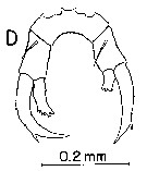
 issued from : A. Fleminger, B.H.R. Othman & J.G. Greenwood in J. Plankton Res., 1982, 4 (2). [Fig.4, I-J). Female: I-J, posterior part of prosome and urosome (lateral and dorsal views).
|
 issued from : A. Fleminger, B.H.R. Othman & J.G. Greenwood in J. Plankton Res., 1982, 4 (2). [Fig.5, D). Female: D, P5 (posterior view).
|
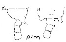 issued from : A. Fleminger, B.H.R. Othman & J.G. Greenwood in J. Plankton Res., 1982, 4 (2). [Fig.6, G-H). Male: G-H, posterior part of prosome and urosomal segments 1 to 3 (dorsal and lateral, rspectively).
|
 issued from : A. Fleminger, B.H.R. Othman & J.G. Greenwood in J. Plankton Res., 1982, 4 (2). [Fig.7, D). Male: D, P5 (posterior view)
|
 issued from : A. Fleminger, B.H.R. Othman & J.G. Greenwood in J. Plankton Res., 1982, 4 (2). [Fig.8, D). Male: D, right A1 (dorsal view).
| | | | | Ref. compl.: | | | Yamazi, 1958 (p.152, Rem.); Anraku & Azeta, 1965 (p.13, Table 2, fish predator); Fleminger, 1986 (p.84, figs. 7, 8, Rem.: geographic vs Wallace's Line); Mizushima, 1990 (fig.2, 3, tab.2); Hirakawa & al., 1990 (tab.3); Hattori, 1991 (tab.1, Appendix); Kotani & al., 1996 (tab.2); Park & Choi, 1997 (Appendix); Ueda & al., 2000 (tab.1); Lan & al., 2004 (p.332, tab.1); Ohtsuka & al., 2008 (p.115, Table 5); Maiphae & Sa-ardrit, 2011 (p.641, Table 2, 3, Rem.); Ohtsuka & Nishida, 2017 (p.565, 578, Table 22.1) | | | | NZ: | 4 | | |
|
Carte de distribution de Labidocera japonica par zones géographiques
|
| | | | | | | Loc: | | | Amur & Poseta Bay, S Kuril Is., Japan Sea, Japan, Honshu: Suruga Bay, Cape of Kinhazan, Nagazaki, Tanabe Bay, off Sanriku, S Korea, China Seas (East China Sea), N Taiwan, G. of Thailand | | | | N: | 19 | | | | Lg.: | | | (22) F: 1,93-1,74; M: 1,94-1,47; (91) F: about 1,93; M: about 1,86; (120) F: 2,06-1,81; M: 1,91-1,67; (269) F: 1,99-1,82; M: 1,84-1,61; {F: 1,74-2,06; M: 1,47-1,94} | | | | Rem.: | inshore
Voir aussi les remarques en anglais | | | Dernière mise à jour : 11/05/2019 | |
|
|
 Toute utilisation de ce site pour une publication sera mentionnée avec la référence suivante : Toute utilisation de ce site pour une publication sera mentionnée avec la référence suivante :
Razouls C., Desreumaux N., Kouwenberg J. et de Bovée F., 2005-2025. - Biodiversité des Copépodes planctoniques marins (morphologie, répartition géographique et données biologiques). Sorbonne Université, CNRS. Disponible sur http://copepodes.obs-banyuls.fr [Accédé le 29 août 2025] © copyright 2005-2025 Sorbonne Université, CNRS
|
|
|
|


;)
;)
;)
;)
;)
;)
;)
;)

