|
|
 |
Fiche d'espèce de Copépode |
|
|
Calanoida ( Ordre ) |
|
|
|
Diaptomoidea ( Superfamille ) |
|
|
|
Pontellidae ( Famille ) |
|
|
|
Labidocera ( Genre ) |
|
|
| |
Labidocera pavo Giesbrecht, 1889 (F,M) | |
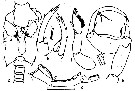
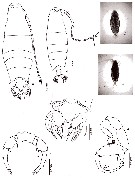
| | | | | | | Ref.: | | | Giesbrecht, 1892 (p.446, 460, 773, figs.F); Giesbrecht & Schmeil, 1898 (p.138, Rem. F); Thompson & Scott, 1903 (p.235, 251); Sewell, 1914 a (p.234, Rem.M, figs.M); 1924 (p.789); Mori, 1932 a (p.171, figs.M); 1937 (1964) (p.92, figs.F,M); Sewell, 1932 (p.365, figs.F,M, juv., Rem.); C.B. Wilson, 1950 (p.248, fig.F); Brodsky, 1950 (1967) (p.409, figs.F,M); Krishnaswamy, 1953 (p.131, fig.F); Kasturirangan, 1963 (p.50, 51, figs.F,M); Sherman, 1964 (p.481); Tanaka, 1964 c (p.255, Rem.F,M); Chen & Zhang, 1965 (p.98, figs.F,M); Saraswathy, 1966 (1967) (p.82); Goswami & Goswami, 1974 (p.109, fig. caryotypes); Silas & Pillai, 1973 (1976) (p.804, Rem., figs.F,M); Greenwood & Othman, 1979 (p.237: Rem.F,M); Lakkis, 1984 (p.293, Rem., figs.F,M); Chihara & Murano, 1997 (p.868, Pl.148,150: F,M); Barthélémy & al., 1998 (p.721, genital area); Barthélémy, 1999 a (p.9, Fig.7, G-H); Mulyadi, 2002 (p.79, figs.F,M, Rem.); Conway & al., 2003 (p.133, figs.F,M, Rem.).; Ferrari & Ueda, 2005 (p.341, figs.juv, F,M); Othman & Toda (2006, p.312, figs.F,M); Vives & Shmeleva, 2007 (p.509, figs.F, Rem.); Phukham, 2008 (p.82, figs.F,M); Abo-Taleb, 2019 (p.367, 369: Key F, M, Table 2, figs.F, M). | 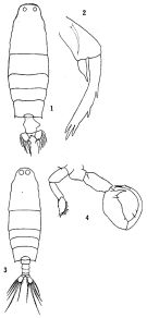 issued from: Q.-c. Chen & S.-z. Zhang in Studia Marina Sinica, 1965, 7. [Pl.41, 1-4]. Female (from E China Sea): 1, habitus (dorsal); 2, left P5 (anterior). Male: 3, habitus (dorsal); 4, P5 (posterior).
|
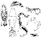
 Issued from : K.A. Brodskii in Calanoida of the Far Eastern Seas and Polar Basin of the USSR. Opred. Fauna SSSR, 1950, 35 (Israel Program for Scientific Translations, Jerusalem, 1967) [p.409, Fig.290]. Female (from NW Pacif.): habitus (dorsal); ADo, A abdomen (dorsal); S5, P5. Male: habitus (dorsal); S5, P5 (Le = left leg).
|
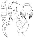
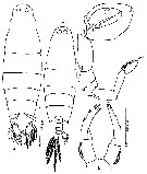
 Issued from : R.B.S. Sewell in Spolia Zeylanica, 1914, 9. [Pl.XXI, Figs.1-3]. Male (from Gulf of Mannar): 1, habitus (dorsal); 2, right A1; 3, P5. Nota: Head and 1st thoracic segment fused, 4th and 5th fused; there is a well-marked groove across the dorsal aspect of the \\\"neck\\\". Head with a pair of large eye lenses, and the ventral lens well-developed. Rostrum bifid, composed of 2 slender spines, no rostral lens. Posterior thoracic margin rounded, and armed with a small spine. Proportional lengths of urosomites and furca 27:32:32:16:9:38. Left A1 22-segmented (segments 7-8 fused, 7-8 partially fused). Right A1 19-segmented, modified to form a grasping organ; segments 11, 12, 13, each bear 1 minute spine on the anterior surface; the 17th segment bears a curved lamellar plate; the 18th segment is armed with a lamellar plate carrying a number of fine acicular spines; the 19th segment carries a raised tooth-plate but only reaching about ¾ the length of the segment (not beyond the distal segment as in L. detruncata. P5: right leg with the proximal process is not so stout as L. detruncata; left leg with the spines on the distal segment somewhat differently arranged that L. detruncata, the 1st spine arises from the middle of the external margin and the remaining three from the extreme tip, the inner margin is beset with fine hairs.
|
 Issued from : R.B.S. Sewell in Mem. Indian Mus., 1932, X (continued). [p.365, Fig.121]. Female (from N Indian): a, urosome (from Sri lanka Pearl Banks); b, idem (from Nicobar Is.); c, P5. Male: grasping A1.
|
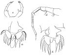
 issued from : E.G. Silas & P.P. Pillai in J. mar. biol. Ass. India, 1973 (1976), 15 (2). [p.805, Fig.14]. Female (from Indian Ocean): a, urosome (dorsal); b, P5. Nota: lateral cephalic hooks. Dorsal eye lenses moderately developed and placed apart. Urosome 2-segmented. Genital segment produced into a conical lobe with rounded tip on its right side; posterior margin is produced ventrally into a lobe resembling a bottle, which extends to middle of caudal rami. Caudal rami broad, arranged perpendicular to the urosome, nearly symmetrical. Male: c, urosome (dorsal, part); d, right A1 (geniculate part); e, right P5; f, left P5.
Scale as in Calanopia minor.
|
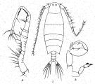
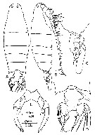
 issued from : T. Mori in The pelagic Copepoda from the neighbouring waters of Japan, 1937 (2nd edit., 1964). [Pl.41, Figs.6-12]. Female: 8, habitus (dporsal); 11, P5. Male: 6, A2; 7, right A1; 9, habitus (dorsal); 10, P5; 12, P1.
|
 issued from : R.-M. Barthélémy in Thèse Doct. Univ. Provence (Aix-Marseille I), 1999. [Fig.7, G-H]. Female (from Banyuls, NW Mediterranean Sea): G, external ventral view urosomal segments; H, genital area (left side double-somite posterior margin);. Note the duck beak-shape of the genital area covering an inferior lip guttering-like (arrows).
|
 issued from : J.G. Greenwood & B.H.R. Othman in J. Plankton Res., 1979, 1 (1). [p.237] Characters in L. pavo compared with other species within the super-species detruncata (see L. cervi, L. farrani, L. caudata, L. detruncata, L. bataviae, L. madurae and L. sinolobata, with reference sources).
|
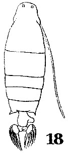 Issued from : W. Giesbrecht in Systematik und Faunistik der Pelagischen Copepoden des Golfes von Neapel und der angrenzenden Meeres-Abschnitte. Fauna Flora Golf. Neapel, 1892. Atlas von 54 Tafeln. [Taf.41 , Fig.18 ]. Female: 18, habitus (dorsal).
|
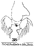 Issued from : W. Giesbrecht in Systematik und Faunistik der Pelagischen Copepoden des Golfes von Neapel und der angrenzenden Meeres-Abschnitte. Fauna Flora Golf. Neapel, 1892. Atlas von 54 Tafeln. [Taf.41 , Fig.38 ]. Female: 38, urosome (ventral).
|
 issued from : Mulyadi in Treubia, 2002, 32; [p.81, Fig.26]. Female (from Indonesian waters): a, habitus (dorsal); b, P5. Male: c, habitus (dorsal); d, P5.
|
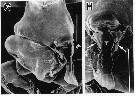
 issued from : B.H.R. Othman & T. Toda in Coastal Mar. Sc., 2006, 30 (1). [p.312, Fig.12]. Female (from Sister's Island, Singapore): A-B, habitus (dorsal and lateral, respectively); C-D, posterior part of prosome and urosome (lateral and dorsal, respectively); E, P5. Nota: Prosome to urosome length ratio 4.95 : 1. - Cephalon without lateral hooks, dorsal eye lenses moderate. - Prosome produced into symmetrical pointed processes. - Urosome 2-segmented. - Genital segment asymmetrical, right margin with 1 conical lobe, left margin with rounded knob-like process. - P5 asymmetrical , right leg exopod with 2 outer spinules and 3 unequal spines at apex, middle one longest; endopod long and produced at apex; left leg exopod with 2 outer spinules and 3 subequal spines at apex, endopod short and rounded.
|
 issued from : B.H.R. Othman & T. Toda in Coastal Mar. Sc., 2006, 30 (1). [p.312, Fig.13]. Male (from Sister's Island, Singapore): A-B, habitus (dorsal and lateral, respectively); C, posterior part of prosome and urosome (dorsal); D, P5. Nota: Prosome to urosome length ratio 4.03 : 1 - Body shape similar to female except dorsal eys lenses larger and contiguous. - Urosome 5-segmented, naked. - Right A1 geniculate, segment 17 rounded anteriorly into arched ridge, slightly sculptured with irregular ribbing, segment 18 with row of denticles on anterior margin which are closely placed, fused segments 19-21 with row of villiform denticles on anterior margin, extending to 3/4 length of segment, segments 24-25 completely fused. - P5 chelate; right leg exopodal segment 1 with well developed thumb, outer margin between thumb and distal end of exopodal segment 1 with 1 seta; exopodal segment 2 elongared, curved with 1 blunt conical projection along inner margin at 1/3 distance from base, inner margin with 2 mid-marginal setae and 1 terminal seta; left leg exopodal segment 1 with distolateral spine; exopodal segment 2 with 1 outer spine and 3 subequal spines at apex, all turned inwards, inner margin of segment irregularly lobular and hirsute
|
 issued from : N. Phukham in Species diversity of calanoid copepods in Thai waters, Andaman Sea (Master of Science, Univ. Bangkok). 2008. [p.163, Fig.37]. Female (from W Malay Peninsula): a, habitus (dorsal); b, urosome (dorsal); c, P5. Male: d, habitus (dorsal); e, P5. Body length after the drawings: F = 2.447 mm; M = 2.006 mm.
|
issued from : R.-M. Barthélémy in Thèse Doct. Univ. Provence (Aix-Marseille I), 1999. [Fig.7, G-H]. Female (from Banyuls, NW Mediterranean Sea): G, external ventral view urosomal segments; H, genital area (left side double-somite posterior margin);. Note the duck beak-shape of the genital area covering an inferior lip guttering-like (arrows).
|
 issued from : R.-M. Barthélémy, C. Cuoc, D. Defaye, M. Brunet & J. Mazza in Phil. Trans. R. Soc. Lond., B, 1998, 353. [p.733, Fig.63]. Schematic representation of external genital area in the species studied; Dashed line, limit of the anterior pad and lateral thickenings; shading, posterior pad; dots, genital operculum.
|
 Issued from : H.A. Abo-Taleb in Egyptian J. Aqua. Res., 2019, 45. [p.369]. Key to species of Labidocera in the Red Sea. Female: 1 - Anterior end of cephalosome rounded without median crest. 2 - Lateral hooks absent. 3 - Urosome 2-segmented. 4 - Caudal rami symmetrical. 5 - P5exopod tip is 3 unequal terminal spines. 6 - P5 endopods asymmetrical; left is rounded, while right is longer with tapered end. Male: 1 - Anterior end of cephalosome with a median spine-like crest in dorsal view. 2 - Last posterolateralm end of metasome almost symmetrical. 3 - Left P5 terminal segment with 3 terminal spines. 4 - Right P5 chela with exopod 1 has short and curved thumb with seta on inner base.
| | | | | Ref. compl.: | | | Gurney, 1927 (p.154); Sewell, 1948 (p.324); Wada, 1953 (p.100, swarms); Chiba & al., 1957 (p.308); 1957 a (p.11); Yamazi, 1958 (p.152, Rem.); Patel, 1975 (p.660); Madhu Pratap & al., 1977 (p.138, Table 3: abundance vs. stations); Grice & Gibson, 1978 (p.23, tab.8); Omori & Hammer, 1982 (p.193, swarms); Ueda & al., 1983 (p.165, Table 1, 2, swarms); Dessier, 1983 (p.89, Tableau 1, Rem., %); Binet, 1984 (tab.3); 1985 (p.85, tab.3); Madhupratap & Haridas, 1986 (p.105, tab.1); Lakkis, 1990 (p.63); Othman & al., 1990 (p.565, Rem.); Dai & al., 1991 (tab.1); Yoo, 1991 (tab.1); Shih & Young, 1995 (p.72); Ramaiah & al., 1996 (p.3); Kotani & al., 1996 (tab.2); Park & Choi, 1997 (Appendix); Sharaf & Al-Ghais, 1997 (tab.1); Ramaiah & Nair, 1997 (tab.1); El-Serehy, 1999 (p.172, Table 1, occurrence); Mauchline, 1998 (tab.8, 78); Padmavati & al., 1998 (p.347); Lenz & al., 2000 (p.338); Osore & al., 2003 (p.69); Lan & al., 2004 (p.332, tab.1); Rakhesh & al., 2006 (p.93, Table 2, spatial distribution); Kovalev, 2006 (p.67: Lessepsian migration); Dur & al., 2007 (p.197, Table IV); Rakhesh & al., 2008 (p.154, Table 5: abundance vs stations); Ayon & al., 2008 (p.238, Table 4: Peruvian samples); Ohtsuka & al., 2008 (p.115, Table 5); Zenetos & al., 2010 (p.397); Shanthi & Ramanibai, 2011 (p.132, Table 1); Maiphae & Sa-ardrit, 2011 (p.641, Table 2, 3, Rem.); Moon & al., 2012 (p.1, Table 1); Varadharajan & Soundarapandian, 2013 (p.2: occurrence vs stations); Jagadeesan & al., 2013 (p.27, Table 3, seasonal distribution); Anjusha & al., 2013 (p.40, Table 3: as pava, abundance & feeding behavior); Terbiyik Kurt & Polat, 2013 (p.1163, Table 2); El Arraj & al., 2017 (p.272, table 2) ? (see remarks); Abo-Taleb & Gharib, 2018 (p.139, Table 5, p.145: occurrence %). | | | | NZ: | 10 + 1 douteuse | | |
|
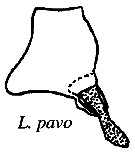
Carte de distribution de Labidocera pavo par zones géographiques
|
| | | | | | | | | | | |  Issued from : H.A. Abo-Taleb in Egyptian J. Aquat. Res., 2019, 45. [p.371, Table 2]. Issued from : H.A. Abo-Taleb in Egyptian J. Aquat. Res., 2019, 45. [p.371, Table 2].
Average values with standard deviation and ranges (individuals/m3) in the different studied habitats (SG: seagrass, CR: coral reef, SSL: sheltered lagoon, and OW: open sea.
See Labidocera euchaeta fig.1: sampling sites and Table 1: measured physical environmental parameters in the northern Red Sea. |
| | | | Loc: | | | E Medit. (Lebanon, Turkey: Iskenderun Bay), Suez Canal, Suez Bay, Hurghada, Red Sea, Arabian Sea, Arabian Gulf, UAE coast, Sri Lanka, Madagascar, Agatti Is., Indian, India (Saurashtra coast, Bombay, Madras, Gulf of Mannar, Palk Bay, Pointcalimere-Manamelkudi, Burhabalanga estuary, Kakinada Bay, Chilka Lake), Bay of Bengal, Andaman Is., W Malay Peninsula, Straits of Malacca (Singapore), Sunda Strait, G. of Thailand, S Java, Flores Sea, Banda Sea, Philippines, Viet-Nam (Cauda Bay), China Seas (Yellow Sea, East China Sea, South China Sea), Taiwan (SW, N), Xiamen Harbour, Korea, Muan Bay, Japan Sea, Japan, Tanabe Bay, Sagami Bay, E Siberia, S Kuril Is., Australia (G. of Carpentaria), New Caledonia, New Hebrides, Tonga Is., Bikini Is., Hawaii, Peru (in IMARPE unpublished) | | | | N: | 79 | | | | Lg.: | | | (22) F: 2,4-1,92; M: 2-1,61; (44) F: 2,304; M: 1,885; (46) F: 2,12; (82) M: 2,2; (120) F: 2,34-2,22; M: 1,92-1,7; (151) M: 1,94; (256) F: 2,52-1,98; M: 2,06-1,79; (290) F: 2,1-2,15; M: 1,7-1,75; (334) F: 1,9; M: 1,9; (351) F: 1,77; M: 1,5; (530) F: 2,5; M: 2; (795) F: 1,7; M: 1,45; (991) F: 1,92-2,52; M: 1,61-2,06; (1086) F: 2,1-2,29; M: 1,6-2,31; (1087) F: 1,95-2,35; M: 1,75-2; {F: 1,70-2,52; M: 1,45-2,31} | | | | Rem.: | inshore, saumâtre.
Cette espèce aurait une origine lessepsienne en Méditerranée. Sa présence sur la côte marocaine nécessite confirmation.
Voir aussi les remarques en anglais | | | Dernière mise à jour : 24/10/2022 | |
|
|
 Toute utilisation de ce site pour une publication sera mentionnée avec la référence suivante : Toute utilisation de ce site pour une publication sera mentionnée avec la référence suivante :
Razouls C., Desreumaux N., Kouwenberg J. et de Bovée F., 2005-2026. - Biodiversité des Copépodes planctoniques marins (morphologie, répartition géographique et données biologiques). Sorbonne Université, CNRS. Disponible sur http://copepodes.obs-banyuls.fr [Accédé le 21 mars 2026] © copyright 2005-2026 Sorbonne Université, CNRS
|
|
|
|


;)
;)
;)
;)
;)
;)
;)
;)
;)
;)
;)
;)
;)
;)
;)
;)

