|
|
 |
Fiche d'espèce de Copépode |
|
|
Calanoida ( Ordre ) |
|
|
|
Diaptomoidea ( Superfamille ) |
|
|
|
Pontellidae ( Famille ) |
|
|
|
Labidocera ( Genre ) |
|
|
| |
Labidocera pseudacuta Silas & Pillai, 1967 (F,M) | |
| | | | | | | Syn.: | Labidocera acutum : Giesbrecht, 1892 (part., p.445, 458, Pl.41, fig.29: F) | | | | Ref.: | | | Silas & Pillai, 1967 (p.347, figs.F,M); 1973 (1976) (p.816); Goswami & Goswami, 1979 a (p.259, figs. caryotypes) | 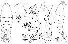 issued from : E.G. Silas & P. Parameswaran Pillai J. Mar. biol. Ass. India, (1967) 1969, 9 (2). [p.350, Text-Fig.1, a-g].
Female (from 09°28'N, 54°52'E): a-b, habitus (dorsal and lateral, respectively); c, right Md (cutting edge of gnathobase); d, part of mandibular gnathobase enlarged to show nature of flagellum; e, Mx1; f, Mxp; g, P5.
Nota : Dorsal cuticular lenses moderately large, separated by about 1.5 times diameter of lens. (in L. acuta, lenses are wider apart and separated by about twice lens diameter).
Lateral hooks absent.
Cephalosome and 1st pediger segment separate, 4th and 5th indistinct ; 4th metasomal segment with 2 short dorsolateral spine-like processes symmetrically placed on either half of segment closer to mid-dorsal line.
Urosome 3-segmented ; its length about 4.3 times in total length.
Genital segment greatly enlarged, its mid-dorsal length distinctly more than half length of urosome ; a well developed right postero-lateral lobe ending in a rudimentary spine-like process present (in L. acuta , urosome 4 times in total length ; genital segment less than half length of urosome ; a stout postero-lateral conical process present) ; genital pores ventro-lateral, situated at middle of segment, but more towards left half (in L. acuta, genital pores mid-ventral, situated in anterior half of segment) ; A short stout and pointed spine present mid-ventrally at posterior margin of genital segment.
2nd urosomal segment distinctly broader than long, its length equalling half length of genital segment (in L. acuta, 2nd urosomal segment about as long as broad, its length equalling ¾ length of genital segment).
Anal segment short, hardly 1/3 length of genital segment, anal lamina rudimentary (in L. acuta, anal segment well developed, and more than half lenth of genital segment).
Caudal rami asymmetrical, right ramus being slightly larger, latter 1.5 to 1.75 times longer than broad and more than ¼ length of urosome (in L. acuta, right caudal ramus 1.6 to 1.8 times longer than broad). Assymetry and size differences in caudal rami of L. pseudacuta and L. acuta. Variations seen typically in enlarged basal part of 3rd and 4th caudal setae from outer margin ; setae of right caudal ramus show more pronounced modifications.
Md : gnathobase with 5 teeth, all bicuspidate, toothed cutting edge with rows of stout spine-like setae in grooves between 3rd, 4th, and 5th teeth in addition to inner side of base of 5th, flagellum moderately long and bent in its distal half.
P5 : exopods asymmetrical, right slightly longer ; endopods almost symmetrical, each terminating in 2 subequal prongs. (In L. acuta, P5 markedly asymmetrical, left leg being relatively stouter and longer ; 2 subterminal prongs on terminal process of exopods conspicuously long, length of longer prongs being about 12.5 per cent length of exopod ; asymmetry in endopods more marked, left being slender and longer ; length of latter about 2.4 times in length of left exopod and 2.3 times in length of right exopod ; right endopod 2.7 and 2.5 times in lengths of left and right exopods respectively).
|
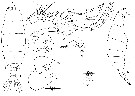
 issued from : E.G. Silas & P. Parameswaran Pillai J. Mar. biol. Ass. India, (1967) 1969, 9 (2). [p.351, Text-Fig.2, a-e, g-i, l, m].
Female: a-e, variability in caudal setae abd caudal rami in five adults; g-i, urosome with spefmatophore (dorsal, ventral and lateral views respectively); m, rostrum.
Male: l, rostrum.
Nota : Spermatophore with coupler ensheathing almost completely genital segment, 2nd urosomal segment and part of anal segment, and expanded as prominent wing-like expansions laterally on both sides of genital segment ; expansion on left side extending to below posterior lobe of last metasomal segment ; sheat above genital segment modified into an irregular elevated ridge. Neck of spermatophore sac short ; sac ventro-lateral to urosome extending to hardly 1/3 its length beyond tip of caudal rami ; sac broadest at its mid-length, its greatest width being about 1/6 its length including neck portion (in L. acuta, coupler sheat less extensive, covering only part of genital segment and 2nd urosomal segment ; slight lateral expansions do not reach to beneath posterior lobes of last metasomal segment ; dorsal surface of coupler sheath smooth ; spermatophore sac cylindrical, its width about 1/8 of its length including neck portion ; sac extends to about 1/6 its length beyond tip of caudal rami.
|
 issued from : E.G. Silas & P. Parameswaran Pillai J. Mar. biol. Ass. India, (1967) 1969, 9 (2). [p.349].
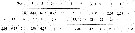
Female: Proportions of the segments of A1.
Nota: A1 23-segmented. Slight differences in relative lengths of segments 3, 6, 7 and 19 are noticeable between the two species L. pseudacuta and L. acuta.
|
 issued from : E.G. Silas & P. Parameswaran Pillai J. Mar. biol. Ass. India, (1967) 1969, 9 (2). [p.356, Text-Fig.4, a, b].
Female: a, urosome (ventral).
Male: b, urosome (dorsal).
|
 issued from : E.G. Silas & P. Parameswaran Pillai J. Mar. biol. Ass. India, (1967) 1969, 9 (2). [p.355, Text-Fig.3, a-f].
Male: a-b, habitus (dorsal and lateral, respectively); c, right A1 with parts of plates enlarged; d, P5; e, terminal part of left P5; f, chela of right P5.
Nota : Dorsal cuticular lenses conspicuously large anf placed close together.
4th and 5th metasomal segments indistinct ; posterior corners modified into an acutely pointed lobe on left side, tip of latter extending to opposite middle of 3rd urosomal segment an douter and inner margins of process provided with few rudimentary hairs (in L. acuta, curved process of right side of last metasomal segment short, not extending beyond 2nd urosomal segment.
Urosome 5-segmented. Genital segment distinctly broader than long, asymmetrical, right postero-lateral margin bearing a conspicuous stout conical process (more than half length of genital segment) which bears on its outer margin a short spine (in L. acuta, right posterior corner of genital segment bears a short spine inner to which is present a conical process which is less than half length of genital segment) ; 2nd urosomal segment 2/3 as long as wide ; 3rd segment longest, as long as wide, and about ¼ longer than 2nd segment ; 4th urosomal segment short , hardly 40 % length of 3rd segment ; anal segment short, bifurcate posteriorly (in L. acuta, 2nd and 3rd urosomal segments of more or less same size and each about ¾ as long as wide ; 4th segment more than 50 % length of 3rd segment).
Caudal rami asymmetrical, right ramus being larger. Caudal setae well developed and spread out ; thickening of basal part of setae as in female is not present.
|
 issued from : E.G. Silas & P. Parameswaran Pillai J. Mar. biol. Ass. India, (1967) 1969, 9 (2). [p.353].
Male: proportions of the segments of the geniculate A1.
Nota : Geniculate A1 23-segmented (segments 7-9, 17-18 and 19-20, indistinct ; presence of an enlarged spine-like process at the distal anterior margin of segment 21, this process extends to 2/3 the length of the distal segment (23th), and has along its outer margin fine backwardly directed serrations (in L. acuta, this spine-like process is shorter and hardly reaches base of segment 23.
|
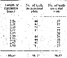 issued from : E.G. Silas & P. Parameswaran Pillai J. Mar. biol. Ass. India, (1967) 1969, 9 (2). [p.353].
Male: Frequency of occurrence of teeth on plates of geniculate A1.
| | | | | Ref. compl.: | | | Madhupratap & Haridas, 1986 (p.105, tab.1); Mauchline, 1998 (tab.8) | | | | NZ: | 1 | | |
|
Carte de distribution de Labidocera pseudacuta par zones géographiques
|
| | | | | | 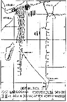 issued from : E.G. Silas & P. Parameswaran Pillai J. Mar. biol. Ass. India, (1967) 1969, 9 (2). [p.359, Text-Fig.7]. issued from : E.G. Silas & P. Parameswaran Pillai J. Mar. biol. Ass. India, (1967) 1969, 9 (2). [p.359, Text-Fig.7].
Map showing the distribution of L. pseudacuta ontained during the Vth cruise of ANTON BRUUN (cruise track indicated). Areas in the Arabian Sea and Central Indian Ocean from where material believed to be L. acuta obtained during R.V. VITYAZ cruise as shown by Voronina (1962) are indicated |
| | | | Loc: | | | Arabian Sea, NW Indian, Laccadive Is. | | | | N: | 2 | | | | Lg.: | | | (457) F: 3,54-3,26; M: 3,37-3,22; {F: 3,26-3,54; M: 3,22-3,37} | | | | Rem.: | océanique
Voir aussi les remarques en anglais | | | Dernière mise à jour : 06/01/2015 | |
|
|
 Toute utilisation de ce site pour une publication sera mentionnée avec la référence suivante : Toute utilisation de ce site pour une publication sera mentionnée avec la référence suivante :
Razouls C., Desreumaux N., Kouwenberg J. et de Bovée F., 2005-2025. - Biodiversité des Copépodes planctoniques marins (morphologie, répartition géographique et données biologiques). Sorbonne Université, CNRS. Disponible sur http://copepodes.obs-banyuls.fr [Accédé le 11 décembre 2025] © copyright 2005-2025 Sorbonne Université, CNRS
|
|
|
|


;)
;)
;)
;)
;)

;)
