|
|
 |
Fiche d'espèce de Copépode |
|
|
Calanoida ( Ordre ) |
|
|
|
Epacteriscoidea ( Superfamille ) |
|
|
|
Ridgewayiidae ( Famille ) |
|
|
|
Ridgewayia ( Genre ) |
|
|
| |
Ridgewayia boxshalli Barthélémy, Ohtsuka & Cuoc, 1998 (F,M) | |
| | | | | | | Ref.: | | | Barthélémy, Ohtsuka & Cuoc, 1998 (p.1303, figs.F,M) | 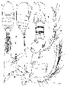 issued from : R. Barthélémy, S. Ohtsuka S. & C.Cuoc in J. Nat. Hist., 1998, 32. [p.1305, Fig.1]. Female (from 28°11.6'N, 129°13.7'E): A-B, habitus (dorsal and lateral, respectively); C, forehead (frontal view); D, urosome (ventral); E, A1: segments 1-17 (I-XIX); F, A1: segments 18-25 (XX-XXVIII); G, A2 (distal endopod segment omitted); H, A2: distal segment of endopod); I, Md. Scale bars in micons. Nota : Head and 1st pedigerous somite separate, 4th and 5th separate . Naupliar eye present. Rostrum sharply pointed witout filament. A1 25-segmented, nearly reaching end of prosome. A2 : coxa with 1 plumose seta ; basis bearing 2 setae of almost equal length ; exopod indistinctly 8-segmented, setal formule 1, 1, 1, 1, 1, 1, 1, 4 ; endopod 2-segmented ; 1st segment with subterminal setae, 2nd segment with 9 middle and 7 terminal setae. Md : gnathobase bearing 8 teeth, 2 dorsalmost of which sharply pointed, and a tuft of minute spinules near base of palp ; basis with 4 inner setae, proximal of which thicker than others and plumose, protruded at inner distal corner ; exopod 5-segmented, setal formula 1, 1, 1, 1, 2 ; endopod 2-segmented, 1st segment with 4 distal inner setae, 2nd segment bearing 11 setae. Urosome 4-segmented, 2nd and 3rd urosomal somites flanged with minute spinules along posterior margin ; 3rd urosomal somite produced dorso-medially into bifurcate tip covering anal somites ; anal somite almost telescoped into preceding somite.; Genital double-somite swollen midlaterally, bearing pair of long hair-sensilla ventrolaterally ; genital operculum located ventromedially. Caudal rami symmetrical, seta I minute, located at anterior third, seta II spiniform with fine setule subterminally, seta III-VI plumose, seta VII located posterodorsally.
|
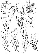 issued from : R. Barthélémy, S. Ohtsuka S. & C.Cuoc in J. Nat. Hist., 1998, 32. [p.1306, Fig.2]. Female: A, praecoxal, coxal and basal endites of Mx1; B, Mx1 (parts of setae omitted); C, Mx2; D, Mxp; E-H, P1 to P4 (anterior views); I, P5 (anterior). Scale bars in microns. Mx1 : praecoxal arthrite bearing 6 spiniform, 2 slender, 3 spinulose and 4 posterior setae ; coxa with 9 setae on epipodite, 2 proximal of which shorter than others, and 5 setae on endite ; basis with 1 short seta on exite, 4 setae on 1st endite and 5 setae on 2nd endite ; exopod unisegmented, bearing 11 setae along outer margin ; endopod 2-segmented, setal formula 4 + 4, 7 ; Mx2 : 1st praecoxal endite bearing 5 spinulose and 1 short spiniform setae ; 2nd oraecoxal and 2 coxal endites each with 3 spinulose setae ; basis with 1 heavily sclerotized spiniform and 3 spinulose setae ; endopod indistinctly 4-segmented, setal formula 3, 2, 2, 3. Mxp : coalescent praecoxa and coxa ; 1st to 4th syncoxal endites bearing 1, 2, 4 and 3 setae, respectively ; basis with 3 setae ; 1st endopod segment incorporated into basis, with 2 setae ; 2nd to 6th endopod segments carrying 4, 4, 3, 3+1 and 4 setae, respectively ; 2nd and 3rd endopod segments each with spinular row, 2nd segment bearing round process with minute prominences near base of setae. P1 with unarmed coxa ; basis bearing curved inner basal seta and an outer tubercule with spinules on the endopod (von Vaupel Klein organ).
|
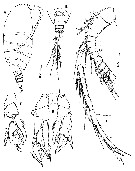 issued from : R. Barthélémy, S. Ohtsuka S. & C.Cuoc in J. Nat. Hist., 1998, 32. [p.1307, Fig.3]. Male: A, habitus (lateral); B, prosomal end and urosome (dorsal); C, A1: segments 1-13 (I-XV); D, A1: segments 14-22 (XVI-XXVIII); E, P5 (anterior); F, left P5 (posterior). Scale bars in microns. Nota : Right A1 22-segmented, weakly geniculate. Urosome 5-segmented ; genital to anal somites flanged distally with minute spinules. variation : The spiniform process at the distal end of the 2nd exopod segment of left P5 was straight in a paratype (fig.E) but curved outward in another paratype (fig. F).
|
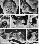 issued from : R. Barthélémy, S. Ohtsuka S. & C.Cuoc in J. Nat. Hist., 1998, 32. [p.1309, Fig.5]. Female (non-inseminated): A-B, genital double-somite (ventral and left lateral view, respectively); C, detail of the genital area. Note the cuticular field (white arrow) under the operculum (go) and the genital atrium (ga); D-E, details of the wall of the genital atrium showing two cuticular expansions (D, E, white arroweds) and the left gonoporal plate (E, gp); F-G internal dorsal views: F, genital area, egg-laying ducts (ed) and shell ducts (sd); G, detail of the left egg-laying duct. as = atrial slit; cf = cuticular fold; iml = zone of insertion of the left opercular muscle. Scale bars in microns.
|
 issued from : R. Barthélémy, S. Ohtsuka S. & C.Cuoc in J. Nat. Hist., 1998, 32. [p.1311, Table 1]. Seta and spine formulae of legs 1-5 female. Spines: Roman numerals; setae: Arabic numerals.
|
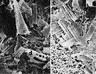 issued from : R. Barthélémy, S. Ohtsuka S. & C.Cuoc in J. Nat. Hist., 1998, 32. [p.1311, Fig.7]. Female: A, B, photomicrographs of gut contents. c: crustacean spine; d: diatom fragments. Scale bars = 0.010 mm.
| | | | | NZ: | 2 | | |
|
Carte de distribution de Ridgewayia boxshalli par zones géographiques
|
| | | | | | | Loc: | | | S Japan (Kakeroma Is and off Mage Is. : Nansei Is.) | | | | N: | 1 | | | | Lg.: | | | (156) F: 0,91-0,71; M: 0,85-0,77; {F: 0,71-0,91; M: 0,77-0,85} | | | | Rem.: | Depths: 4-39 m.
According to Barthélémy & al. (1998, p.1314) the large serrate process on the distal outer corner of the 2nd exopod segment of P1 is found in both sexes of R. boxshalli. This process seems to be unique to the genus. Althought its functions are unknown, it may be a prehensile organ to cling to substrata (algae and animals). Also according to von Vaupel Klein (1972), the organ on the basis is assumed to play a role in grooming ; the A1 are drawn across the Mxp at the bend created by the proximal segment. Perhaps the organ on P1 groom antennules together with maxillipeds because the syncoxa and basis of maxillipeds bear a structure resembling that on leg 1 : curved setae and tuft of minute spinules at the distal corner of the sybncoxa, and one or more spinular rows along the inner margin of the basis (cf. Nishida & Ohtsuka, 1997) | | | Dernière mise à jour : 10/01/2020 | |
|
|
 Toute utilisation de ce site pour une publication sera mentionnée avec la référence suivante : Toute utilisation de ce site pour une publication sera mentionnée avec la référence suivante :
Razouls C., Desreumaux N., Kouwenberg J. et de Bovée F., 2005-2026. - Biodiversité des Copépodes planctoniques marins (morphologie, répartition géographique et données biologiques). Sorbonne Université, CNRS. Disponible sur http://copepodes.obs-banyuls.fr [Accédé le 29 mars 2026] © copyright 2005-2026 Sorbonne Université, CNRS
|
|
|
|


;)
;)
;)
;)
;)

