|
|
 |
Fiche d'espèce de Copépode |
|
|
Calanoida ( Ordre ) |
|
|
|
Clausocalanoidea ( Superfamille ) |
|
|
|
Scolecitrichidae ( Famille ) |
|
|
|
Lophothrix ( Genre ) |
|
|
| |
Lophothrix frontalis Giesbrecht, 1895 (F,M) | |
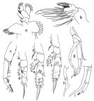
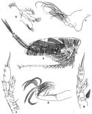
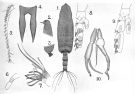
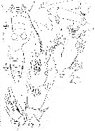
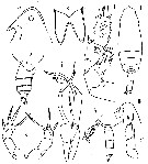
| | | | | | | Syn.: | Scolecithrix frontalis : Giesbrecht & Schmeil, 1898 (p.49, Rem.F, fig.F); Esterly, 1906 a (p.65, figs.F); in CalCOFI regional list (MDO, Nov. 2013; M. Ohman, comm. pers.) | | | | Ref.: | | | Giesbrecht, 1895 c (p.254, Descr.F, pl.2, figs.1-5,9-12:F); Farran, 1908 b (p.58); A. Scott, 1909 (p.99, figs.F,M); Wolfenden, 1911 (p.268); Sewell, 1913 (p.354); With, 1915 (p.211, figs.F,M); Lysholm & Nordgaard, 1921 (p.21); Sars, 1925 (p.162, figs.F,M); Farran, 1926 (p.267); Rose, 1929 (p.27); Sewell, 1929 (p.193, figs.F,M, Rem.: 2 forms); Rose, 1933 a (p.145, figs.F,M); Jespersen, 1934 (p.87); 1940 (36); Lysholm & al., 1945 (p.26); Sewell, 1947 (p.148, figs.F,M, Rem.); Davis, 1949 (p.40, figs.F, Rem.F,M); Brodsky, 1950 (1967) (p.245, figs.F,M, Rem.); Marques, 1953 (p.105, figs.F); Tanaka, 1961 a (p.150, figs.F,M, Rem.); Fagetti, 1962 (p.23); Paiva, 1963 (p.49, figs.F: anormalité); Vervoort, 1965 (p.59, Rem.); Owre & Foyo, 1967 (p.67, figs.F,M); Tanaka, 1969 (p.275, fig.juv.M); Silas, 1972 (p.647); Bradford, 1973 (p.144); Björnberg & al., 1981 (p.637, figs.F,M); Gardner & Szabo, 1982 (p.278, figs.F,M); Bradford & al., 1983 (p.88, Rem., figs.F,M); Park, 1983 (p.178, Redescr.F,M, figs.F,M, Rem.); Nishida & al., 1991 (p.527, midgut structure, feeding); He & al., 1992 (p.250); Chihara & Murano, 1997 (p.899, Pl.169,170: F,M); Bradford-Grieve & al., 1999 (p.881, 932, figs.F,M); Boxshall & Halsey, 2004 (p.187, 189: figs.F); Vives & Shmeleva, 2007 (p.745, figs.F,M, Rem.) | 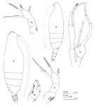 Issued from : J.M. Bradford, L. Haakonssen & J.B. Jillett in Mem. N.Z. oceanogr. Inst., 1983, 90. [p.89, Fig.51]. Female: A, habitus (lateral right side); B, endopod of P2; C, D, P5. Male: E, habitus (lateral left side); F, P5. The south-west Pacific specimens show minor differences from the original description. The fusion line of pediger segments 4 and 5 is evident dorsally in the female; the A1 is slightly shorter and some of the female specimens have 4 spines on P5. Tanaka (1961) figures a pair of P5 which has 3 spines on one leg and 4 on the other, which is very like the limb Wolfenden (1911) figures for L. quadrispinosa.
|
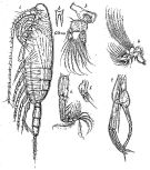
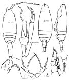
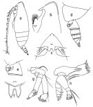 issued from : T. Park in Antarct. Res. Ser. Washington, 1983, 38 (3). [p.179, Fig.7]. Female: a, habitus (lateral left side); b, forehead (lateral); c, idem (ventral); d, last thoracic segment and urosome (lateral left side); e, genital segment (lateral left side); f, rostrum (anterior); g, A2; h, Md. Nota: 4th and 5th metasomal segment fused without a line of demarcation. Spermatheca elongate and fingerlike. Urosome about 22/100 length of prosome.
|
 issued from : T. Park in Antarct. Res. Ser. Washington, 1983, 38 (3). [p.180, Fig.8]. Female: a, Mx1; b, Mx2; c, Mxp; d, P1 (anterior); e, P2 (posterior); f, P3 (posterior); g, P4 (posterior); h, P5 (posterior).
|
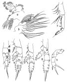
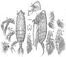
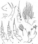
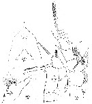
 issued from : T. Park in Antarct. Res. Ser. Washington, 1983, 38 (3). [p.182, Fig.9]. Male: a, habitus (lateral right side); b, forehead (lateral); c, idem (dorsal); d, idem (ventral); e, last thoracic segments and urosome (lateral right side); f, idem (dorsal); h, Md. Nota: 1st metasomal segment incompletely fused with cephalosome with a short line of joint laterally, 4 th and 5th separate. Urosome about 37/100 length of prosome.
|
 issued from : T. Park in Antarct. Res. Ser. Washington, 1983, 38 (3). [p.183, Fig.10]. Male: a, Mx1; b, Mx2; c, Mxp; d, P1 (anterior); e, P2 (posterior); f, P3 (posterior); g, P5 (right anterior); h, distal part of right exopod of P5 (anterior); i, distal part of left P5 (medial).
|
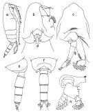
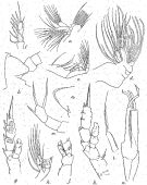
 Issued from : G.O. Sars in Résult. Camp. Scient. Prince Albert I, 69, pls.1-127 (1924). [Pl.XLVI, figs.1-7]. Male: 1, habitus (lateral left side); 2, rostrum; 3, Md; 4, Mx1; 5, Mx2; 6, Mxp; 7, P5.
|
 Issued from : G.O. Sars in Résult. Camp. Scient. Prince Albert I, 69, pls.1-127 (1924). [Pl.XLV, figs.9-21]. Female: 9, habitus (dorsal); 10, idem (lateral left side); 11, forehead (lateral); 12, rostrum (frontal view); 13, A2; 14, Md; 15, Mx1; 16, Mx2; 17, Mxp; 18, P1; 19, P2; 20, P3; 21, P5.
|
 issued from : R.B.S. Sewell in The John Murray Expedition, 1933-34, Scientific Reports, VIII (1), 1947. [p.148, Fig.37]. Female (from Arabian Sea): A, genital segment and 2nd urosomal segment in forma minor (ventral); B, proximal segments of A1 in forma minor; C, genital segment and 2nd urosomal segment in forma major; D, proximal segments of A1 in forma major. Nota: Apart from size the only valid difference between the two forms is the presence in f. minor of a row of spines on the ventro-posterior surface of the 1 st segment of the A1 near the distal margin. The number of setae in Mx1 on the various lobes: inner lobe 1 (13), inner lobe 2 (2), inner lobe 3 (4); basal segment 2 (5); endopod (3, 5); exopod (9); outer lobe (9). The number of setae on lobes of Mx2: lobe 1 (4 + 1 very small), lobe 2 (3), lobe 3 (3), lobe 4 (3), lobe 5 (3); the terminal portion bears 8 setae that are modified.
|
 issued from : R.B.S. Sewell in The John Murray Expedition, 1933-34, Scientific Reports, VIII (1), 1947. [p.150, Fig.38]. Male: A, habitus (dorso-lateral right side); B, Mx1; C, Mx2; D, Mxp; E, P2; F, P4. Nota: The proportional lengths of the cephalothorax and abdomen as 74 to 26. The proportional lengths of the various segments of the body (cephalon to caudal rami) as 510:82:63:46:39:28:79:57:57:9:30 = 1000. The muscular development of the cephalothoracic region is far more powerful than in the female, notably in the musculature of the mouth-parts and especially of Md. In order to provide attachment for the posterior muscle an \"apodeme\" has been developed on the inner aspect of the dorsal surface, a little in front of the hinge-joint between the 1st and 2nd thoracic segments, probably this infolding of the chitin is situated along the line of fusion of the cephalon and the 1st thoracic segment. This increase in the musculature of the mouth-parts is peculiar because generally the male mouth-parts are often degenerate. Right A1 19-segmented (segments 8-13 and 20-21 fused); left A1 20-segmented (segments 8-12 and 20-21 fused, 12th and 13th a trace of fusion); In both A1 segments 24-25 partly fused. In A2 the setae of the row on the inner margin of the 1st basal segment are much stronger than in female. The Mx1 is less strongly developed than in female, with a reduction in the number of setae on the various lobes.
|
 Issued from : K.A. Brodskii in Calanoida of the Far Eastern Seas and Polar Basin of the USSR. Opred. Fauna SSSR, 1950, 35 (Israel Program for Scientific Translations, Jerusalem, 1967) [p.245, Fig.154]. Female (from NW Pacif.): habitus (dorsal and lateral left side); R, rostrum; S1, P1; S5, P5 (another specimen). Male: habitus (dorsal and lateral right side); S5, P5.
|
 issued from : R.B.S. Sewell in Mem. Indian Mus., 1929, X. [p.197, Fig.72]. As Lophothrix frontalis forma minor.
Female (from Laccadive Sea): a, forehead and rostrum (lateral); b, posterior thoracic margin and genital segment (lateral left side); c, Mx1; d, Mx2; e, P1; f, P2; g, P3; h, P4; j, P5.
Remarks: Sewell (1929) called attention to the presence of two size groups in the female. Specimens of the large form measured 6.250 and 6.178 mm; the small form 5.633-4.750 mm. The difference between these 2 forms, apart the size, is the presence in forma minor of a row pf spines on the ventro-posterior surface of the 1st segment of A1 near the distal margin
|
 issued from : R.B.S. Sewell in Mem. Indian Mus., 1929, X. [p.195, Fig.71]. As Lophothrix frontalis forma major. Female (from Laccadive Sea): a, forehead and rostrum (lateral); b, posterior thoracic margin and genital segment (lateral left side); c, Mx1; d, Mx2; e, Mxp; f, P1; g, P2; h, P3; k, P4; l, seta on basal 2 of P4 (enlarged); m, P5. Male: j, P3; n, P5.
|
 issued from : A. Scott in Siboga-Expedition, 1909, XIX a. [Plate XXIX, Figs.1-10]. Male (from Indonesia-Malaysia): 1, habitus (dorsal); 2, forehead (lateral); 3, last thoracic and genital segments (left side); 4, rostrum; 5, A1; 6, A2 (distal part of exopodite); 7, Mx2 (distal portion); 8, P2; 9, P4; 10, P5.
|
 issued from : A. Scott in Siboga-Expedition, 1909, XIX a. [Plate XXVI, Figs.11-20]. Female: 11, habitus (dorsal); 12, forehead (lateral); 13, last thoracic and genital segments (left side); 14, rostrum; 15, A1; 16, A2 (distal part of exopodite); 17, Mx2 (distal portion); 18, P2; 19, P4; 20, P5.
|
 issued from : C.O. Esterly in Univ. Calif. Publs Zool., 1906, 3 (5). [Pl.9, Fig.14]. As Scolecithrix frontalis. Female (from San Diego, California): 14, forehead (lateral). Nota: A1 24-segmented, reaching about to the end of the body. Exopod and endopod of A2 about equal in length. 2nd basal of Mx1 with 5 bristles, endopod with 8, exopd with 9. Mx2 with penicillate appendages. 1st segment of exopod of P1 without marginal spine.
|
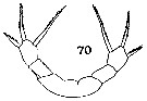 issued from : C.O. Esterly in Univ. Calif. Publs Zool., 1906, 3 (5). [Pl.13, Fig.70]. As Scolecithrix magna. Female: 70, P5.
|
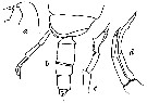
 issued from : W. Giesbrecht in Bull. Mus. comp. Zool. Harv., 25 (12). [Taf. II, Figs.1-5]. Female (from 35°N, 125°W): 1, urosome (ventral); 2, idem (lateral, left side); 3, A2 ( witout setae); 4, P2; 5, Mx1.
|
 issued from : W. Giesbrecht in Bull. Mus. comp. Zool. Harv., 25 (12). [Taf. II, Figs.9-12]. Female (from 35°N, 125°W): 9, anterior part of head (ventral); 10, idem (lateral); 11, P5; 12, P1.
|
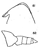 issued from : C.C. Davis in Univ. Wash. Publs Biol., 1949, n. ser. 14. [Pl.6, Figs.81-82]. Female (from NE Pacific): 81, forehead (lateral); 82, last thoracic segment and urosome (lateral). Nota : The head projects anteriorly, and bears a prominent crest ; viewed from above the head appears pointed.Rostrum very strong, bifurcated, with its apical points extremely thick. Thoracic segments 4 and 5 fused ; lateral corners of posterior prosome rounded and not pointed.Genital segment as long as the following two segments together. A1 24-segmented, reach a short distance posterior to the genital segment.
|
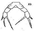 issued from : C.C. Davis in Univ. Wash. Publs Biol., 1949, n. ser. 14. [Pl.7, Fig.83]. Female: P5. Nota : P5 3-segmented, 1st segment with group of spinules on the posterior surface ; apical spines fringed with fine spinules. Male : No distinct crest. Rostrum with the apical points considerably elongated. Thoracic segments 4 and 5 separated, 5th segment very short. Urosome 5-segmented, the anal segment very short. A1 shorter than in female, barely reach the posterior margin of prosome. Masicatory portion of Md reduced, but the mandibular pal pis much more strongly developed than in the female., with the basal portion considerably enlarged. Mx1 and Mx2 somewhat degenerate.
|
 issued from : O. Tanaka in Publ. Seto Mar. Biol. Lab., 1961, IX (1). [p.151, Fig.110]. Female (from Izu region): a, forehead (lateral); b, last thoracic segment and urosome (lateral, right side); c, rostrum (frontal view); d, P2; e, P5 (abnormal). Male (from Izu region): f, habitus (dorsal); g, forehead (lateral); h, last thoracic segment and urosome (lateral); i, exopod of P1; j, P5. Nota Female: - Proportional lengths of the cephalothorax and abdomen 4.15-4.26 (81 : 19 or 5.56 : 1.34). - Head and 1st pediger segment fused, 4th and 5th fused. - Abdominal segments and caudal rami in the proportional lengths 37 : 20 : 17 : 9 :17 = 100. - Genital segment about as long as wide, slightly contracts proximally; lateral margin carries a small tuft of hairs on the either side near the base in lateral view. - 1st to 3rd urosomal segments fringed with fine teeth on the distal border. - Caudal rami about as long as wide. - A1 extends to the end of the abdomen (segments 24 and 25 well separated, the segment 24 is 1.87 times as long as the 25th). - The spinulation on the posterior surface of P2 and P3 seems to be variable in authors, and differs even in the right and left legs of the same individual. - P5 3-segmented, carries 3 spines on the distal segment; the 1st segment furnished with rows of spinules on the posterior surface. An individual had and abnormal right P5 carrying 4 spines on the distal segment (fig.e) Nota Male: - Cephalothorax about 2.74 times the abdomen length (3.95 : 1.44). - Head without median crest, but highly arched forehead. - Rostrum consists of 2 fairly long spines. - Abdominal segments and caudal rami in the proportional lengths 13 : 30 : 22 : 22 : 4 : 9 = 100. - A1 extends to the end of the 2nd abdominal segment; segments 8 to 12 fused, 12 and 13 fused posteriorly. - A2 exopod slightly shorter than endiopod (27 : 29). - Md with a robust basis. - Mx1 with 9 setae on the outer large lobe; exopod with 9 serae. - Mx1 somewhat reduced. - P1 as in the female (fig.i). - Left P5 with a fairly long endopod, reaching the distal end of the basis of the right leg.
|
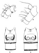
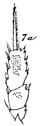 issued from : C. With in The Danish Ingolf-Expedition, Copepoda I, 1915, III, 4. [Pl. VII, Fig. 7a]. Female (from 57°52'N, 9°53'W): 7a, exopod of left P3.
|
 issued from : C. With in The Danish Ingolf-Expedition, Copepoda I, 1915, III, 4. [Pl. VII, Fig. 7 b-d]. Female: b, labrum (anterior view); c, same (oral surface); d, lamina labialis and serrula 6-dentata.
|
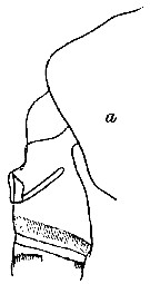 issued from : C. With in The Danish Ingolf-Expedition, Copepoda I, 1915, III, 4. [p.212, Text-fig.66, a]. Female: a, corner of last thoracic segment and genital segment (left lateral).
|
 issued from : C. With in The Danish Ingolf-Expedition, Copepoda I, 1915, III, 4. [p.213, Text-fig.67]. Male: a, forehead (lateral); b, last thoracic segment with P5 and urosome (left lateral); c, exopodal segments 2 & 3 of right P5; d, left P5.
|
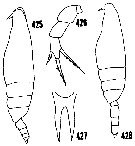 issued from : H.B. Owre & M. Foyo in Fauna Caribaea, 1, Crustacea, 1: Copepoda. Copepods of the Florida Current. [p.67, Figs.425-428]. After Sars (1925). Female: 425, habitus (lateral); 426, P5; 427, rostrum. Nota: Forehead with a crest; distal segment of P5 with 3 spines, the internal spine much longer than the external; Male: 428, habitus (lateral).
|
 issued from : H.B. Owre & M. Foyo in Fauna Caribaea, 1, Crustacea, 1: Copepoda. Copepods of the Florida Current. [p.24, Fig.104]. After Sars (1925). Male: 104, P5.
|
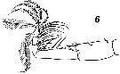 Issued from : V.N. Andronov in Russian Acad. Sci. P.P. Shirshov Inst. Oceanol. Atlantic Branch, Kaliningrad, 2014. [p.89, Fig.23: 6]. Lophothrix frontalis after Sars, 1924. Mxp male.
| | | | | Ref. compl.: | | | Pearson, 1906 (p.19); Wilson, 1942 a (p.191); Sewell, 1948 (p.329, 521, 530, 532, 534, 539, 546, 557, 566); C.B. Wilson, 1950 (p.250); Wickstead, 1962 (p.550, food & feeding); Grice, 1963 a (p.495); Gaudy, 1963 (p.23, Rem.); De Decker & Mombeck, 1964 (p.13); Furuhashi, 1966 a (p.295, vertical distribution in Oyashio/Kuroshio transitional area, Table 7); Fleminger, 1967 a (tabl.1); Grice & Hulsemann, 1967 (p.16); 1968 (tab.2); Morris, 1970 (p.2301); Roe, 1972 (p.277, tabl.1, tabl.2); 1972 b (p.531); Björnberg, 1973 (p.333, 387); Vives & al., 1975 (p.43, tab.II, XII); Deevey & Brooks, 1977 (p.256, tab.2, Station "S"); Carter, 1977 (1978) (p.35); Pipe & Coombs, 1980 (p.223, vertical occurrence); Vives, 1982 (p.292); Roe, 1984 (p.357); Brenning, 1984 (p.4, Rem.); Guangshan & Honglin, 1984 (p.118, tab.); Brenning, 1985 a (p.28, Table 2); Rudyakov, 1986 (tab.1); Lozano Soldevilla & al., 1988 (p.58); Cervantes-Duarte & Hernandez-Trujillo, 1989 a (tab.1); Heinrich, 1990 (p.17); Suarez & al., 1990 (tab.2); Suarez & Gasca, 1991 (tab.2); Hattori, 1991 (tab.1, Appendix); Hernandez-Trujillo, 1991 (1993) (tab.I); Suarez, 1992 (App.1); Gopalakrishnan & Balachandran, 1992 (p.167, fig.7, Table 1, 2); Shih & Young, 1995 (p.73); Suarez-Morales & Gasca, 1998 a (p.111); Padmavati & al., 1998 (p.347); Lapernat, 2000 (tabl. 3, 4); Yamaguchi & al., 2002 (p.1007, tab.1); Razouls & al., 2000 (p.343, Appendix); Holmes, 2001 (p.58); Park, W & al., 2004 (p.464, tab.1); Lavaniegos & Jiménez-Pérez, 2006 (tab.2, 4, Rem.); Kuriyama & Nishida, 2006 (p.299: Tab.II; fig.7, 10, vertical distribution); Ikeda & al., 2006 (p.1791, Table 2); Fernandes, 2008 (p.465, Tabl.2); Gaard & al., 2008 (p.59, Table 1, N Mid-Atlantic Ridge); Galbraith, 2009 (pers. comm.); Park & Ferrari, 2009 (p.143, Table 5, Appendix 1, biogeography); Hidalgo & al., 2010 (p.2089, Table 2); Tutasi & al., 2011 (p.791, Table 2, abundance distribution vs La Niña event); Pillai H.U.K. & al., 2011 (p.239, Table 3, vertical distribution); in CalCOFI regional list (MDO, Nov. 2013; M. Ohman, pers. comm.); Bonecker & a., 2014 (p.445, Table II: frequency, horizontal & vertical distributions) ; Fierro Gonzalvez, 2014 (p.1, Tab. 3, 5, occurrence, abundance); Dias & al., 2015 (p.483, Table 2, abundance, biomass, production); Belmonte, 2018 (p.273, Table I: Italian zones); Bode & al., 2018 (p.66, Rem.: p.75); Dias & al., 2019 (p.1, Table II, IV, fig.5, occurrrence, vertical distribution, Rem.: p.12). | | | | NZ: | 20 | | |
|
Carte de distribution de Lophothrix frontalis par zones géographiques
|
| | | | | | | | | | | | | | | 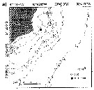 Issued from : C.O. Dias, A.V. de Araujo & S.L.C. Bonecker in An. Acad. Bras. Ciênc., 2019, 91 (1). [p.11, Fig.5, a] Issued from : C.O. Dias, A.V. de Araujo & S.L.C. Bonecker in An. Acad. Bras. Ciênc., 2019, 91 (1). [p.11, Fig.5, a]
Study area in The Campos basin (Brazil) showing the seasonal variation of Lophothrix frontalis (ind.m3) at 1 m depth, during the dry season.
Nota: This species has been considered as epipelagic-abyssopelagic, in the Campos Basin was found mainly in the subsurface water during the dry season. In the brasilian study, all samples were collected during the night and explain the higher abundance in 1-m depth samples compared to the deeper samples. |
| | | | Loc: | | | sub-Antarct. (Indian, SW & SE Pacif.), South Africa (E), off Tristan da Cunha, Namibia, Angola, off da Trindade Is., G. of Guinea, Senegal, Cape Verde Is., off NW Cape Verde Is., off Mauritania, Canary Is., off Morocco-Mauritania, off Madeira, Brazil, off Rio de Janeiro, Campos Basin, off Amazon, Caribbean, G. of Mexico, Florida, off Cape Hatteras, off Bermuda: Station S (32°10N, 64°30W), Sargasso Sea, off S Iceland, Madeira, Portugal, off W Cabo Finisterre, Bay of Biscay, Ibero-morocacan Bay, W Medit. (Tyrrhenian Sea), off Nova Scotia, S Strait of Davis, Iceland, Wyville Thomson Ridge, Faroe Is., off W Ireland, Indian (G. of Aden, Arabian Sea, Maldive Is., Natal, Indian, Bay of Bengal, 10° N - 10° S in Gopalakrishnan & Balachandran, 1992), Andaman Sea (Batten Island), Indonesia-Malaysia, China Seas (East China Sea, South China Sea), Japan, Sagami Bay, off Sanriku, Station Knot, G. of Alaska, Icy Strait, Pacif. (central subarctic), NE Pacif., off British Columbia, California, W Baja California, Pacif. (W equatorial), New Zealand, Fiji Is., SE Easter Is., Pacif. (SE tropical), off Juan Fernandez Is., Galapagos-Ecuador, Chile (N-S) | | | | N: | 87 | | | | Lg.: | | | (1) F: 6,3; M: 5,5; (5) F: 7,4; M: 5,75; (6) F: 7,25-5,83; M: 5,91-5,58; (7) F: 6,45; M: 5,66; (9) F: 7,4-5,6; M: 5,75; 5,65; (16) F: 6,25-5,3; M: 5,73-4,52; (17) F: 6; (22) F: 6,4; M: 6; (29) F: 6,125-4,875; (38) F: 6-5,65; (47) F: 6,6; (59) F: 7,4-6; M: 5,5; (73) F: 5,87-5,51; (75) ?F: 5,11; (108) F: 6,9; M: 5,39; (199) F: 6,24-5,32; M: 5,4; (800) F: 6,7-5,64; (1000) F: 6,2 ± 0,5; M: 5,3; (1111) F: 5,3; {F: 4,875-7,400; M: 4,520-6,000} | | | | Rem.: | Méso-bathy-abyssopélagique (mésopélagique in Roe, 1984).
Lophotrix frontalis forma minor Sewell, 1929 (p.196-200, figs.70 part.)
Loc.: Gulf of Aden, Arabian Sea
Voir aussi les remarques en anglais | | | Dernière mise à jour : 19/01/2021 | |
|
|
 Toute utilisation de ce site pour une publication sera mentionnée avec la référence suivante : Toute utilisation de ce site pour une publication sera mentionnée avec la référence suivante :
Razouls C., Desreumaux N., Kouwenberg J. et de Bovée F., 2005-2025. - Biodiversité des Copépodes planctoniques marins (morphologie, répartition géographique et données biologiques). Sorbonne Université, CNRS. Disponible sur http://copepodes.obs-banyuls.fr [Accédé le 30 août 2025] © copyright 2005-2025 Sorbonne Université, CNRS
|
|
|
|


;)
;)
;)
;)
;)
;)
;)
;)
;)
;)
;)
;)
;)
;)
;)
;)
;)
;)
;)
;)
;)
;)
;)
;)
;)
;)
;)
;)

;)
