|
|
 |
Fiche d'espèce de Copépode |
|
|
Calanoida ( Ordre ) |
|
|
|
Clausocalanoidea ( Superfamille ) |
|
|
|
Scolecitrichidae ( Famille ) |
|
|
|
Scolecithricella ( Genre ) |
|
|
| |
Scolecithricella dentata (Giesbrecht, 1892) (F,M) | |
| | | | | | | Syn.: | Scolecithrix dentata Giesbrecht, 1892 (p.266, 775, Descr.F, figs.F); Giesbrecht & Schmeil, 1898 (p.44, Rem. F); Rose, 1924, p.5, 7, 8, 10); Kotani & al., 1996 (tab.2); Hattori, 1991 (tab.1, Appendix);
Scolecithrix dubia (M) Giesbrecht, 1892 (p.266, 775, fig.M); Giesbrecht & Schmeil, 1898 (p.44, Rem. M); Sewell, 1948 (p.516); Tanaka, 1937 (p.261, figs.M); De Decker & Mombeck, 1964 (p.14); Bainbridge, 1972 (p.61, Appendix Table I: vertical distribution vs day/night);
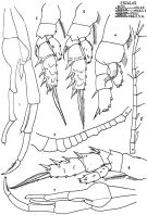
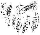
no Scolecithricella dentata (M): C.B. Wilson, 1950 (p.333, figs.M) | | | | Ref.: | | | Farran, 1908 b (p.51); Pesta, 1920 (p.518); Sars, 1925 (p.191, figs.,F); Farran, 1926 (p.259); 1929 (p.209, 247); Rose, 1933 a (p.158, figs.F); Farran, 1936 a (p.96); Rose, 1942 (p.144, Redescr.F, figs.F, p.154, Redescr.M n°2, figs.M); Lysholm & al., 1945 (p.29); C.B. Wilson, 1950 (p.333, fig.F); Tanaka, 1962 (p.42, figs.F,M); Owre & Foyo, 1967 (p.61, figs.F,M); Mazza, 1967 (p.163, 172, figs.F,M, juv.); Vilela, 1968 (p.19, figs.F,M); Park, 1968 (p.555, figs.F, Rem. F); Corral Estrada, 1970 (p.160, figs.F,M, Rem.); Bradford, 1970 a (p.356, figs.M); 1972 (p.44, figs.F, Rem.); Razouls, 1972 (p.94, Annexe: p.54, figs.F); Bradford, 1973 (p.142); Park, 1980 (p.42, figs.F, Rem.); Dawson & Knatz, 1980 (p.6, figs.F); Björnberg & al., 1981 (p.639, figs.F); Bradford & al., 1983 (p.103,104, figs.F,M, Rem.); Campaner, 1984 a (p.176, figs.F, Rem.); He & al., 1992 (p.250); Kim & al., 1993 (p.270); Chihara & Murano, 1997 (p.901, Pl.173,176: F,M); Mazzocchi & al., 1995 (p.200, figs.F,M, Rem.); Bradford-Grieve & al., 1999 (p.881, 932, figs.F,M); Vyshkvartzeva, 1999 (2000) (p.234); Avancini & al., 2006 (p.88, Pl. 57, figs.F,M, Rem.); Vives & Shmeleva, 2007 (p.778, figs.F,M, Rem.) | 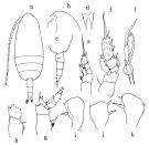 issued from : O. Tanaka in Publ. Seto Mar. Biol. Lab., 1962, X (1). [p.43, Fig.130]. Female (Izu Region): a, habitus (dorsal); b, head (lateral left lside); c, last thoracic segment and urosome (lateral left side); d, rostrum; e, P1; f, P2; g, P3; h, P4; i, P5; j, k, P5 (from different specimens). Male: l, 5th pair of legs. Nota Female: - Cephalothorax about 3.13 times the length of urosome; or 3 times the length of the abdomen (75 to 25). - Posterolateral corners of the last thoracic segment notched on each side. - Rostrum with a strong base to which rather short filaments. - Urosome 4-segmented; abdominal segments and caudal rami in the proportional lengths 37 : 24 : 20 : 7 : 12 = 100. - Genital segment not swollen ventrally. - Caudal rami as long as wide. - A1 22-segmented, extends about to the distal end of the genital segment. - A2 exopod 1.3 times as long as endopod.. - Mx1 exopod with outer lobe with 7+2 setae; 9 setae on exopod; 7 setae on endopod. - Mx2 with both amalliform and vermiform filaments on the distal segments. - Mxp with the segments in the proportions 18 : 14 : 9 (endopod). - P1 without outer edge spine on the 1st exopodal segment. - P2 with a long and curved spine on the 1st segment reaching the distal margin of the 2nd segment. The 1st basal segment finely serrated on the proximal outer margin; 2nd segment with 3 small spines on the inner margin about the middle. The terminal spine of the exopod has 45 teeth. - Coxa of P3 and P4 with 1 inner seta and any hairs (see figs. g, h). - P5 1-segmented and plate-like; the inner marginal spine is the longest; the outer marginal one is small; the distal spine is shorter than the inner marginal one. Spinulation variable, in some specimen the outer marginal spine is absent (see figs. i, J, k). Nota Male: - Cephalothorax and abdomen in the proportional lengths 72 : 28. - Posterolateral corners of the last thoracic segment rounded. - Abdominal segments and caudal rami in the proportional lengths 15 : 30 : 21 : 21 : 4.9 = 100. - Caudal rami divergent, about as long as wide. - A1 extends to the distal margin of the 3rd abdominal segment. - P5 extends to the distal margin of the 4th abdominal segment. Right leg with a short endopod extending to the middle of the 1st segment of the exopod; terminal spine of exopod short. Left leg with endopod reaching only the distal margin of the 1st segment of exopod; distal segment of exopod finely haired on the inner margin. Remarks: The female specimen of S. dentata Giesbtrcht resembles quite well with Scolecithrix dubia Giesbrecht, but can be disyinguished from the latter by the length of A1, and by the terminal segment of the exopod of the left leg of P5.
|
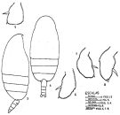
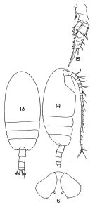 Issued from : T.S. Park in Fishery Bull. Fish Wild. Serv. U.S., 1968, 66 (3). [p;553, Pl.8, Figs.13-16]. Female: 13, habitus (dorsal); 14, idem (lateral right side); 15, P2; 16, P5.
|
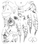
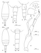
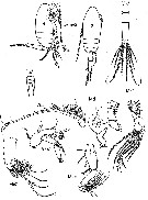
 Issued from : J.M. Bradford, L. Haakonssen & J.B. Jillett in Mem. N.Z. oceanogr. Inst., 1983, 90. [p.106, Fig.63]. Female: A, habitus (lateral left side); C, P5; B, endopod of P2 (another specimen). Male: D, habitus (lateral left side); E, P2; ; F, P5; G, terminal part of left P5 exopod. Nota Female: - Basis of Md with 1 seta. - Mx2 with 3 of 5 brush-like filaments on endopod, long and thin, while other short and thick. - P2 basis with group of small spines on inner margin; endopodal segment 2 elongated, with 5 spines on posterior surface; exopodal segment 1 outer spine long and strongly curved. - P5 terminal segment rounded, terminal spine very small, inner spine longer than terminal spine, outer margin spine small and may be absent. Nota Male: - A1 19-segmented, extends to distal end of urosomal segment 3. - Mouthparts as in female but Mx1 smaller. - P5 extends to distal end of urosome. Right P5 basis swollen; endopod 1-segmented, very short, with distal spine; exopod 2-segmented, segment 1 long, with projection at middle of inner margin, segment 2 much shorter, with terminal spine. left leg endopod 1-segmented, with terminal spine; exopod 3-segmented, terminal segment spatula-shaped with small spines more or less aligned and stronger spines on one surface.
|
 issued from : T. Park in Antarct. Res. Ser. Washington, 1980, 31 (2). [p.41, Fig.7]. Female: a, b, forehead (lateral); c, last thoracic segmentand urosome (lateral left side); d, genital segment (lateral left side); e, rostrum (anterior); f, A2; g, Md; h, Mx1; i, distal part of Mx2; j, Mxp; k, P1 (anterior); l, P2 (posterior); m, P3 (posterior); n, P4 (posterior); o, P5 (posterior). Nota: A1 reaching posterior end of 2nd urosomal segment. Caracterized by a large incision on the distal margin of the prosome and squarish P5.
|
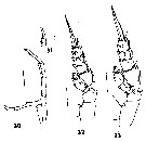
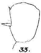
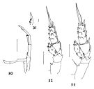 issued from : J.M. Bradford in N.Z. Jl Mar. Freshw. Res., 1970, 4 (4). [p.356, Figs 30-33]. Male (off Kaikoura, New Zealand): 30, P5; 31, terminal exopod segment of left P5; 32, P2; 33, P3. Scale bars represent 0.1 mm in 30, 32, 33 and 0.01 mm in 31.
|
 Issued from: M.G. Mazzocchi, G. Zagami, A. Ianora, L. Guglielmo & J. Hure in Atlas of Marine Zooplankton Straits of Magellan. Copepods. L. Guglielmo & A. Ianora (Eds.), 1995. [p.201, Fig.3.36.1]. Female: A, habitus (dorsal); B, urosome (dorsal); C, idem (lateral right side); D, rostrum; E, P5. Nota: A1 almost reaching distal end of genital somite. Proportional lengths of urosomites and furca 37:22:22:8:11 = 100. P5 may be variable in spinulation, with outer marginal spine absent in some specimens (See Tanaka, 1962). Male: F, habitus (dorsal); G, urosome (dorsal); H, P5. Nota: Proportional lengths of urosomites and furca 15:30:21:23:3:8 = 100.
|
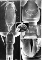 Issued from: M.G. Mazzocchi, G. Zagami, A. Ianora, L. Guglielmo & J. Hure in Atlas of Marine Zooplankton Straits of Magellan. Copepods. L. Guglielmo & A. Ianora (Eds.), 1995. [p.202, Fig.3.36.2]. Female (SEM preparation): A, habitus (dorsal); B, forehead (ventral); C, urosome (dorsal); D, genital somite (ventral); E, inner notches on posterior margins of last thoracic somite (dorsal); F, P5. Bars: A 0.100 mm; B, C 0.050 mm; D, E, F 0.010 mm.
|
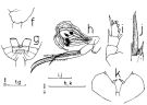 issued from : A.F. Campaner in Bolm. Zool., Univ. S. Paulo, 1984, 6 . [p.177, Fig.6, f-k]. Female (from off Rio de Janeiro): f, last segment of prosome (latera lleft side); g, idem (ventral); h, Mx2 (endopodite and inner lobe 5); i, P1; j, distal part of exopodal segment 3 of P2; k, P5.
|
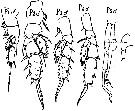
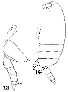
 issued from : J. Corral Estrada in Tesis Doct., Univ. Madrid, A-129, Sec. Biologicas, 1970. [Lam.44]. Male (from Canarias Is.): 1, P5; 2, P2; 3-3', A1; 4, P1; 5, P2; 6, P3; 7, P5 (another individual).
|
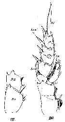
 issued from : J. Corral Estrada in Tesis Doct., Univ. Madrid, A-129, Sec. Biologicas, 1970. [Lam.43, figs.3-8]. Female: 3, habitus (lateral right side); 4, idem (dorsal); 5, P5; 6, left P5 (another individual); 7, right P5 (idem); 8, P5 (another specimen).
|
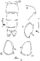
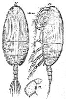 Issued from : G.O. Sars in Résult. Camp. Scient. Prince Albert I, 69, pls.1-127 (1924). [Pl.LII, figs.21-23]. As Scolecithrix dentata. Female: 21, habitus (dorsal); 22, idem (lateral left side); 23, P5. - Nota Female: - Rostrum little prominent, of the same structure than Scolecithricella vittata. - Head and 1st pedigerous segment fused, 4th and 5th pedigers thoracic fused. - Posterolateral corners of the last thoracic segment regularly obtusely rounded and define above with a prominent tooth in lateral and dorsal views. - Genital segment very slightly swollen, the length slightly longer than the two following segments together. - Caudal rami slightly longer than wide. - A1 reaching barely the prosome length. - P5 lamella-shape irregularly rounded, with 1 short, straight spine on the middle inner margin, 1 minute spine at apex and the external margin naked, but slightly angular.
|
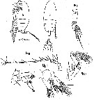 issued from : M. Rose in Annls Inst. océanogr., Monaco, 1942, XXI (3). [p.145, Figs.38, 39]. . Female (from Alger Bay, Algeria): habitus (lateral and dorsal, respectively); Ur, urosome; A1; A2; Mx1 (as Mx); Mxp (as Mxp2). A1 22-segmented. Exopodite of A2 about 2/3 length endopodite.
|
 issued from : M. Rose in Annls Inst. océanogr., Monaco, 1942, XXI (3). [p.146, Fig.40, 41]. Female: Mx2 (as Mxp1); Md; R, rostrum; P1 to P5.
|
 issued from : M. Rose in Annls Inst. océanogr., Monaco, 1942, XXI (3). [p.155, Figs.46, 47]. As Mâle n°2. Male: habitus (lateral and dorsal, respectively); Ur, urosome; A1; A2; Mx1 (as Mx); Mx2 (as Mxp1); Md; Mxp (as Mxp2); R, rostrum. Nota: A1 19-segmented (8th and 9th fused; sharply bended at the end of 14th segment from particular form. Endopodite of A2 = 3/4 exopodite.
|
 issued from : M. Rose in Annls Inst. océanogr., Monaco, 1942, XXI (3). [p.156, Fig.48]. As Mâle n°2. Male: P1 to P5.
|
 issue from : J.M. Bradford in N. Z. Jl mar. freshw. Res., 1970, 4 (4). [p.356, Figs.30-33]. Male (from Kaikoura): 30, P5; 31, terminal exopod segment of left P5; 32, P2; 33, P3. Scale bars: 0.01 mm (31); 0.1 mm (others).
|
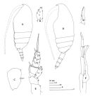
 issued from : J.M. Bradford in Mem. N. Z. Oceonogr. Inst., 1972, 54. [p.43, Fig.10, (4-5)]. Female (from Kaikoura, New Zealand): 4, habitus (dorsal); 5, P5. Scale bars: 1 mm (4); 0.1 mm (5). Nota: The most notable characteristic is the notched posterolateral borders of the cephalothorax, which indicates the line of fusion between segments 4 and 5.
|
 issued from : C. Razouls in Th. Doc. Etat Fac. Sc. Paris VI, 1972, Annexe. [Fig.45, B, E, F-H]. Female (from Banyuls, G. of Lion): B, urosome (dorsal); R, forehead (lateral); F-H, P5 (from different specimens).
|
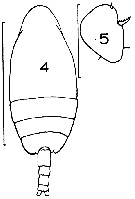
 Issued from : W. Giesbrecht in Systematik und Faunistik der Pelagischen Copepoden des Golfes von Neapel und der angrenzenden Meeres-Abschnitte. Fauna Flora Golf. Neapel, 1892. Atlas von 54 Tafeln. [Taf. 37 , Figs.13, 14 ]. As Scolecithrix dentata. Female: 13, last thoracic segment and urosome (lateral, left side); 14, habitus (lateral).
|
 Issued from : W. Giesbrecht in Systematik und Faunistik der Pelagischen Copepoden des Golfes von Neapel und der angrenzenden Meeres-Abschnitte. Fauna Flora Golf. Neapel, 1892. Atlas von 54 Tafeln. [Taf. 13 , Figs.12, 20 ]. As Scolecithrix dentata. Female: 12, P4 (basipod only, posterior view); 20, P2 (posterior view).
|
 Issued from : W. Giesbrecht in Systematik und Faunistik der Pelagischen Copepoden des Golfes von Neapel und der angrenzenden Meeres-Abschnitte. Fauna Flora Golf. Neapel, 1892. Atlas von 54 Tafeln. [Taf. 13 , Fig.33 ]. As Scolecithrix dentata. Female: 33, P5.
|
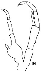 issued from : H.B. Owre & M. Foyo in Fauna Caribaea, 1967, 1, Crustacea, 1: Copepoda. [p.23, Fig.94]. Male (from Florida Current): 94, P5. Nota: P5 length great enough to reach the distal margin of the urosmal segment 4. Right end one-half the length eof exopod 1. Distall segment of left exopod finely hirsute on their inner margin, without an apical spine.
|
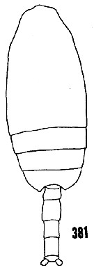 issued from : H.B. Owre & M. Foyo in Fauna Caribaea, 1967, 1, Crustacea, 1: Copepoda. [p.60, Fig.381]. Male (from Florida Current): 381, habitus (dorsal).
|
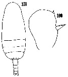 issued from : H.B. Owre & M. Foyo in Fauna Caribaea, 1967, 1, Crustacea, 1: Copepoda. [p.60, Figs.379-380]. Female (from Florida Current): 379, habitus (dorsal); 380, P5. Nota: Genital segment longer than wide.
| | | | | Ref. compl.: | | | Pearson, 1906 (p.18); Rose, 1925 (p.152); Sewell, 1948 (p.345, 502, 508, 546, 550, 551); Østvedt, 1955 (p.15: Table 3, p.70); Grice & Hart, 1962 (p.287, 293: Rem.); Gaudy, 1962 (p.93, 99, Rem.: p.107); Duran, 1963 (p.16); V.N. Greze, 1963 a (tabl.2); Björnberg, 1963 (p.41, Rem.); Ahlstrom & Thrailkill, 1963 (p.57, Table 5, abundance); De Decker & Mombeck, 1964 (p.14); Grice & Hulsemann, 1965 (p.224); Shmeleva, 1965 b (p.1350, lengths-volume-weight relation); Pavlova, 1966 (p.43); Furuhashi, 1966 a (p.295, vertical distribution in Kuroshio region, Table 10) ; Mazza, 1966 (p.70); 1967 (p.367); Furuhashi, 1966 a (p.295, vertical distribution in Oyashio/Kuroshio transitional area, Table 8, 9); Grice & Hulsemann, 1967 (p.16); Fleminger, 1967 a (tabl.1); De Decker, 1968 (p.45); Vinogradov, 1968 (1970) (p.79, 261); Park, 1970 (p.476); Deevey, 1971 (p.224); Carli, 1971 (p.373, tab.2); Binet & al., 1972 (p.53, 69); Apostolopoulou, 1972 (p.327, 350); Roe, 1972 (p.277, tabl.1, tabl.2); 1972 b (p.538); Björnberg, 1973 (p.332, 389); Corral Estrada & Pereiro Muñoz, 1974 (tab.I); Vives & al., 1975 (p.43, tab.II, III); Deevey & Brooks, 1977 (p.256, tab.2, Station "S"); Carter, 1977 (1978) (p.35); Dessier, 1979 (p.205); Pipe & Coombs, 1980 (p.223, vertical occurrence); Vaissière & Séguin, 1980 (p.23, tab.1); Vives, 1982 (p.292); Kovalev & Shmeleva, 1982 (p.84); Scotto di Carlo & Ianora, 1983 (p.150); Dessier, 1983 (p.89, Tableau 1, Rem., %); Guangshan & Honglin, 1984 (p.118, tab.); Scotto di Carlo & al., 1984 (1042); Roe, 1984 (p.357); Regner, 1985 (p.11, Rem.: p.32); Madhupratap & Haridas, 1986 (p.105, tab.1); Rudyakov, 1986 (tab.1); Ambler & Miller, 1987 (tab.2,3); Lozano Soldevilla & al., 1988 (p.59); Hopkins & Torres, 1988 (tab.1, as dentipes); Herman, 1989 (p.247); Madhupratap & Haridas, 1990 (p.305, fig.5: vertical distribution night/day; fig.7: cluster); Hirakawa & al., 1990 (tab.3); Scotto di Carlo & al., 1991 (p.270); Gopalakrishnan & Balachandran, 1992 (p.167, figs.1, 4, Table 1, 2); Hirakawa & al., 1992 (p.1); Kouwenberg, 1994 (tab.1); Hajderi, 1995 (p.542); Shih & Young, 1995 (p.73); Gopalakrishnan & Saraledevi, 1998 (p.132, fig.7, chart); Hure & Krsinic, 1998 (p.101); Suarez-Morales & Gasca, 1998 a (p.111); Siokou-Frangou, 1999 (p.476); Dolganova & al., 1999 (p.13, tab.1); Voronina & Kolosova, 1999 (p.71); Seridji & Hafferssas, 2000 (tab.1); Razouls & al., 2000 (p.343, Appendix); d'Elbée, 2001 (tabl. 1); Holmes, 2001 (p.59); Bode & al., 2003 (p.85, Table 1, abundance); Vukanic, 2003 (139, tab.1); Hsiao & al., 2004 (p.326, tab.1); Kang & al., 2004 (p.1524); Lo & al., 2004 (p.89, tab.1); Shimode & al., 2005 (p.113 + poster); Isari & al., 2006 (p.241, tab.II); Lavaniegos & Jiménez-Pérez, 2006 (p.155, tab.2, 4, Rem.); Lopez-Ibarra & Palomares-Garcia, 2006 (p.63, Tabl. 1, seasonal abundance vs El-Niño); Kuriyama & Nishida, 2006 (p.293, 300: Tab.II; p.309: Tab.III, fig.7, 10, vertical distribution, Rem.: p.313); Zervoudaki & al., 2006 (p.149, Table I); Valdés & al., 2007 (p.104: tab.1); Dur & al., 2007 (p.197, Table IV); Khelifi-Touhami & al., 2007 (p.327, Table 1); Morales-Ramirez & Suarez-Morales, 2008 (p.522); Ayon & al., 2008 (p.238, Table 4: Peruvian samples); Fernandes, 2008 (p.465, Tabl.2); Lan Y.-C. & al., 2008 (p.61, Table 1, % vs stations); C.-Y. Lee & al., 2009 (p.151, Tab.2); Galbraith, 2009 (pers. comm.); Park & Ferrari, 2009 (p.143, Table 5, Appendix 1, biogeography); Licandro & Icardi, 2009 (p.17, Table 4); Brugnano & al., 2010 (p.312, Table 3); Hafferssas & Seridji, 2010 (p.353, Table 2, 3); Hafferssas & al., 2010 (p.1281, Table III, abundance vs spatial distribution); Schnack-Schiel & al., 2010 (p.2064, Table 2: E Atlantic subtropical/tropical); Mazzocchi & Di Capua, 2010 (p.427); Medellin-Mora & Navas S., 2010 (p.265, Tab. 2); Hsiao S.H. & al., 2011 (p.475, Appendix I); Brugnano & al., 2012 (p.207, Table 2); in CalCOFI regional list (MDO, Nov. 2013; M. Ohman, comm. pers.); Lidvanov & al., 2013 (p.290, Table 2, % composition); Bonecker & a., 2014 (p.445, Table II: frequency, horizontal & vertical distributions); Benedetti & al., 2016 (p.159, Table I, fig.1, functional characters); Ohtsuka & Nishida, 2017 (p.565, Table 22.1); El Arraj & al., 2017 (p.272, table 2); Benedetti & al., 2018 (p.1, Fig.2: ecological functional group); Belmonte, 2018 (p.273, Table I: Italian zones); Hure M. & al., 2018 (p.1, Table 1: abundance, % composition); Dias & al., 2019 (p.1, Table II, IV, occurrrence, vertical distribution). | | | | NZ: | 20 | | |
|
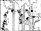
Carte de distribution de Scolecithricella dentata par zones géographiques
|
| | | | | | | | | | | | | | |  issued from : Gopalakrishnan & Saraledevi in Pelagic Biogeography ICoPB II. Proceedings of the 2nd International Conference. Final report of SCOR/IOC working group 93. Noordwijkerhout, The Netherlands 9-14 July 1995. Workshop Report No.142. UNESCO, 1998. [p.133, Fig.7]. issued from : Gopalakrishnan & Saraledevi in Pelagic Biogeography ICoPB II. Proceedings of the 2nd International Conference. Final report of SCOR/IOC working group 93. Noordwijkerhout, The Netherlands 9-14 July 1995. Workshop Report No.142. UNESCO, 1998. [p.133, Fig.7].
Distribution and abundance in number of specimens/standard haul of Scolecithricella dentata in the Indian Ocean. |
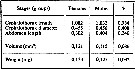 issued from : A.A. Shmeleva in Bull. Inst. Oceanogr., Monaco, 1965, 65 (n°1351). [Table 6: 22 ]. Scolecithricella dentata (from South Adriatic). issued from : A.A. Shmeleva in Bull. Inst. Oceanogr., Monaco, 1965, 65 (n°1351). [Table 6: 22 ]. Scolecithricella dentata (from South Adriatic).
Dimensions, volume and Weight wet. Means for 50-60 specimens. Volume and weight calculated by geometrical method. Assumed that the specific gravity of the Copepod body is equal to 1, then the volume will correspond to the weight. |
 Issued from : M. Madhupratap & P. Haridas in J. Plankton Res., 12 (2). [p.312, Fig.5]. Issued from : M. Madhupratap & P. Haridas in J. Plankton Res., 12 (2). [p.312, Fig.5].
Vertical distribution of calanoid copepod (mean +1 SE), abundance No/100 m3. 51- Scolecithricella dentata.
Night: shaded, day: unshaded.
Samples collected from 6 stations located off Cochin (India), SE Arabian Sea, November 1983, with a Multiple Closing Plankton Net (mesh aperture 300 µm), in vertical hauls at 4 depth intervalls (0-200, 200-400, 400-600, 600-1000 m). |
| | | | Loc: | | | Antarct. (Weddell Sea), Sub-Antarct. (Indian, SW & SE Pacif.), South Africa (E & W), Tristan da Cunha, Congo, G. of Guinea, Ivorian shelf, S Brazil, off Rio de Janeiro, The Campos Basin, off Amazon, off Morocco-Mauritania, Canary Is., off Madeira, Portugal, Coruña, off W Cabo Finisterre, Azores, Caribbean Sea, Caribbean Colombia, G. of Mexico, Florida, off Bermuda: Station S (32°10N, 64°30W), Sargasso Sea, Bay of Biscay, W Ireland, Porcupine Bank, off W Scotland, Wyville Thomson Ridge, Norwegian Sea, Ibero-moroccan Bay, Medit. (Alboran Sea, Gulf of Annaba, Castellon, Banyuls, Marseille, Ligurian Sea, Corsica, Tyrrhenian Sea, Strait of Messina, Adriatic Sea, Ionian Sea, Aegean Sea, Lebanon Basin, Marmara Sea), Natal, Indian, Bay of Bengal, Philippines, China Seas (East China Sea, South China Sea), Taiwan (SW, Mienhua Canyon, NW, Kuroshio Current), Korea (S & E), Japan Sea, Japan, Honshu (Toyama Bay), off Sanriku, Pacif. (W equatorial), Australia (Great Barrier), New Zealand (Kaikoura), Hawaii, off NE Hawaii, Pacif. (central N), off British Columbia (rare), California, W BaJa California, Bahia Magdalena (rare), Central America, Costa Rica (Dome), W Costa Rica, off SW Galapagos, Peru, Chile, Straits of Magellan | | | | N: | 159 | | | | Lg.: | | | (1) F: 1,6; (9) F: 1,5-1,3; M: 1,85-1,3; (34) F: 1,35; (36) F: 1,96-1,84; M: 1,94-1,76; (38) F: 1,7-1,5; (72) F: 1,68-1,48; (116) F: 1,55; (117) F: 2,07-1,43; M: 1,75-1,4; (128) M: 1,48; (147) F: 1,65; M: 1,7-1,5; (180) F: 1,42-1,28; M: 1,59-1,37; (199) F: 1,98-1,29; M: 1,44-1,37; (207) F: 1,56-1,47; M: 1,57-1,5; (237) F: 1,2; (313) M: 1,5; (327) F: 1,53; M: 1,49; (449) F: 1,45-1,3; (523) F: 1,62-1,32; (532) F: 1,4-1,21; 1,68-1,62; (1000) F: 1,6 ± 0,1; M: 1,7 ± 0,2; {F: 1,20-2,07; M: 1,30-1,94} | | | | Rem.: | épi-mésopélagique.
Sampling depth (sub-Antarct.) : 0-2500 m.
Voir remarque à Scolecithrix dubia.
Voir aussi les remarques en anglais | | | Dernière mise à jour : 25/10/2022 | |
|
|
 Toute utilisation de ce site pour une publication sera mentionnée avec la référence suivante : Toute utilisation de ce site pour une publication sera mentionnée avec la référence suivante :
Razouls C., Desreumaux N., Kouwenberg J. et de Bovée F., 2005-2026. - Biodiversité des Copépodes planctoniques marins (morphologie, répartition géographique et données biologiques). Sorbonne Université, CNRS. Disponible sur http://copepodes.obs-banyuls.fr [Accédé le 07 janvier 2026] © copyright 2005-2026 Sorbonne Université, CNRS
|
|
|
|


;)
;)
;)
;)
;)
;)
;)
;)
;)
;)
;)
;)
;)
;)
;)
;)
;)
;)
;)
;)
;)
;)
;)
;)

;)
;)
;)
