|
|
 |
Fiche d'espèce de Copépode |
|
|
Misophrioida ( Ordre ) |
|
|
|
Misophriidae ( Famille ) |
|
|
|
Benthomisophria ( Genre ) |
|
|
| |
Benthomisophria palliata Sars, 1909 (F,M) | |
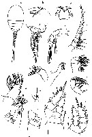
| | | | | | | Syn.: | Misophria japonica Tanaka, 1966 a (p.51, figs.F,M) | | | | Ref.: | | | Sars, 1909 (p.1, fig.F); Sars, 1938 (p.36, fig.F); Boxshall & Roe, 1980 (p.12, figs.F,M, juv.); Boxshall, 1982 (p.126, figs.); 1983 (p.120); 1984 (p.32, figs.,Rem.); 1986 (p.159); 1989 (p.524); Chihara & Omori, 1997 (p.933, Pl.191: F,M); Boxshall & Huys, 1998 (p.768, figs.juv., M, fig.5); Boxshall & Halsey, 2004 (p.217: fig.F); Ferrari & Dahms, 2007 (p.71, Rem.); Vives & Shmeleva, 2010 (p.135, figs.F,M, Rem.) | 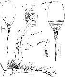 issued from : G.A. Boxshall & H.S.J. Roe in Bull. Br; Mus. Hist. (Zool.), 1980, 38 (1). [p.19, Fig.5]; Female (from W portugal): B, urosome (ventral); C, habitus (dorsal); D, P5; F, A1. Male: A, habitus (dorsal); E, P6. Scale bars 0.200 mm unless otherwise indicated. Nota: The numerous large aesthetes on A1 probably have a chemosensory function, necessary for the location of food, and suggests that they also have a reproductive function in male to locate a female.
|
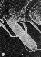
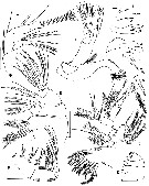 issued from : G.A. Boxshall & H.S.J. Roe in Bull. Br; Mus. Hist. (Zool.), 1980, 38 (1). [p.21, Fig.6]. Female: A, A2; B, Md (showing armature elements 1-7 enlarged); C, Mx1 (with rami separated from limb); D, Mx2 (showing segmentation only); E, Mx2 (with segments separated from each other); F, Mxp. Scale bars 0.200 mm unless otherwise indicated.
|
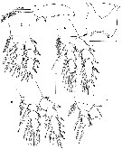 issued from : G.A. Boxshall & H.S.J. Roe in Bull. Br; Mus. Hist. (Zool.), 1980, 38 (1). [p.22, Fig.7]. Female: A, P6; B, labrum (ventral); C-F, P1 to P4. Scale bars 0.5 mm unless otherwise indicated.
|
 issued from : G.A. Boxshall & H.S.J. Roe in Bull. Br; Mus. Hist. (Zool.), 1980, 38 (1). [p.18, Fig.4, J]. Male: J, A1 (with setae omitted). Scale bar 0.200 mm.
|
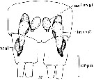
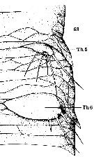 issued from : G.A. Boxshall in Phil. Trans. R. Soc. Lond., 1982, B- 297 (1086). [p.175, Fig.63]. Male (from 20°N, 21°W): Vental view of half genital somite (from body length 4.2 mm); Th5 : thoracic leg 5; Th6: thoracic leg 6. Nota: Each genital aperture is closed off by a plate formed from the fused P6, which extends across it, parllel to the vental body surface. Each aperture is situated dorsal to the P6 and posterior to its base. The dorsal surface of the P6 is flexible and slightly convex.
|
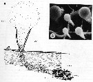
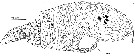 issued from : G.A. Boxshall in Phil. Trans. R. Soc. Lond., 1982, B- 297 (1086). [p.156, Fig.25]. 25, Female (from 20°N, 21°W): lateral view, drawn from serial s.e. micrographs. co area : area occupied by cone organs. Nota 1: The primary lamellae ornamentating the body surface can be seen, as can the positions of pores in the integument. The stippled area represent sites of muscle attachments marked externally by the pattern of secondary lamellae. Ornamentation is omitted from the area occupied by cone organs (co area). Nota 2: The cone organs , which are clearly secretory, are situated on either side of the cephalosome in such a position as to allow the long setae of A2 and mandibular palp to sweep dorsoventrally across them. In the course of this sweeping movement these setae presumably come into contact with the raised globules of secretion. As they do some or all of the secretion presumably adheres to them. The secretion is a continuous process. The secretion could be either retained on the setae, smeared over the posterolateral of the cephaloossible interpretations are numeroussome, or dispersed in the surrounding water. It is difficult to determine the role of the cone organs: 1- The secretion may be used in the feeding process by being retained on the setae. 2- The secretion may be smeared over part of the body surface as a means of protection or of preventing the establishment and growth of micro-organisms. 3- The secretion may possess an odour repellent to predators. 4- The secretion may serve as a chemical signal attracting potential mates. 5- The secretion may be bioluminescent (this is regarded as unlikely in an organism apparently devoid of photoreceptors). For Boxshall, the secretion is probably defensive or protective.
|
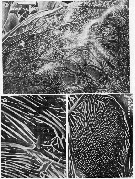 issued from : G.A. Boxshall in Phil. Trans. R. Soc. Lond., 1982, B- 297 (1086). [Plate 2, Figures 26-28]. 26: Dorsal view of lateral portion of cephalosome, showing the surface ornamentation, which comprises a system of primary lamellae delimiting area containing numerous more-or-less parallel secondary lamellae. Scale bar 0.075 mm. 27, detail from figure 26 of an area of closely packed fine lamellae marking externally the site of origine of a dorsal longitudinal trunk muscle fibre. Scale bar 0.010 mm. 28, Detail from figure 26, showing an integumetal pore and an associated sensilla. Scale bar 0.010 mm.
|
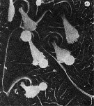
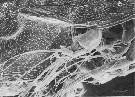 issued from : G.A. Boxshall in Phil. Trans. R. Soc. Lond., 1982, B- 297 (1086). [Plate 4, Figure 34]. 34, Lateral view of the area of cone organs on the cephalosome. Most of the cone organs have been damaged and have lost their globules of secretion. Scale bar 0.100 mm.
|
 issued from : G.A. Boxshall in Phil. Trans. R. Soc. Lond., 1982, B- 297 (1086). [Plate 5, Figure 38]. 38, Detail from cone organ area in which the cone organs have cbeen damaged and lost their globules of secretion or had them displaced. Scale bar 0.002 mm. Nota: The chemical nature of the secretion has not yet been ascertained. The lobules are not membrane-bound. When fixed the secretion is rather fibrous.
|
 issued from : G.A. Boxshall in Phil. Trans. R. Soc. Lond., 1982, B- 297 (1086). [Plate 1, Figure 3]. 3, Aesthetasc on the female A1. Scale bar 0.020 mm.
|
 issued from : G.A. Boxshall in Phil. Trans. R. Soc. Lond., 1982, B- 297 (1086). [p.170, Fig. 57]. 57, Vental view of anal somite and uropods, showing secretory glands; Nota: In the urosome a pair of glands is situated near the ventral surface of each of the 3rd to 5th somites. These appear to be absent in males. In both sexes there are two pairs of glands in the anal somite, one opening via pores on the ventral surface of the anal somite (med an gl), the other via pores on the ventral surface of the uropods (caudal rami) (lat an gl). The lateral gland is divided into two areas, an anterodorsal area, and a posteroventral area. The more medial gland is bladder-like, having a large lumen. Another small pear-shaped gland is founf within each uropod (ur gl).
|
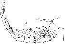
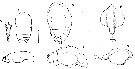 issued from : G.A. Boxshall & H.S.J. Roe in Bull. Br; Mus. Hist. (Zool.), 1980, 38 (1). [p.36, Fig.16]. 16, Variation in body shape. Slim specimen (Body length 3.22 mm) A, gut; B-C, prosome (dorsal and lateral, respectively). Swollen specimen (Body length 4.27-4,5 mm) D-E, prosome (dorsal and lateral, respectively); both with gut visible through body wall); F, gut (dorsal and lateral, respectively). Scale bars 1 mm. Nota: The small and large morphs differ only in size, no other morphological differences were observed between them. In addition to size dimorphism there is also considerable variation in the body shape. Some specimens have a bloated, inflated appearance whereas others are much thinner. Swollen specimens are inflated both laterally and dorsally and have a distinct hump-backed appearance in lateral view. Slim individuals have a much flatter dorsal profile. These changes in body proportions occur in both small and large individuals and do not correspond to changes in body length. The gut is very distended in swollen specimens and is visible through the thin carapace virtually filling the entire prosome. In slim individuals the gut is much smaller and cannot be seen through the carapace. The combination of a very elastic gut and a thin flexible integument enables to gorge untill its entire prosome is distended. The ornate pattern of surface markings on the exosqueleton of this species may facilitate this streching. The ability to eat as much as possible when the opportunity arises is presumably a considerable advantages in areas, such as deep oceans, where food is scarce.
|
 issued from : O. Tanaka in Sympos. Ser. Mar. Biol. Ass. India, 1966, 1 (2). [p.52, Fig.1]. As Misophria japonica. Female (from Izu Region, Japan): a, habitus (dorsal); b-c, forehead (lateral and frontal aspects); d, last thoracic segment and urosome (lateral); e, A1; f, A2; g, Md; h, Mx1; i, Mx2; j, Mxp; k, P1; l, P2; m, P4 (incomplete); n, P5. Nota : A1 18-segmented, short, extending to the distal margin of the head ; the proximal segments fringed with minute spinules only along the posterior margin. Ptoportional lenhths of urosomal segments and caudal rai : 16 : 16 :18 :17 :12 :10 :11 = 100. Caudal rami slightly longer than wide. : A2 : two basal segments ; exopod 6-segmented ; endopod 3-segmented, 2.5 times as long as the exopod and carries 5 setae on the disytal margin of the 2nd segment .Md palp large ; exopod 5-segmented ; endopod 2-segmented ; masticatory gnathobase slender, with 8 teeth and an inner marginal spine.Mx1 1st inner lobe with 10 strong spines, 3 delicate ones ; 2 setae arise from the anterior margin ; 2nd inner lobe with 3 setae on the distal margin ; 3rd inner lobe with 2 setae on the distal margin ; 2nd outer lobe with 4 marginal and 4 terminal setae ; 1st outer lobe with 5 setae. Mx2 with 4 marginal and 4 terminal setae ; 5th outer lobe is not modified into a strong claw as figured in by Sars.
|
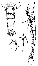 issued from : O. Tanaka in Sympos. Ser. Mar. Biol. Ass. India, 1966, 1 (2). [p.53, Fig.2]. As Misophria japonica. Male: a, last thoracic segment and urosome (dorsal); b, A1; c, P5; d, P6. Nota : A1 13-segmented, modified into a clasping organ, the proximal 5 segments broad, the middle section is 6-segmented, the hinge jointi is situated betxween segments 11 and 12. Proportional lengths of urosomal segments and caudal rami 16 :22 :16 :16 :10 :10 :10 = 100. Caudal rami slightly longer than wide. The mouth parts and swimming legs as in the female ; P5 is just as in female.
|
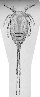 issued from : G.O. Sars in Res. Camp. Scient. Prince Albert 1er Monaco, 1938, 97. [Pl. III, fig.5]. Female (30°-44° N, 02°-42°W): 5, habitus (dorsal).
|
 issued from : G.A. Boxshall in Microscopic Anatomy of Invertebrates. 9: Crustacea. 1992. Wiley-Liss, Inc. [p.357, Fig.7, A-B]. A: Cone organs, showing continuous variation in size. B: Schematic reconstruction of section through a cone organ showing secretory gland integumental pore, and epicuticular nature of cone walls. co = cone organ; se = secretion globule; e = epidermis; p = procuticle; ep = epicuticle; sc = secretory gland cell; sv = secretory vesicle; st = secretory tubule.
| | | | | Ref. compl.: | | | Kazmi, 2004 (p.231) | | | | NZ: | 4 | | |
|
Carte de distribution de Benthomisophria palliata par zones géographiques
|
| | | | | | | Loc: | | | Atlant. (S equatorial), off Cape Verde Is., off W Canary Is., Portugal, off Azores, Bay of Biscay, Pakistan, S Indian, Japan (Izu) | | | | N: | 6 | | | | Lg.: | | | (95) F: 2,67; (610) F: 5,7-2,88; M: 4,72-2,7; {F: 2,67-5,70; M: 2,70-4,72} | | | | Rem.: | bathy-abyssopélagique.
Voir aussi les remarques en anglais | | | Dernière mise à jour : 30/12/2014 | |
|
|
 Toute utilisation de ce site pour une publication sera mentionnée avec la référence suivante : Toute utilisation de ce site pour une publication sera mentionnée avec la référence suivante :
Razouls C., Desreumaux N., Kouwenberg J. et de Bovée F., 2005-2025. - Biodiversité des Copépodes planctoniques marins (morphologie, répartition géographique et données biologiques). Sorbonne Université, CNRS. Disponible sur http://copepodes.obs-banyuls.fr [Accédé le 24 décembre 2025] © copyright 2005-2025 Sorbonne Université, CNRS
|
|
|
|


;)
;)
;)
;)
;)
;)
;)
;)
;)
;)
;)
;)
;)
;)
;)

