|
|
 |
Fiche d'espèce de Copépode |
|
|
Calanoida ( Ordre ) |
|
|
|
Diaptomoidea ( Superfamille ) |
|
|
|
Pseudodiaptomidae ( Famille ) |
|
|
|
Pseudodiaptomus ( Genre ) |
|
|
| |
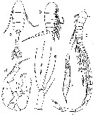
Pseudodiaptomus hessei (Mrazek, 1895) (F,M) | |
| | | | | | | Ref.: | | | Sewell, 1924 (p.785, Rem.); Marsh, 1933 (p.Dussart & Defaye, 1983 (p.29); Grindley, 1984 (p.219, fig.M); Walter, 1986 (p.131); 1986 a (p.503); ; 1987 (p.367); Dussart, 1989 (p.8, figs.F,M); Bradford-Grieve & al., 1999 (p.884, 953, figs. F,M, p.876: chart); Boxshall & Halsey, 2004 (p.173, figs.F) | 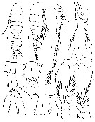 issued from : J.R. Grindley in Ann. S. Afr. Mus., 1963, 46 (15). [p.376, Fig.1]. Female (from W & South coasts of South Africa): a-b, habitus (dorsal and lateral, respectively; c, A1; d, P5; e, distal part of P5 (enlarged); f, genital segment (ventral); g, anal segment and caudal ramus; h, genital segment (lateral); i, comb-like seta (enlarged); j, P4; k, P1. Nota : Rostrum well developed with 2 strong filaments. A1 31-segmented, reaching the 2nd urosomal segment ; segments 1+2, 3+4, 8+9 and 24+25 apparently fused (Giesbrecht & Schmeil, 1898, and Marsh, 1933 reported 22 segments). A specialized comb-like set is present on the third last segment, it lies parallel to the antennula, its distal en dis curved and slightly expanded and bears about 3 distinct recurved hooks and a number of much smaller ones which grade into a series of minute comb-like teeth (3 micron long and 1 micron apart) along the shaft of the seta, there is a bulbous swelling near the base of this seta. ; there is also an unspecialized plumose seta on this segment. Cephalosome and pedigerous segment 1 partly separate ; pedigerous segments 4 and 5 fused. Posterior angles of metasome with a sharp spine on each side on the postero-dorsal margin. P5 uniramous, 4-segmented and asymmetrical, particularly as regards the hyaline projections of the inner margin of the 2nd basal segment (there seems to be some individual variation in the forms of these projections ; Marsh, 1933, suggests that these might be regarded as rudimentary indications of an endopod ; such rudiments are present also in P. stuhlmanni). The outer distal angles of the 1st and 2nd basal segments are produced into very small spines ; the 1st segment of the ramus bears 1 spine near the distal end of the outer margin ; the 2nd segment bears 1 spine on the outer distal angle an dis produced distally on the medial side into a large curved spiniform process bearing short bristles on the anterior and posterior margins and reaching about the midpoint of the terminal spine ; the terminal spine has a small distinct basal portion bearing a slender plumose seta on its posterior face, while the long terminal blade-like portion is furnished with minute bristles on its anterior and posterior margins ; terminal spine, including the basal portion, slightly longer than the 3rd segment (marsh, 1933, points out that the distinct basal portion of the terminal spine found in some species is not a real segment but a modification of the terminal spine or hook). Urosome 4-segmented and almost 2/3 as long as prosome. Genital segment asymmetrical, with irregular swellings laterally and a prominent genital boss ventrally ; a number of setae present mainly on the lateral swellings, and there is a patch of fine hairs in the middle of the dorsal surface. The genital boss bears a row of spinules anteriorly and the genital flaps each have a distal seta. The egg sac is single, containing about 25 eggs. The first three urosomal segments are furnished with rows of coarse teeth on their postero-dorsal margins. Caudal rami divergent, more than 3 times as long as wide and with fine hairs on their inner margins ; the 3rd or middle caudal seta (not counting the sensory bristle between the 4th and 5th setae) is broad and flattened in a characteristic blade-like form, it is about 3 times as wide as the other setae.
|
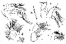 issued from : J.R. Grindley in Ann. S. Afr. Mus., 1963, 46 (15). [p.377, Fig.2]. Female: a, A2; b, divided seta from Mxp; c, Md; d, Mx1; e, Mxp; f, Mx2. A2 with basipod partially divided by an incomplete suture, bearing 1 lateral and 2 terminal setae. Exopod 3-segmented, 3rd segment only partially separated, bearing 2 setae on the 1st segment, 7 setae on the 2nd segment, and 6 terminal setae and a lateral fringe of fine hairs on the distal segment. Endopod apparently 4-segmented, 3rd segment small and indistinct, with setal formula 1, 2 +3, 1, 2 +3, respectively. Md with gnathal blade heavily chitinized and bearing about 10 fine teeth and a plumose spinule. Basipod of palp with 4 inner marginal setae. Exopod indistinctly 3-segmented, bearing 1 lateral and 5 terminal setae. Endopod 2-segmented, bearing 4 setae on segment 1 (1 separate) and 8 setae distally on segment 2 (1 jointed and crooked), this last segment is narrowly triangula rand bears a row of tiny bristles. Mx1 with 1st inner lobe bearing about 10 strong spines, 2nd inner lobe bearing ?3 setae and 3rd inner lobe bearing 3 terminal setae ; outer lobe bearing 8 long setae ; exopod with 9 marginal setae ; endopod 3-segmented bearing 4 setae medially on the basal portion andgroups of 4 setae medially on segments 1 and 2 and approximately 7 termi,nal setae on segment 3. Mx2 with 5 large medial lobes (endites) and 2 smaller terminal lobes each bearing 3 or 4 setae. Distal portion indistinctly segmented. Mxp 6-segmented, 2 basal segments large and 4 distal segments small and decreasing in size distally ; 1st segment with 3 medial lobes bearing 2, 3 and 2 setae +1 spine, respectively ; there are short fine hairs medially and tiny hooked spinules on the 3rd lobe. 2nd segment expanded medially with 3 setae and a fringe of short fine hairs on the medial margin. 3rd segment bearing 2 + 3 setae. 4th segment with 2 setae. 5th segment with 2 setae. 6th segment with 2 setae medially and 6 on an outer lobe. Among the setae on segments 3, 4 and 5 are four with a peculiar branched structure, these branced setae have a thickened basal portion and divide near their middle, one branch continuing as a thinner seta forming the extension of the proximal part while the other is short, broad and spatulate, fringed with short curved spinules (well noted by Mrazek, 1894).
|
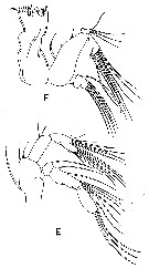 issued from : G.A. Boxshall & S.H. Halsey in The Ray Society, 2004, 166. [p.173, Fig.40, E-F]. Redrawn after Grindley (1963). Female: E, A2; F, Md.
|
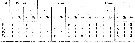 issued from : J.R. Grindley in Ann. S. Afr. Mus., 1963, 46 (15). [p.379]. Female: Setal formulae of swimming legs P1-P4. Si, St, Sr = inner, terminal and outer setae or spines (setae: Arabic numerals; spines: Roman numerals. Nota : Swimming legs P1-P4 similar in both sexes.
|
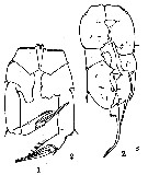
 issued from : J.R. Grindley in Ann. S. Afr. Mus., 1963, 46 (15). [p.380, Fig.3]. Male: a-b, habitus (dorsal and lateral, respectively); c, right A1; d, P5; e, caudal ramus; f, spermatophore. Nota : Posterior angles of the metasome without spines. Left A1 as in female (including setation and arrangement of aesthetascs ; right A1 geniculate with 21 segments, reaching the 2nd urosomal segment ; the last 4 long segments are apparently formed by the fusion of two segments ; there is a hooked spine on segment 10 ; there is 1 aesthetasc on segments 2, 5, 7, 9-16, and the second last and last segments. P5 biramous, 4-segmented. In the right leg the 1st basal segment is naked except for some rugosity on the medial surface ; 2nd basal segment larger, with some small spinose processes on the inner margin, and bears a small rod-like endopodite with 1 short terminal seta ; on the outer margin there are a row of minute spinules and 1 fine seta near the distal angle. Exopodite 2-segmented, the 1st segment produced into a curved spine at its outer distal angle, which just reaches the base of the spine on the following segment, and bears some spinules near the inner distal angle ; the 2nd segment bears a long, partly plumose spine on the distal part of the outer margin and a rugose swelling on the proximal part of the inner margin ; the terminal hook has a triangular basal portion, broadest distally where it bears a row of small spinules and is produced at the outer corner into a slightly curved hook as long as the two exopodal segments; the tip is recurved and minutely serrate. In the left leg the 1st basal segment is naked and the 2nd basal segment bears a few spinules near the outer margin, some rugosity on the inner margin and a long hyaline endopodite ; this has an expansion basally, is apically acute, and bears a fringe of about 8 fine hairs near the apex. Exopodite 2-segmented, the 1st segment short, bearing 1 short spine at its outer distal angle ; the 2nd segment is expanded and flattened, somewhat convex posteriorly and emarginate medially ; the outer distal margin is strongly convex and the inner distal margin nearly straight ; there is 1 spine near the middle of the outer margin, and a number of small setae and spinules on the posterior surface and margins and a ; the left leg reaches the base of the terminal hook of the right leg.round the apex Urosome 5-segmented, first and last segments shorter than the remainder ; posterior margins of the 2nd, 3th and 4th segments fringed partially (2nd) or completely (3rd and 4th) with rows of coarse teeth. Spermatophore fusiform, stalked, and 250-400 microns. Caudal setae distinctly different from those of the female, for the middle seta is not expanded and all the setae are jointed about 1/3 of their length from the proximal end.
|
 Issued from : C.D. Marsh in Proc. U.S. natn. Mus., 1933, 82 (18) (2959). [Pl. 19, figs.1-2]. After Mrazek, 1894. Female: 1, P5. Male: 2, P5.
| | | | | Ref. compl.: | | | Grindley, 1977 (p.341, Table 2, fig.2, 3, 4, 7, 8, 14); Hart, 1977 (p.1, feeding); Wooldridge & Erasmus , 1980 (p.107); , tidal utilization); Jerling & Wooldridge, 1991 (p.121, population dynamics, production); 1992 (p.309, lunar light effect); Mauchline, 1998 (tab.33, 47, 48, 51); Froneman, 2000 (p.543, grazing); Kibirige & Perissinotto, 2003 (p.727, Table 1, 2, fig.5, seasonal distribution); Pagano & al., 2003 (p.433, feeding); Isla & Perissinotto, 2004 (p.579, metabolism vs t°C, salinity and sex); Miller C.B., 2008 (p.332, fig.3: biomass vs. development time); Barton & al., 2013 (p.522, Table 1: metabolism, biogeo); Noyon & Froneman, 2013 (p.306, egg production vs environmental factors) | | | | NZ: | 4 | | |
|
Carte de distribution de Pseudodiaptomus hessei par zones géographiques
|
| | | | | | 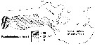 issued from : J.R. Grindley in Trans. roy. Soc. S. Afr., 42, 3 & 4. [p.353, Fig.4]. issued from : J.R. Grindley in Trans. roy. Soc. S. Afr., 42, 3 & 4. [p.353, Fig.4].
Distribution of Pseudodiaptomus hessei from Saldanha Bay to Schrywershoek area (33°S, 18° E).
Compare this distribution with the species in the same site: Paracartia longipatella, Tortanus capensis, Oithona similis. |
| | | | Loc: | | | South Africa (west and south coasts, Mpenjati Estuary, Sunday River estuary, Algoa Bay, Saldanha Bay, Kariega estuary, Kleinmond estuary, Natal), Congo (mouth of the Congo River), Senegaouth, Ivory Coast (Ebrié Lagoon) | | | | N: | 15 | | | | Lg.: | | | (1148) F: 1,2; { F: 1,2; } | | | | Rem.: | saumâtre, estuaire.
Incomplete data.
Voir aussi les remarques en anglais | | | Dernière mise à jour : 09/03/2023 | |
|
|
 Toute utilisation de ce site pour une publication sera mentionnée avec la référence suivante : Toute utilisation de ce site pour une publication sera mentionnée avec la référence suivante :
Razouls C., Desreumaux N., Kouwenberg J. et de Bovée F., 2005-2025. - Biodiversité des Copépodes planctoniques marins (morphologie, répartition géographique et données biologiques). Sorbonne Université, CNRS. Disponible sur http://copepodes.obs-banyuls.fr [Accédé le 20 août 2025] © copyright 2005-2025 Sorbonne Université, CNRS
|
|
|
|


;)
;)
;)
;)
;)

