|
|
 |
Fiche d'espèce de Copépode |
|
|
Calanoida ( Ordre ) |
|
|
|
Diaptomoidea ( Superfamille ) |
|
|
|
Pseudodiaptomidae ( Famille ) |
|
|
|
Pseudodiaptomus ( Genre ) |
|
|
| |
Pseudodiaptomus serricaudatus (T. Scott, 1894) (F,M) | |
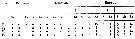
| | | | | | | Syn.: | Heterocalanus serricaudatus T. Scott, 1894 b (p.40, figs.F,M); Sewell, 1947 (p.164);
Pseudodiaptomus nudus Tanaka, 1960 (p.47, figs.F,M); De Decker, 1962 (p.112); Gindley, 1963 (p.384); Tanaka, 1964 (p.9); De Decker, 1964 (p.16, 24, 29); 1968 (p.45); Grindley, 1977 (p.341, Table 2, fig.2); De Decker, 1984 (p.316); Hutchings, 1985 (p.1); Verheye & al., 1994 (p.155); Bradford-Grieve & al., 1999 (p.884, 953, figs.F,M); Jerling, 2008 (p.55, Tabl.1);
Schmackeria serricaudatus : Marsh, 1933 (p.46, figs.F,M);
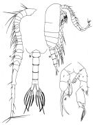
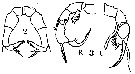
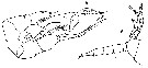
Schmackeria serricaudatus : Sewell, 1947 (p.164); Krishnaswamy, 1953 (p.123); Ganapati & Shanthakumari, 1962 (p.8, 16); Saraswathy, 1966 (1967) (p.79) | | | | Ref.: | | | A. Scott, 1902 (p.404, fig.F); Thompson & Scott, 1903 (p.234, 248); Sewell, 1912 (p.365: Rem.); 1914 a (p.226); 1924 (p.785); 1932 (p.235); Mori, 1942 (p., figs.F,M); Marques, 1951 a (p.14); 1953 (p.117), 1955 (p.18); 1958 (p.215); 1973 (p.240); 1974 (p.16); Vervoort, 1965 (p.92, figs.F,M, Rem.); Wellershaus, 1969 (p.258, figs.F,M, Rem.); Pillai, 1976 (1980) (p.252, figs.F,M); Goswami & Goswami, 1978 (p.11, figs.); Grindley, 1984 (p.220, Rem.); Walter, 1986 (p.133, 162); 1986 a (p.503); 1987 (p.368); Dussart, 1989 (p.7, figs.F, M); Bradford-Grieve & al., 1999 (p.884, 953, figs.F,M, p.876: chart); Altaff, 2018 (p.387, figs.: oviducal glands) | 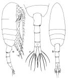 Issued from : W. Vervoort in Atlantide Report, 1965, 8. [p.93, Fig.20] Female (from off Port Marshall, Liberia): a, habitus (lateral right side); b, idem (dorsal); c, posterior part cephalothorax and urosome (dorsal).
|
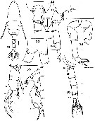
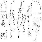
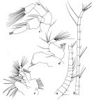 Issued from : W. Vervoort in Atlantide Report, 1965, 8. [p.94, Fig.21] Female: a-b, A1; c, A2; d, Md; e, Mx1. Nota: Mx1 with praecoxal arthrite with 8 spiniform setae; 3 setae on the coxal endite and 3 setae on the basal endite; the basipodite (2nd basal segment) with 5 setae; endopodite distinctly 3-segmented, with 4, 4, 5 setae, respectively; exopodite with 9 setae; basal exite acsent; coxal exite (epipodite) with 7 strong and 2 small setae. Mx2 with praecoxa and coxa completely fused, each has 2 endites with 3 setae, one of which is a spinulose appendicular seta; basis has 2 endites, the big proximal has 3 setae, the much smaller distal carries 2; the small endopodite 3-segmented with a total of 5 setae. Mxp has on its coxa 3 groups of 2, 3, 3 setae, respectively; the swollen median part of the basis has 3 setae, 2 setae occur on a small apical lobe; endopodite 4-segmented, with 3, 3, 3, 7 setae, respectively; Two of the setae on segment 1, and 1 of the setae on segments 2 and 3 have the branched structure characteristic of this genus; the branched seta on segment 3 is very strong; the 4th (apical) segment apparently results from the fusion of two segments.
|
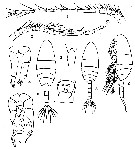
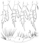 Issued from : W. Vervoort in Atlantide Report, 1965, 8. [p.95, Fig.22] Female: a, P1; b, P2; c, P3; d, P4; e, Mx2; f, Mxp; f', branched seta of endopodite of Mxp.
|
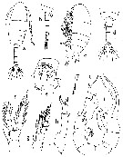
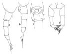 Issued from : W. Vervoort in Atlantide Report, 1965, 8. [p.96, Fig.23] Female: a, posterior part cephalothorax and urosome (lateral right side); b, idem (lateral left side); c, genital complex (ventral); d, P5.
|
 Issued from : W. Vervoort in Atlantide Report, 1965, 8. [p.98, Fig.24] Male: a, habitus (lateral right side); b, posterior part cephalothorax and urosome (dorsal); c, right A1; d, P5.
|
 Issued from : S. Wellershaus in Veröff. Inst. Meeresforsch. Bremerh., 1969, 11 (2). [p.261, Figs.31-36]. Female (Cochin Backwater, S. India): 31, habitus (dorsal); 32, thoracic segment 5 and urosomal segments 1-2 (dorsal) and thoracic segment 5 and and urosome (right side), respectively; 33, posterior margin of anal segment (dorsal); 34, P5. Nota: Ratio cephalothorax: urosome = 2.0; Proportions of the urosomal segments 23, 21, 24, 13; caudal rami 19%). Genital segment prolonged into a sheath which overlaps a part of the urosomal segment 3; this overlapping is more expressed on the right side,and its posterior margin bears a row of spines which extends into a group of bigger and scattered spines on the right angle of the sheath; on the left side there is no trace of a \"low cup-shaped swelling\" as in P. nudus (see Tanaka 1960, p.47). Lengths of 3 females (and ratio thorax : abdomen): 1.10 (1.6); 1.26 (1.5); 1.32 (1.6). Male: 35, P5 (anterior); 36, urosome (left side; caudal rami (ventral). Nota: Ratio cephalothorax:urosome 12, 21, 18, 20, 11; caudal rami 18 %.
Scale : single line = 0.1 mm ; double line = 0.05 mm.
|
 issued from : T. Mori in Palao trop. biol. Stn Stud., 1942, 2 (3). [Pl.IX, Figs. 1-9]. Female (from Palao Island): 1, A1; 3, rostrum; 4, P5; 6, habitus (dorsal); 7, genital segment (ventral); 9, habitus (lateral). Male: 2, right A1; 5, P5 (anterior view); 8, habitus (dorsal).
|
 issued from : J.R. Grindley in Ann. S. Afr. Mus., 1963, 46 (15). [p.385, Fig.5]. As Pseudodiaptomus nudus. Female (from off The Cape, Agulhas bank): c, habitus (lateral); d, urosome (dorsal); e, genital segment (ventral); f, P4; g, P1; h, P5. Nota: P5 uniramous; 4-segmented; the terminal spine is shorter than the spiniform process of the 4th segment, and bears a slender plumose seta in place of the short, stout, serrate spine of serricaudatusMale: a, habitus (dorsal); b, urosome (lateral); j, P5. Nota: A1 21-segmented, right geniculate. P5 are identical with those of P. serricaudatus, but in the right leg the 2nd marginal spine reaches as far as the terminal hook does; the endopod is more complex than in serricaudatus.Urosome 5-segmented and similar to that of P. hessei; 2nd urosomal segment with a mid-ventral patch of hairs.
|
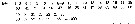
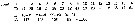
 issued from : J.R. Grindley in Ann. S. Afr. Mus., 1963, 46 (15). [p.385, Fig.5]. As Pseudodiaptomus nudus. Female (from South Africa): Setal formula from the swimming legs P1-P4. Si = inner setae; Se = outer setae. Arabic numeral = seta; Roman numerals = spines.
|
 Issued from : C.D. Marsh in Proc. U.S. natn. Mus., 1933, 82 (18) (2959). [Pl. 22, figs.2-3]. As Schmackeria serricaudatus. After T. Scott, 1893. Female : 2, P5. Male: 3, P5 (L = left leg, R = right leg).
|
 Issued from : O. Tanaka in Spec. Publs. Seto mar. biol. Lab., 10, 1960 [Pl. XXI, 1-7]. As Psedodiaptomus nudus. Female (from 35°09'S, 20°13'E): 1, habitus (dorsal); 2, head and A1 (lateral); 3, last thoracic segment and abdomen (lateral); 4, genital segment (ventral); 5, P1; 6, P2; 7, P5. Same scale for 5 and 6. Nota: Cephalothorax and abdomen in the proportional lengths 66 to 34. Rostrum composed of 2 strong spines reaching the anterior margin of the proximal segment of A1. Abdominal segments and caudal rami in the proportional lengths 28 : 18 : 22 : 14 : 18 = 100. Genital segment asymmetrical; the right side inflated and left side depressed; the distal lateral corner of the right margin furnished with a group of small spines; the left side with a low cup-shaped swelling furnished with 2 long hairs and minute spinules on the distal margin when viewed from the left side; the ventral surface of the segment swollen; the genital boss furnished with spinules on the periphery, and with 2 whip-like spines on each side of the distal margin. 2nd and 3rd abdominal segments furnished with coarse teeth on the distal margin. Caudal rami 5.5 times as long as broad.
|
 Issued from : O. Tanaka in Spec. Publs. Seto mar. biol. Lab., 10, 1960 [p.48]. As Psedodiaptomus nudus. Female: Proportional lengths of segments of A1. Nota: A1 21-segmented, extends about to the level of the posterior margin of the 2nd abdominal segment.
|
 Issued from : O. Tanaka in Spec. Publs. Seto mar. biol. Lab., 10, 1960 [Pl. XXI, 8, 9]. As Psedodiaptomus nudus. Male: 8, last thoracic segment and abdomen (lateral); 9, P5. Nota: Abdomen 5-segmented; segments and caudal rami in the proportional lengths 11 : 20 : 20 : 22 : 11 : 16 = 100. 2nd abdominal segment with hairs on the ventral surface about the middle of the segment; 2nd, 3rd and 4th segments furnished with coarse teeth on the distal margin. For Tanaka, P5 are in the main as those of P. serricaudatus , but the distal exopodal segment of left leg is prolonged into a long curved spine, and the outer edge spine on the 1st exopodal segment of the same leg is so long that it reaches the distal margin of the terminal spine of the 3rd segment.
|
 Issued from : O. Tanaka in Spec. Publs. Seto mar. biol. Lab., 10, 1960 [p.48]. As Psedodiaptomus nudus. Male: Proportional lengths of segments of A1. Nota: A1 21-segmented
|
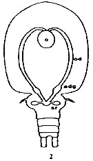 Issued from : K. Altaff in International Journal Zoology and Applied Biosciences, 2018, 3 (3). [p.389, Fig.2]. Female reproductive system of Pseudodiaptomus serricaudatus. 0: ovary; odg: oviducal gland; sr: seminal receptacle.
Nota: Compare with P. annandalei.
| | | | | Ref. compl.: | | | Cleve, 1904 a (p.196); Sewell, 1948 (p.323, 367); Neto & Paiva, 1966 (p.26, Table III, annual cycle); Subbaraju & Krishnamurphy, 1972 (p.25, 26 ); Binet & al., 1972 (p.69); Bainbridge, 1972 (p.61, Appendix Table IV: seasonal abundance); Dessier, 1979 (p.95, 201, 206); Stephen, 1984 (p.161); Brenning, 1985 (p.5, Rem.: p.9); 1985 a (p.16, 28, Table 2); Madhupratap & Haridas, 1986 (p.105, tab.2); Diouf & Diallo, 1987 (p.260); Godhantaraman, 1994 (tab.7); Ramaiah & Nair, 1997 (tab.1); Achuthankutty & al., 1998 (p.1, Table 2, seasonal abundance vs monsoon); Mauchline, 1998 (tab.8); Dalal & Goswami, 2001 (p.22, fig.2); Kazmi, 2004 (p.229); Madhu & al., 2007 (p.54, Table 4, abundance vs monsoon); Shanthi & Ramanibai, 2011 (p.132, Table 1: as P. serricauda); Rakhesh & al., 2013 (p.7, Table 1, 4, abundance vs stations); Jagadeesan & al., 2013 (p.27, Table 3, 4, 5, 6, fig.11, seasonal variation); Anjusha & al., 2013 (p.40, Table 3, abundance & feeding behavior) | | | | NZ: | 6 | | |
|
Carte de distribution de Pseudodiaptomus serricaudatus par zones géographiques
|
| | | | | |  Carte de 1996 Carte de 1996 | |
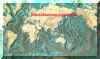
 issued from : C.T. Achuthankutty, N. Ramaiah & G. Padmavati in Pelagic biogeography ICoPB II. Proc. 2nd Intern. Conf. Final report of SCOR/IOC working group 93, 9-14 July 1995. Workshop Report No. 142, Unesco, 1998. [p.8, Fig.6]. issued from : C.T. Achuthankutty, N. Ramaiah & G. Padmavati in Pelagic biogeography ICoPB II. Proc. 2nd Intern. Conf. Final report of SCOR/IOC working group 93, 9-14 July 1995. Workshop Report No. 142, Unesco, 1998. [p.8, Fig.6].
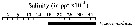
Salinity ranges for P. serricaudatus in coastal and estuarine waters of Goa (India).
Shaded area indicates the range of higher abundance. |
| | | | Loc: | | | South Africa (E & W), Agulhas Current, Saldanha, S Benguela Current, Angola, Congo, G. of Guinea, off Lagos, Ivorian shelf, Casamance, Dakar, Cape Verde Is., Red Sea, G. of Aden, Arabian Sea, Ceylan, India (W, S, Porto Novo, Bombay, off Muttukadu & Ennore Station, Kerala, Goa, Lawson's Bay, Madras, Gulf of Mannar, Palk Bay, Godavari estuary, Chilka Lake), Bay of Bengal, Andaman Is., Palau Is. | | | | N: | 47 | | | | Lg.: | | | (16) F: 1,3-1,03; M: 1,11; (66) F: 1,38-1,31; M: 1,2-1,18; (114) F: 1,21; (172) F: 1,10-1,32; M: 1,10; (530) F: 1,3; M: 1,2; (786) F: 1,52-1,22; M: 1,29-1,2; (795) F: 1,2; M: 1,15; (1005) F: 1,25-1,32: M: 1,02-1,09; (1308) F: 1,34-1,44; M: 1,1-1,15; {F: 1,03-1,52; M: 1,02-1,29} | | | | Rem.: | euryhaline; estuaire, côtière.
Observé off Cap de Bonne Espérance. Pour Sewell (1947) cette forme d'origine indienne aurait conquis les côtes ouest africaines en contournant le sud de l'Afrique.
Bradford-Grieve & al. (1999) n'admettent pas la synonymie avec P. nudus, contrairement à Vervoort (1965, p.94-96).
Voir aussi les remarques en anglais | | | Dernière mise à jour : 07/02/2020 | |
|
|
 Toute utilisation de ce site pour une publication sera mentionnée avec la référence suivante : Toute utilisation de ce site pour une publication sera mentionnée avec la référence suivante :
Razouls C., Desreumaux N., Kouwenberg J. et de Bovée F., 2005-2025. - Biodiversité des Copépodes planctoniques marins (morphologie, répartition géographique et données biologiques). Sorbonne Université, CNRS. Disponible sur http://copepodes.obs-banyuls.fr [Accédé le 01 janvier 2026] © copyright 2005-2025 Sorbonne Université, CNRS
|
|
|
|


;)
;)
;)
;)
;)
;)
;)
;)
;)
;)
;)

;)
