|
|
 |
Fiche d'espèce de Copépode |
|
|
Calanoida ( Ordre ) |
|
|
|
Clausocalanoidea ( Superfamille ) |
|
|
|
Aetideidae ( Famille ) |
|
|
|
Pseudochirella ( Genre ) |
|
|
| |
Pseudochirella hirsuta (Wolfenden, 1905) (F,M) | |
| | | | | | | Syn.: | Euchirella hirsuta Wolfenden, 1905 a (p.17, Descr.F, figs.F); 1911 (p.240, figs.F); Farran, 1929 (p.208, 237, Rem.: juv.); Hardy & Gunther, 1935 (1936) (p.159, Rem.); ; Sewell, 1948 (p.569);
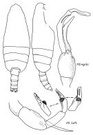
Pseudeuchirella hirsuta: Björnberg, 1973 (p.324, 388) | | | | Ref.: | | | Vervoort, 1957 (p.69, Rem.); Park, 1978 (p.163, figs.F); Markhaseva, 1989 (p.36,38,41, Descr.M, figs.M); Razouls, 1994 (p.80, figs.F,M); Markhaseva, 1996 (p.266, figs.F,M); Bradford-Grieve & al., 1999 (p.879, 923, figs.F,M); | 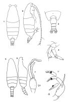 Female: 1, 2, habitus (dorsal, lateral); 3, Urosome (dorsal); 4, A2; issued from : Park in Antarct. Res. Ser. Washington, 1978, 27: 163. Male: 5, 6, habitus (dorsal, lateral); 7, 7', P5 (Dt: right, G: left); issued from : Markhaseva E.L., in Explorations of the Fauna of Seas. M.G. Petrushevska & S.D. Stepanjants (eds). Marine Plankton, 1989, 41 (49): 33-60.
|
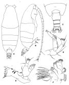 issued from : T. Park in Antarctic Res. Ser. Washington, 1978, 27. [p.164, Fig.41]. Female (54°17'S and 68°17'S in Pacific): : A, habitus (dorsal); B, idem (lateral); C, posterior part of metasome and urosome (dorsal); D, A2; E, Md; F, Mx1. Nota: Proportional lengths of prosome and urosome about 83 : 17. Rostrum single, pointing straight downward. Frontal eminence bearing suprafrontal sensilla. Cephalosome fused with 1st metasomal segment, with line of segmentation visible; 4th and 5th metasomal segments almost completely fused. Posterolateral corner of metasome produced distally into blunt lappet covering anterior 2/5 of genital segment. Genital segment (dorsally) symmetrical, with lateral sides markedly bulging at middle; genital swelling located midway; Spermatheca digitiform (viewed laterally). A1 extending beyond distal end of caudal ramus by last 2 segments. Exopod of A2 longer than endopod by about 1/3 its length, 2nd segments with 3 setae of equal length. Mx1 with 14 setae on first, 5 on second, and 4 on third inner lobe; 5 on basis, 16 on endopod, 11 on exopod, and 9 on outer lobe. Coxopodite of P4 with comb of 10-13 strong spines across mediodistal portion of posterior surface.
|
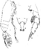
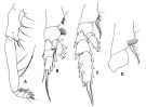 issued from : T. Park in Antarctic Res. Ser. Washington, 1978, 27. [p.165, Fig.42]. Female A, Mxp; B, P1 (anterior); C, P2 (idem); D, basipod of P4 (posterior). Nota: Coxa of Mxp with conical process distally on anterior surface. Basis longer than coxa by about 2/5 its length.
|
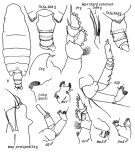 issued from : E.L. Markhaseva in Trudy Zool. Inst. RAN, St. Petersburg, 1996, 268. [p.269, Fig.214]. Female (from Antarct.: S Pacif.). Ce: forehead (lateral); CP4: coxopod of P4 (part.). Male (from Antarct.: S Pacif.). P.md: mandibular palp. Nota: Cephalothorax 4.0-4.2 times longer than urosome. Cephalon and 1st thoracic segment indistinctly separated, 4th and 5th segments virtually completely fused. Posterior corners of last thoracic segment symmetrical, of smooth triangular shape (lateral view), covering nearly the first third of genital segment, with hairs; in dorsal view corners with somewhat bifid tops. Genital segment symmetrically widened in its midlength (dorsal view); ventral swelling not prominent. Urosomal segments 1-3 surface with hairs. A1 by 1-3 last segments longer than body. Exopodite of P1 3-segmented. Endopodite of P2 2-segmented. Coxopodite of P4 with15 robust spines (10-13 spines in Park, 1978).
|
 issued from : E.L. Markhaseva in Proc. Zool. Inst. RAN, St. Petersburg, 1996, 268. [p.270, Fig.215]. Male (from Markhaseva, 1989). Nota: Cephalothorax 3.4 times longer than urosome. Cephalon and 1st thoracic segment fused, 4th and 5th fused. A1 reaching nearly urosomal segment 2. Oral parts reduced in comparison to those in females. Exopodite of P1 3-segmented; exopodal segment 1 with external spine vert small. Exopodal segment 2 of left P5 with tooth, exopodal segment 3 of left P5 nearly 4 times longer than wide.
|
 issued from : R.N. Wolfenden in Die Marinen Copepoden der Deutschen Südpolar-Expedition 1901-1903, 1911. [Pl.XXVIII, Figs.7-9]. As Euchirella hirsuta. Female: 7, habitus (lateral); 8, posterior part cephalothorax and urosome (dorsal); 9, P4.
|
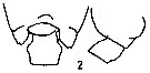 Issued from : E.L. Markhaseva in USSR Acad. Sci., Zool. Inst., Explor. Fauna Seas. Marine Plankton, 1989, 41 (49). p.52, Fig. 2]. Female: Thoracic segment 5 and genital segment. Nota from the key:
1 - Genital segment symmetrical.
3 - Exopodite 2 of A2 with 3 setae.
3 - Thoracic posterior corners symmetrical.
4 - Exopodite 2 of A2 with 3setae.
5 - Thoracic segments 4 and 5 not well separate.
5 - Genital segment (in lateral view) with ventral protuberance forward, 1.2 times large than the following segment.
6 - Body length > 6 mm.
|
 Pseudochirella hirsuta Pseudochirella hirsuta Male: 1 - Exopodite 2 of left P5with 1 tooyh in its distal part. 2 - Posterior coprners of last toracic segment without spines, rounded. 3 - Posterior corners without teeth. 4 - Exopodite 3 of left P5 triangular shape apically with knob. 5 - Body length > 7 mm.
|
 Issued from : C. Razouls in Ann. Inst. océanogr., Paris, 1994, 70 (1). [p.80]. Caractéristiques morphologiques de Pseudochirella hirsuta femelle et mâle adultes. Terminologie et abbréviations: voir à Calanus propinquus.
| | | | | Ref. compl.: | | | Grice & Hulsemann, 1967 (p.15); 1968 (tab.2); Hopkins & Torres, 1988 (tab.1); Razouls & al., 2000 (p.343, tab. 3, 5, Appendix); Park & Ferrari, 2009 (p.143, Table 4, Appendix 1, biogeography from Southern Ocean); Kouwenberg & al., 2014 (p.290, biogeography, Map 7) | | | | NZ: | 5 | | |
|
Carte de distribution de Pseudochirella hirsuta par zones géographiques
|
| | | | | | | | | 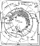 issued from : W. Vervoort in B.A.N.Z. Antarctic Reseach Expedition, Reports - Ser. B, Vol. III, 1957 [Fig.54] issued from : W. Vervoort in B.A.N.Z. Antarctic Reseach Expedition, Reports - Ser. B, Vol. III, 1957 [Fig.54]
Chart showing the geographical distribution (white triangle) in the seas surrounding the Antarctic continent.
Nota: In this chart the area frequented by whaling vessels has been hatched. The Antarctic circle (66°.5 S) has been drawn as a broken line. The numbers I to VI refer to the sectors into which the Antarctic seas are divided according to Mackintosh (1942) (after Vervoort, 1951). |
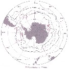
 Issued from : E.L. Markhaseva in Issled. Fauny Morei, 1989, 41 (49). [p.60, Fig.19]. Issued from : E.L. Markhaseva in Issled. Fauny Morei, 1989, 41 (49). [p.60, Fig.19].
Geographical distribution of P. hirsuta.
4: from literature; 5: present occurrences. |
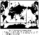
 Issued from : J.H.M. Kouwenberg, C. Razouls & N. Desreumaux in Biogeographic Atlas of the Southern Ocean, Scient. Comm. Antarct. Res., Cambridge, 2014, 6.6. [p.293, Map 7]. Issued from : J.H.M. Kouwenberg, C. Razouls & N. Desreumaux in Biogeographic Atlas of the Southern Ocean, Scient. Comm. Antarct. Res., Cambridge, 2014, 6.6. [p.293, Map 7].
Distribution of Pseudochirella hirsuta. |
| | | | Loc: | | | Antarct. (Croker Passage, Peninsula, Weddell Sea, SW Atlant., Indian, SW & SE Pacif.), South Georgia, sub-Antarct. (SW Atlant., SE Pacif.), S Atlant. (off E Tristan da Cunha Is.), W Indian, SE Pacif., off Juan Fernandez Is., S Chile
Nota: Female specimens obtained by Wolfenden (1905, 1911) in the South Atlantic (35°10'S, 02°33'E and 35°39'S, 08°16'E and the Indian Ocean sector of Antarctic (61°58'S, 95°01'E).
Type locality: region of 35°S, 2-8°E | | | | N: | 15 | | | | Lg.: | | | (10) F: 9-8,5; (20) F: 9,41-8,66; (25) F: 9,68; (37) F: 9,41-8,5; M: 7,2; {F: 8,50-9,68; M: 7,20} | | | | Rem.: | méso & bathypélagique.
Sampling depth (Antarct., sub-Antarct.) : 500-1000-4000 m.
Voir aussi les remarques en anglais | | | Dernière mise à jour : 24/01/2017 | |
|
|
 Toute utilisation de ce site pour une publication sera mentionnée avec la référence suivante : Toute utilisation de ce site pour une publication sera mentionnée avec la référence suivante :
Razouls C., Desreumaux N., Kouwenberg J. et de Bovée F., 2005-2026. - Biodiversité des Copépodes planctoniques marins (morphologie, répartition géographique et données biologiques). Sorbonne Université, CNRS. Disponible sur http://copepodes.obs-banyuls.fr [Accédé le 18 mars 2026] © copyright 2005-2026 Sorbonne Université, CNRS
|
|
|
|


;)
;)
;)
;)
;)
;)
;)

;)
;)
;)
