|
|
 |
Fiche d'espèce de Copépode |
|
|
Calanoida ( Ordre ) |
|
|
|
Clausocalanoidea ( Superfamille ) |
|
|
|
Aetideidae ( Famille ) |
|
|
|
Pseudochirella ( Genre ) |
|
|
| |
Pseudochirella obesa Sars, 1920 (F,M) | |
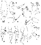
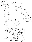
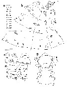
| | | | | | | Syn.: | Pseudochirella tuberculata Tanaka, 1957 b (p.195, figs.F); Grice & Hulsemann, 1967 (p.15); 1968 (p.324, tab.2); Tanaka & Omori, 1969 a (p.166, Rem.); Roe, 1975 (p.307, figs.M); Vives, 1982 (p.291) | | | | Ref.: | | | Sars, 1920 c (p.6, Rem.F); 1925 (p.94, figs.F); Jespersen, 1934 (p.66, Rem.F); Sewell, 1948 (p.500); Owre & Foyo, 1964 (p.351, Redescr.F, figs.F); 1967 (p.49, figs.F); Bradford & Jillett, 1980 (p.66, 71, figs. F,M, fig.73, distribution chart); Vaupel Klein, 1984 a (p.52, figs.F, Table II: characters); Markhaseva, 1989 (p.39, 41, figs.F,M); 1996 (p.276, figs.F,M); Vaupel Klein, 1996 (p.451, Redescr.F, Rem.); Vaupel Klein & Rijerkerk, 1996 (p.567, figs.F); 1997 (p.394, figs.F); Koomen & Vaupel Klein, 1997 (p.527, figs.F); Vives Shmeleva, 2007 (p.599, figs.F,M, Rem.) | 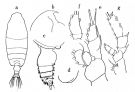 issued from : Tanaka O. in Publ. Seto mar. Biol., Lab., 1957, 6 (2). [Fig.56, p.196]. As Pseudochirella tuberculata. Female (Suruga and Sagami): a, habitus (dorsal aspect); b, head (lateral aspect); c, last thoracic segment and urosome (lateral aspect); d, last thoracic segment (lateral aspect, left side); e, P1; f, endopodite of P2; g, P4. Nota Female: - Cephalothorax about 4.55 times the abdomen length (5.0 : 1.1). - Head and 1st pediger segment separate; 4th and 5th imperfectly separate. - Posterolateral corners of last thoracic segment asymmetrical, left side evenly rounded, right side with a rounded process on the posterolateral margin; lateral corners furnished with short hairs. - Rostrum strong, directs somewhat posteriorly. - Abdomen 4-segmented; segments and caudal rami in ptoportional lengths 49 : 16 : 11 : 6 : 18 = 100. - Genital segment wider than long, slightly asymmetrical, left side more inflated than the right. - Abdominal segments and caudal rami very setose. - Caudal rami about as long as wide. - A1 23-segmented, extends to distal end of genital segment. - A2 endopod slightly longer than half the length of exopod (19 : 10). - Md gnathobase very strong. - Mx1 with 5 setae on 2nd basal segment; 15 setae on endopod; 11 setae on exopod; 7 long and 2 short setae on outer lobe. - Mxp with strong setae on 4th and 5th lobes on 1st basal segment; basal segments 1 and 2, and exopod in proportional lengths 42 : 74 : 23. - P1 with the exopodal segments fused between the 1st and 2nd; outer edge spines slender and long. - P2 endopod 2-segmented articulated on the anterior surface. - P4 with 7 long spines on the inner margin on coxa at the base of inner marginal seta
|
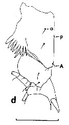
 issued from : E.L. Markhaseva in Proc. Zool. Inst. RAN, St. Petersburg, 1996, 268. [p.279, Fig.223]. Female (from SE Pacif.). Ce: forehead; CP4: coxopod of P4 (partial). Nota: Cephalothorax about 4 times longer than urosome. Posterior corners of last thoracic segment slightly asymmetrical, rounded, with knob on the right arranged ventrally (lateral view); this part of segment covered with hairs. Genital segment slightly asymmetrical, ore prominent on the left than on the right (dorsal view) and covered with hairs; genital segment slightly wider than long. A1reaching the end of genital segment. Exopodite of P1 3-segmented, separation between segments 1 and 2 incomplete. Endopodite of P2 2-segmented. Coxopodite of P4 with 7-8 or 7-10 spines.
|
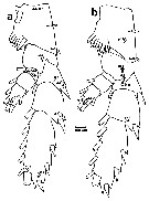
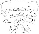
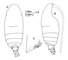 issued from : J.M. Bradford & J.B. Jillett in Mem. N.Z. oceanogr. Inst., 86, 1980. [p.72, Fig.48]. Female: A, habitus (dorsal); B, idem (lateral right side); C, basipod 1 of P4.
|
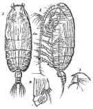 Issued from : G.O. Sars in Résult. Camp. Scient. Prince Albert I, 69, pls.1-127 (1924). [Pl.XXVI, figs.1-4]. Female: 1, habitus (dorsal); 2, idem (lateral left side); 3, forehead (lateral); 4, basal segment of P4. Nota: Head and 1st thoracic segment with suture weakly visible; 4th and 5th segments i partly separate. Posterior thoracic lateral lobes short and obtuse. Occurrences: 38°02'N, 10°44'W; 46°31'N, 5°13'W.
|
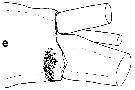 issued from : J.C. von Vaupel Klein in Crustaceana, Supplt 9, Studies on Copepoda, III, 1984. [p.74, Fig.12, e]. Female: e, spinules on the distal margin of 5th endite of Mx2 (right appendage, antero-lateral view). Nota: with a group of more 20 spinules (about 30).
|
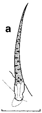 issued from : J.C. von Vaupel Klein in Crustaceana, Supplt 9, Studies on Copepoda, III, 1984. [p.63, Fig.5, a]. Female: a, condition of the distal small seta on A1 segment 5. Nota: unmodified, normal type II seta. Scale bar 0.05 mm.
|
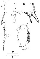 issued from : J.C. von Vaupel Klein in Crustaceana, Supplt 9, Studies on Copepoda, III, 1984. [p.84, Fig.18, c, g, i]. Female: number and structure of spines along the lateral margin of exopodal segments 1, 2 and 3 of P1 (right leg, anterior aspect) (note: 3 long, setiform, coarsely bipectinate spines); g, anterior view of the hinge-joint exopodal segments 2/3 of left P2 (note: showing contiguous row of 18 spinules, inserting on exopodal segment 2). Scale bars: q = 0.2 mm for c, g and i.
|
 issued from : J.C. von Vaupel Klein in Crustaceana, Supplt 9, Studies on Copepoda, III, 1984. [p.78, Fig.15, j-k]. Female: j-k, the partly fused condition of exopodal segments 1 and 2 of P1 (comprises a combination of largely to fully coalesced stretches, i. e., posteriorly and antero-medially, and a (still) completely free part of the suture, antero-laterally (j: left appendage, posterior view); k: right one, anterior aspect). Scale bar 0.1 mm.
|
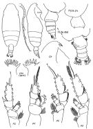
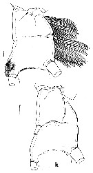
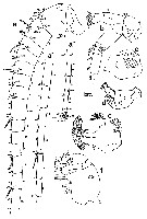
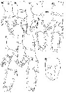
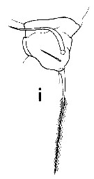
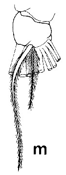
 issued from : H.S.J. Roe in Bull. British Mus. (Natural History) Zool., London, 1975, 28 (7). [p.308, Fig.7]. As Pseudochirella tuberculata. Male (from Cape Verde Islands): a, habitus (dorsal); b, A2; c, Md; d, Mx1; e, Mx2; f, Mxp; g, P1; i, P4; j, P5; k, left exopodite of P5; l, P5 (right 3rd expopodite segment). Bar scale 0.1 mm unless indicated. Nota: Rostrum as a single spike directed ventrally. Cephalothorax 3.1 times as long as the 5-segmented abdomen. Head and 1st thoracic segment fused, 4th and 5th separate. 5th thoracic segment rounded.laterally, without spines. A1 21-segmented, reaches to the hind edge of the 3rd abdominal segment. Exopodite of A2 1.4 times as long as the endopodite. Mandibular blade, Mx1 and Mx2 reduced. On the left side of P5 (Fig.7k) the 2nd exopodite segment has 2 strong points, and the 3rd segment is kidney-shaped with brunches of hairs; the 3rd segment of the right exopodite (Fig.7l) has 2 lamellae, one on a central swelling and one terminating the segment.
|
 issued from : J.C. von Vaupel Klein in Crustaceana, Supplt 9, Studies on Copepoda, III, 1984. [p.82, Fig.17, d]. Female: arrangement of hair-sensilla on the posterior face of the proximal segments of right P4. Scale bar 0.4 mm.
|
 issued from : J.C. von Vaupel Klein in Crustaceana, Supplt 9, Studies on Copepoda, III, 1984. [p.80, Fig.16, i, l, m]. Female: i, number of teeth on the serrate lateral mrgin of the terminal spines of exopodal segmnt 3 of P2-P4 (medial plumosy omitted); l, P3 with 44 teeth; m, P4 with 67 reeth; n, arrangement of medial spines on the posterior face of basipodal segment 1 of P4.. Scale bar 0.2 mm (i, l, m); 0.1 mm (n). Nota: Spine of P2 with 66 teeth.
|
 issued from : P. Koomen & J.C. von Vaupel Klein in Crustaceana, 1997, 70 (5). [p.532, Fig.3]. In situ mapping of the integumental organs of the female: a, a', a'', left A1 (lateral view); b, left A2 (lateral view); c, proximal part of the left mandibular gnathobasis (ventral view; hatched area marks insertion of palpus mandibularis); d, left palpus mandibularis (postero-medial view); e, right Mx1 (anterior view); f, right Mx2 (antero-lateral aspect). Structures drawn in dotted lines are situated on the reverse side of the appendage. Pictogram coding see fig. 1a.
|
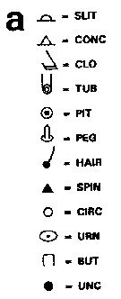 issued from : P. Koomen & J. C. von Vaupel Klein in Crustaceana, 1997, 70 (5). [p.530, Fig.1, a]. Abbreviations used for integumental organs Pictogram coding), studied by differential interference contrast microscope/yy (DIC) and scanning electron microscope/y (SEM). SLIT = regular slit-shaped glandular pore. CONC = (partly) concealed slit-shaped glandular pore. CLO = large closing-flap glandular pore. TUB = tubular glandular pore. PIT = pit-sensillum. PEG = peg-sensillum. HAIR = hair-sensillum. SPIN = spine-sensillum and/or spinular pore. CIRC = circular pore. URN = urn-shaped glandular pore. BUT = button-shaped pore. UNC = integumental organ of uncertain nature. Possibly, someofthe PITs, as oberved by DIC, may be damaged PEGs (see in KOOmen, 1992). For this reason, only in in situ drawings of specified specimens (e.g. figs 1-6) distinction has been made between PITs and PEGs (as observed by DIC). In overall schemes such as the present fig.7, both types of integumental organs have provisionally been represented by PEG symbols. Nota: The slit-shaped glandular pores are small (external opemning diameter 0.001 mm diameter or less). By DIC a small circular structure with a central dot is visible, connected to a canal. These structures have been called SLITs see fig.1a, b), because fhey occupy the same site as the SLITs established in Euchirella mssinensis (except for some intraspecific differences), and may thus be expected to be homologous of last-mentioned species. Peg-sensilla (PEG), long and slender (length up to 0.022 mm) (see fig. 1c, d), sometimes difficult to discriminate from small sensilla. The shape of the canal through the inteegument has been considered decisive (see Vaupel Klein, 1992c ; Koomen, 1992). Most PEGs are situated in depressions of the integument, while their basal discs meet the surface of the integument at an angle (see Pl. 1c, 1d). Spine-sensilla or spinular pore (SPIN), rather long and slender in comparison with those of Euchirella messinensis, and in fact come close to the PEGs of this species. However, distinction between SPINs and PEGs of P. obesa is possible, because a SPIN is shorter and stouter than the average PEG of the same species, an dits basal disc is in line with the surface of the integument. Circular integumental structure (TUB), resembling a disc (0.0035 mm) with a cenrtral knob (see Pl. 1g).
|
 issued from : P. Koomen & J.C. von Vaupel Klein in Crustaceana, 1997, 70 (5). [p.533, Fig.4]. In situ mapping of the integumental organs of the female: a, right Mxp (medial aspect); b, right caudal ramus (ventral view); c, left caudal ramus (dorsal aspect); d, internal face of the upper lip (top = distal; bottom = proximal), rows of organs have been numbered from ''a'' through ''e'', patches/rows of hairs are indicated by numerals from ''1'' through ''5'' inclusive. Pictogram coding see fig. 1a.
|
 issued from : P. Koomen & J.C. von Vaupel Klein in Crustaceana, 1997, 70 (5). [p.534, Fig.5]. In situ mapping of the integumental organs of the female: a, left P1 (anterior view); b, right P1 (posterior aspect); c, left P2 (anterior; the PEG drawn in dotted lines was present, unilaterally, in 1 out of 3 specimens only); d, right P2 (posterior); e, left P3 (anterior; the Babasipodite 1-1e-2 PEG, drawn here in dotted lines, was absent in 1 out of 3 specimens); f, right P3 (posterior face); g, ventral aspect of anteriormost sternal keel (top = rostral; bottom = caudal edge). Pictogram coding see fig. 1a.
|
 issued from : P. Koomen & J.C. von Vaupel Klein in Crustaceana, 1997, 70 (5). [p.535, Fig.6]. In situ mapping of the integumental organs of the female: a, left P4 (anterior view); b, right P4 (posterior aspect). The PEGs drawn in dotted lines were bilaterally present in 2 out of 3 specimens. Pictogram coding see fig. 1a.
|
 issued from : P. Koomen & J.C. von Vaupel Klein in Crustaceana, 1997, 70 (5). [p.530, Fig.1]. In situ mapping of the integumental organs of the female: a, pictogram coding (dee Abbreviations used); b, ceohalic pleurotergal integument, given four cuts to make a flat spread possible and shown in dorsal aspect, with line ''m'' representing the dorsal midline; c-f, Urogenital somite (double-somite), urosomal somites 3, 4 and 5, respectively, (left figures, ventral halves, right figures, dorsal halves, top = anterior, bottom = posterior, ''m'' represents the dorsal midline); g, ventral aspect of sternal integument of the cephalothorax (''oral field'' and adjacent regions), from its anterior limitation in-between the antennules (top) up to the level of insertion of the maxillipeds (bottom). Structures drawn in dotted lines are seen through the (hyaline) integument of parts in the foreground. Integumental organs within the oral cavity (hidden under the flattened upper lip) have not been depicted (see also fig.4d). Pictogram coding see fig. 1a.
|
 issued from : P. Koomen & J.C. von Vaupel Klein in Crustaceana, 1997, 70 (5). [p.531, Fig.2]. In situ mapping of the integumental organs of the female: Pleuro-tergites of the thoracic somites spread out flatly; a-c, thoracic somites 1 - 3; d, thoracic somirtes 4 and 5 together. Top = rostral; bottom = caudal line; line ''m'' represents the dorsal midline. Pictogram coding see fig. 1a.
|
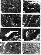 issued from : P. Koomen & J.C. von Vaupel Klein in Crustaceana, 1997, 70 (5). [p.538, Pl.1]. Female: a, irrugular SLIT (at site thoracic somite 3); b, very small SLIT (at site endopodite segment 1 of P4); c, PEG (at site thoracic somites 4+5); d, PEG (at sitethoracic somites 4+5), note the integumental depression at its base; e, SPIN (at site basipodite segment 2 of P1); f, integumental organ (at site endopodite segment 1 of P4), turning out to be a TUB; g, circular integumental structure on the lateral (external) face of the segment 6 of A2 (as seen by DIC); h, same (as seen by SEM), the straight structure is debis and no part of the integument. Scale bars = 0.001 mm. a--f, h by SEM.
|
 issued from : H.B. Owre & M. Foyo in Fauna Caribaea, 1, Crustacea, 1: Copepoda. Copepods of the Florida Current. 1967. [p.28, Fig.131]. Female: 131, basipodite 1 of P4.
|
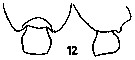 Issued from : E.L. Markhaseva in USSR Acad. Sci., Zool. Inst., Explor. Fauna Seas. Marine Plankton, 1989, 41 (49). [p.52, Fig.12]. Female: Thoracic segment 5 and genital segment. Nota from the key: 1 - Genital segment slightly asymmetrical. 2 - Thoracic posterior corners rounded. 3 - Thoracic posterior corners without spines, slightly asymmetrical with small rounded protuberance backward on the right (lateral view). 4 - Genital segment broadened in its middle (dorsal view). Coxopodite of P4 with 7-10 spines.
|
 Pseudochirella obesa Pseudochirella obesa Male: 1 - Exopodite 2 of left P5 with tooth. 2 - Exopodite 2 of left P5 2 teeth distally. 3 - Posterior corners of last thoracic segment without spines. 4 - Exopodite 2 of right P5 with excavation and rounded swelling proximally. Complement after Markhaseva (1996, p.276): Cephalothorax about 3 times longer than urosome. Cephalon and thoracic segment 1, as 4th and 5th segments separated. Posterior corners of last thoracic segment rounded, without spines. A1 21-segmented, reaching urosomal segment 3. Oral parts and their setation reduced in comparison with those in females. Exopodite of P1 3-segmented; external spine of exopodal segment 1 small. Endopodite of P2 1-segmented, but line of fusion poorly visible. Coxopodite of P4 without spines. Exopodal segment 2 of left P5 with excavation and rounded projection in proximal part of segment.
|
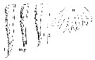
 issued from : Tanaka O. in Publ. Seto mar. Biol., Lab., 1957, 6 (2). [p.196]. As Pseudochirella tuberculata. Female A1: proportional lengths of segments. -A1 23-segmented, extends ro distal end of genital segment.
|
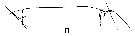 issued from : J.C. von Vaupel Klein in Crustaceana, Supplt 9, Studies on Copepoda, III, 1984. [p.66, Fig.7, n]. Pseudichirella obesa: Structural feature of exopodite 1 and exopodite 2 of A2 (lateral aspects of left appendages and medial views of right antennal parts). n, medial.
|
 issued from : J.C. von Vaupel Klein in Crustaceana, Supplt 9, Studies on Copepoda, III, 1984. [p.68, Fig.8, i]. Pseudochirella obesa: Structure and setal complement of endopod 1 of Md (left appendage, anterior face drawn from posterior aspect). 3 setae condition, similar to Batheuchaeta lamellata.
|
 issued from : J.C. von Vaupel Klein in Crustaceana, Supplt 9, Studies on Copepoda, III, 1984. [p.68, Fig.8, m]. Pseudochirella obesa: Accessory setae on the endopodite 2 of Md (left, posterior). 2 unequal setae.
|
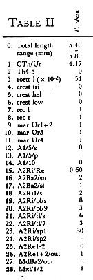 Issued from : J.C. von Vaupel Klein in Crustaceana, (Supplement) 9, 1984. [p.93, Table II ]. Pseudochirella obesa Female: Datamatrix stating observed states of characters from Table I (p.87-90) presently examined; nos. refer to the input nos. used in Table I (see to the family Aetideidae).
|
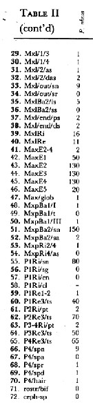 Issued from : J.C. von Vaupel Klein in Crustaceana, (Supplement) 9, 1984. [p.94, Table II (cont' d) ]. Pseudochirella obesa Female: Datamatrix stating observed states of characters from Table I (p.87-90) presently examined; nos. refer to the input nos. used in Table I (see to the family Aetideidae).
|
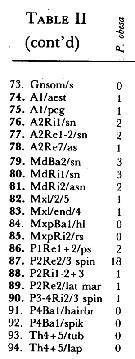 Issued from : J.C. von Vaupel Klein in Crustaceana, (Supplement) 9, 1984. [p.94, Table II (cont' d) ]. Pseudochirella obesa Female: Datamatrix stating observed states of characters from Table I (p.87-90) presently examined; nos. refer to the input nos. used in Table I (see to the family Aetideidae).
| | | | | Ref. compl.: | | | Deevey & Brooks, 1977 (p.256, tab.2, Station "S"); Razouls & al., 2000 (Appendix) | | | | NZ: | 11 | | |
|
Carte de distribution de Pseudochirella obesa par zones géographiques
|
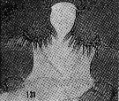
| | | | | | | | | 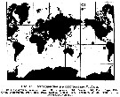 Issued from : E.L. Markhaseva in Issled. Fauny Morei, 1989, 41 (49). [p.58, Fig.16]. Issued from : E.L. Markhaseva in Issled. Fauny Morei, 1989, 41 (49). [p.58, Fig.16].
Geographical distribution of P. obesa .
1: from literature; 2: present occurrences.
Nota (1996, p.276): Tropical-borreal species, panoceanic. The northernmost locality in the region of the Strait of Davis (Jespersen, 1934), the southernmost in the region of New Zeland (Bradford & Jillett, 1980). The Chjna Sea (original data). |
| | | | Loc: | | | G. of Guinea, Cape Verde Is., off Morocco-Mauritania, off Portugal, Bay of Biscay, Barbados Is., Florida, Sargasso Sea, off Bermuda: Station "S" (32°10'N, 64°30'W), S Davis Strait, W Indian, Celebes Sea, China Seas, Japan (Izu), Pacif. (equatorial), off Peru, New Zealand, off Juan Fernandez Is.
NE Atlantic (Bay of Biscay). | | | | N: | 15 | | | | Lg.: | | | (1) F: 5; (8) M: 5,76; (37) F: 6,2-5; (60) F: 6-5,2; (56) F: 6,1; (111) F: 6-5,7; (159) F: 5,85-5,55; (201) F: 5,5; (1257) F: 5,4-5,8; {F: 5,00-6,20; M: 5,76} | | | | Rem.: | méso-bathypélagique.
Voir aussi les remarques en anglais | | | Dernière mise à jour : 22/02/2021 | |
|
|
 Toute utilisation de ce site pour une publication sera mentionnée avec la référence suivante : Toute utilisation de ce site pour une publication sera mentionnée avec la référence suivante :
Razouls C., Desreumaux N., Kouwenberg J. et de Bovée F., 2005-2025. - Biodiversité des Copépodes planctoniques marins (morphologie, répartition géographique et données biologiques). Sorbonne Université, CNRS. Disponible sur http://copepodes.obs-banyuls.fr [Accédé le 26 août 2025] © copyright 2005-2025 Sorbonne Université, CNRS
|
|
|
|


;)
;)
;)
;)
;)
;)
;)
;)
;)
;)
;)
;)
;)
;)
;)
;)
;)
;)
;)
;)
;)
;)
;)
;)
;)

;)
