|
|
 |
Fiche d'espèce de Copépode |
|
|
Calanoida ( Ordre ) |
|
|
|
Epacteriscoidea ( Superfamille ) |
|
|
|
Ridgewayiidae ( Famille ) |
|
|
|
Ridgewayia ( Genre ) |
|
|
| |
Ridgewayia stygia Ohtsuka, Kase & Boxshall, 2000 (F,M) | |
| | | | | | | Ref.: | | | Ohtsuka, Kase & Boxshall, 2000 (p.202, figs.F,M) | 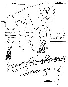 issued from : S. Ohtsuka, T. Kase & G.A. Boxshall in Species Diversity, 2000, 5. [p.203, Fig.1]. Female (from 07°7.185'N, 134°14.147'E): A-B, habitus (dorsal and lateral, respectvely); C, forehead (ventral) showing rostrum and labrum; D, ventrolateral corners of 2nd to 4th pedigers (pointed posterior corners arrowed); Eurosome (dorsal); F, genital operculum; G, A1 (segments I to XIX; H, A1 (segments XX to XXVII-XXVIII). Nota : Cephalosome separate from 1st pediger, 4th and 5th separate. Rostrum remarkably produced ventrally, rounded at tip, with pair of secretory gland openings subterminally. Labrum protruded ventrally, bearing tuft of fine setules on median swelling ; medial papilla present anterior to labrum, beneath rostrum. Urosome 4-segmented. Genital double-somite slightly asymmetrical, protruding laterally more on left side than on right ; seminal receptacle ( ?) visible by transparency only in left dorsal view ; genital operculum located nearly at posterior margin. Posterior margin of first 3 urosomites striated ; anal somite small with operculum convex at tip. Caudal rami symmetrical, almost entirely covered by minute spinules and granules ; seta I apparently lacking ; seta II spiniform ; seta V longest ; seta VII hirsute, located dorsally, near bases of setae V and VI. A1 26-segmented, with 9th (X) and 10th (XI) segments partly fused. Patch of spinules present at anterodistal corner of compound second segment (II-III) ; transverse row of spinules on each segment from 13th (XIV) to 22nd (XXIII).
|
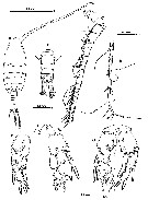
 issued from : S. Ohtsuka, T. Kase & G.A. Boxshall in Species Diversity, 2000, 5. [p.204, Fig.2]. Female: A, A2; B, Md; C, Mx1; D, Mx2; E, Mxp. Nota : A2 with 2-segmented endopod and indistinctly 8-segmented exopod. Coxa bearing spinulose seta and patch of setules; basis with 2 setae of unequal length. Setal formula of exopod 1, 1, 1, 1, 1, 1, 1, 4. First endopod segment with 2 setae subterminally; 2nd segment bilobed, proximal and distal lobes with 9 and 7 setae, respectively. Mx1 well developed ; praecoxal arthrite bearing 1 anterior and 4 posterior setae and 1 minute, 4 spinulose, and 5 heavily sclerotized setae along inner margin ; coxal epipodite with 2 short proximal and 7 long distal setae ; basal exite bearing seta ; coxal and 2 basal endites having 5, 4, and 5 setae, respectively ; exopod unisegmented, with 11 setae along outer margin ; endopod 2-segmented, setal formula (4+4), 7. Mx2 with praecoxa and coxa incompletely fused ; 1st praecoxal endite with 5 long and 1 proximally directed setae in addition to one rudimentary element ; setal formula of 2nd praecoxal to basal endites 3, 3, 3, 4 ; endopod indistinctly 4-segmented, setal formula 3, 2, 2, 3. Mxp with setal formula of syncoxal endites 1, 2, 4, 3, each endite ornamented with patch of setules ; basis bearing 3 inner setae and row of setules along entire inner margin ; 1st endopod segment distinctly separate from basis, setal formula of endopod segments 2, 4, 4, 3, (3+1), 4.
|
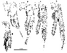 issued from : S. Ohtsuka, T. Kase & G.A. Boxshall in Species Diversity, 2000, 5. [p.205, Fig.3]. Female: A-C, P1 to P4, respectively (anterior surface); E, P5 (posterior surface).
|
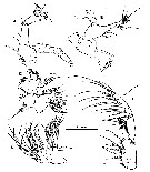
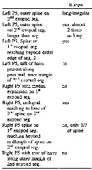
 issued from : S. Ohtsuka, T. Kase & G.A. Boxshall in Species Diversity, 2000, 5. [p.206, Fig.4]. Male: A, habitus (dorsal); B, urosome (dorsal); C, rostrum and antennulary segments I to XVIII; D, A1 (segments XIX to XXVII-XXVIII); E, right P5 (anterior surface); F, left P5 (anterior surface; G, P5 (posterior surface). Nota : Right A1 weakly geniculate, 24 free segments. Geniculation between 19th (XX) and 20th (XXI-XXIII) segments. Patch of spinules present anteriorly on compound second segment ; transverse row of spinules on each segment from 14th (XV) to 18th (XIX) ; 20th segment compound (XXI-XXIII), bearing 3 transverse spinular rows (these presumably indicating margins of original segments.P5 with intercoxal sclerite and both coxae completely fused to form common base on posterior surface, incompletely fused on anterior surface. Right leg with 2-segmented exopod and unisegmented endopod. Left leg with 3-segmented exopod and 1-segmented endopod
|
 issued from : D.F. Figueroa & K.L. Hoefel in J. Crustacean Biol., 2008, 28 (1). [p.145, Table 3]. Characters male of R. stygia.
|
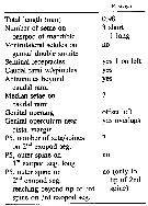 issued from : D.F. Figueroa & K.L. Hoefel in J. Crustacean Biol., 2008, 28 (1). [p.145, Table 2]. Characters female of R. stygia.
| | | | | NZ: | 1 | | |
|
Carte de distribution de Ridgewayia stygia par zones géographiques
|
| | | | Loc: | | | W Pacif. (Ngemelis Island, Palau Is.) | | | | N: | 1 | | | | Lg.: | | | (1078) F: 0,98; M: 0,90-0,98; {F: 0,98; M: 0,90-0,98} | | | | Rem.: | In submarine cave.
For Ohtsuka & al. (2000, p.201), this species is similar to R. fosshageni, but can be distinguished from the latter by the location of the female genital operculum and the structure of P5 of both sexes. The affinities of the new species appear to lie with the North Atlantic/Mediterranean R. marki species-group rather than the Indo-West Pacific R. typica species-group. This suggests that, within the Tethys Sea, westward dispersal of marine copepods from the Caribbean Region extended as far as the western Central Pacific rather than only to the eastern Central Pacific.
After the authors (p.208) the gut of the female was packed with fragments of small crustaceans and unidentified remains. This cavernicolous species is tinged with reddish brown. | | | Dernière mise à jour : 30/01/2015 | |
|
|
 Toute utilisation de ce site pour une publication sera mentionnée avec la référence suivante : Toute utilisation de ce site pour une publication sera mentionnée avec la référence suivante :
Razouls C., Desreumaux N., Kouwenberg J. et de Bovée F., 2005-2024. - Biodiversité des Copépodes planctoniques marins (morphologie, répartition géographique et données biologiques). Sorbonne Université, CNRS. Disponible sur http://copepodes.obs-banyuls.fr [Accédé le 27 avril 2024] © copyright 2005-2024 Sorbonne Université, CNRS
|
|
|
|


;)
;)
;)
;)
;)
;)
