|
|
 |
Fiche d'espèce de Copépode |
|
|
Calanoida ( Ordre ) |
|
|
|
Calanoidea ( Superfamille ) |
|
|
|
Paracalanidae ( Famille ) |
|
|
|
Parvocalanus ( Genre ) |
|
|
| |
Parvocalanus leei Moon, Youn & Soh, 2014 (F,M) | |
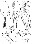
| | | | | | | Ref.: | | | Moon, Youn & Soh, 2014 (p. 32, Descr. F, M, figs. F,M, Rem., Table I, p.45: Comparison of female species) | 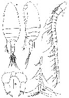 Issued from : S. Y. Moon, S.-H. Youn & H.Y. Soh in ZooKeys, 2014, 456. [p.33, Fig.2]. Female (from SW Korea): A-B, habitus (dorsal and lateral, respectively); C, A1; D, P5. Scale bars: 0.1 mm (A, B); 0.005 mm (C); 0.025 mm (D). Nota: - Prosome length 2.7 times as long as urosome - Cephalosome and 1st pedigerous somite completely fused, 4th and 5th pedigers completely separated. - Proportional length (%) of prosomites 68.2 : 11.6 : 10.4 : 5.5 : 4.3 = 100. - Rostrum short, broad. - Urosome 4-segmented. - Genital double-somite symmetrical, swollen anterolaterally, 1.12 times wider than long - Caudal rami nearly symmetrical, 2.4 times longer than wide, each with row of hairs on anterior inner margin; 5 caudal setae: seta II and VI spiniform, III, IV and VII setiform and plumose. - Proportional length (%) of urosomites and caudal rami 28.3 : 9.5 : 10.4 : 27.2 : 24.6 = 100. - A1 25-segmented, extending to midlength of anal somite; ancestral segments II to IV and XXVII-XXVIII completely fused. - P5 2-segmented, proximal segment smooth, unarmed; distal segment 2.15 times as long as wide with row of spinules subdistally and with 2 unequal terminal spines, inner longest, denticulated along distal part of outer margin.
|
 Issued from : S. Y. Moon, S.-H. Youn & H.Y. Soh in ZooKeys, 2014, 456. [p.35, Fig.3]. Female: A, rostrum (ventral view); B, urosome (dorsal); C, genital double-somite and urosomite 2 and 3 (ventral); D, A2; E, Md; F, gnathobase (cutting edge); G, Mx1; H, Mx2. Scale bars: 0.05 mm (A-C); 0.025 mm (D-H). Nota: - Genital double-somite with genital system remarkably symmetrical with paired gonopores located eavh side, genital operculum midventrally, rounded, about 1/3 as long as genital double-somite. - A2 biramous, coxa, basis, endopod 2-segmented, 1st endopodal segment with 2 setae, 2nd endopodal segment with 8 setae about midway of inner margin, 7 setae terminally, and oblique row of tiny spinules midway and subdistally on outer margin; exopod 7-segmented, setal formula 1, 3, 1, 1, 1, 1, 4. - Md gnathobase well developed, cutting edge with short teeth and dorsal single seta; mandibular palp biramous, basis with 4 setae, exopod 5-segmented, setal formula 1, 1, 1, 1, 2; endopod 2-segmented, proximal and distal segments with 4 and 11 setae, respectively; oblique row of tiny spinules subterminally on distal segment. - Mx1: praecoxa and coxa completely fused; praecoxal arthrite (inner lobe 1) with 14 elements, and with several rows of spinules on anterior surface; coxal endite with 3 setae; coxal epipodite with 9 setae; proximal basl endite with 3 setae, distal basl endite with 4 setae; endopod 3-segmented, setal formula 3, 3, 7; exopod unsegmented with 11 marginal setae. - Mx2: praecoxa and coxa completely fused, each with 2 endites, posteromedial surface furnished with setules; proximal praecoxal endite with 6 setae, distal endite with 3 setae; coxal endites each with 3 setae; coxal epipodite seta present; basis with 4 setae and row of spinules subterminally; endopod 4-segmented, 1st and 2nd segments incompletely separated with setal formula 1, 2, 2, 3.
|
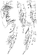 Issued from : S. Y. Moon, S.-H. Youn & H.Y. Soh in ZooKeys, 2014, 456. [p.38, Fig.4]. Female: A, Mxp; B, P1 (dorsal view); C, P2 (dorsal view); D, P3 (dorsal view); E, P4 (dorsal view). Scale bars: 0.05 mm. (A-E) .
Nota:
- Mxp: stncoxa robust with setal formula 1, 2, 3, 4 and oblique rows of spinules on anterior surface; basis with 3 setae and setules on medial surface; endopod 6-segmented, 1st and 2nd segments completely separated with setal formula 2, 3, 4, 3, 3+1, 4.
|
 Issued from : S. Y. Moon, S.-H. Youn & H.Y. Soh in ZooKeys, 2014, 456. [p.33, Fig.2]. Female: Armature formula of swimming legs 1-4 (P1-P4). Roman numerals indicate spines, Arabic numerals indicate setae.
|
 Issued from : S. Y. Moon, S.-H. Youn & H.Y. Soh in ZooKeys, 2014, 456. [p.39, Fig.2]. Male: A-B, habitus (dorsal and lateral, respectively) ; C, A1; D, A2; E, Md; F, Mx1; G, Mxp. Scale bars: 0.1 mm (A, B); 0.025 mm (C-G). Nota: - Body plumper than female. - Prosome 5-segmented, length 2.6 times as long as urosome (including caudal rami). - Cephalosome without dorsal hump; 1st pedigerous somite completely fused, 4th and 5th pedigers completely separated. - Proportional length (%) of prosomites 60.5 : 13.5 : 13.5 : 12.5 = 1400 - Rostrum as in female. - Urosome 5-segmented, 1st urosomal somite longest; proportional length (%) of urosomites 25.2 : 20.3 : 16.6 : 14.6 : 23.3 = 100. - Caudal rami nearly symmetrical, abouy 2.2 times longer than wide, each with 5 setae (ancestral setae I and II wanting; see in Huys & Boxshall, 1991: Copepod evolution, p.28). - A1 19-segmented, extending to distal part of 3rd urosomite; ancestral segments I-IV, V-VIII, IX-X, XI-XII, and XXVII-XXVIII completely fused. - A2 biramous but vestigial; coxa and basis completely fused, both unarmed; endopod 2-segmented, proximal naked, distal with 5 setae about midway of inner margin and 6 terminal setae; exopod 5-segmented, setal formula 0, 1, 1, 1, 2. - Md coxal gnathobase lacking; basis unarmed; exopod 5-segmented, setal formula 1, 1, 1, 1, 2; endopod 2-segmented, 1st segment with single seta, 2nd segment with 8 setae. - Mx1 vestigial ( coxal epipodite with 5 setae). - Mxp: comprising robust syncoxa, basis, and 3-segmented endopod; syncoxa with 1 seta and row of tiny spinules on inner distal edge; basis medially with 1 stout seta; prpximal endopodal segment with 6 setae, of whoch distal seta robust; 2nd segment with 1 seta; distal segment with 3 setae
|
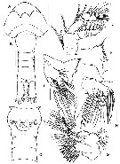
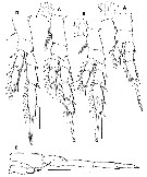 Issued from : S. Y. Moon, S.-H. Youn & H.Y. Soh in ZooKeys, 2014, 456. [p.40, Fig.2]. Male: A-D, P1 to P4 respectively (all dorsal view); E, P5 (dorsal view). Scale bars: 0.05 mm.
Nota:
- In male, swimming legs generally as in female, but with some differences: P1 laks posterior spinules on coxa, basis and endopod unadorned and the 3rd exopodal segment laks of row of spinules on posterior surface; P2 has the 2nd and distal endopodal segments with denticles on outer edge; distal endopodal segment without row of spinules on mediolateral margins; P3 has the 2nd and distal endopodal segments with denticles on outer edge; 2nd exopodal segment without row of spinules on the posterodistal margin; P4 has the 2nd and dital endopodal segments with denticles on outer edge; and 1st exopodal segment without row of spinules on the anterodistal margin.
- P5 strongly asymmetrical and uniramous.
- P5 strongly asymmetrical, uniramous; Right leg 5-segmenyed and longer than 2nd urosomal segment; basis and 1st exopodal segment unarmed; 2nd exopodal segment with pointed process on distomedial angle; distal segment with 2 pointed processes, inner tiny. Left leg 3-segmented; distal segment with tiny outer apical spine, inner apocal spine long, 9 times as long as outer spine.
|
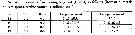
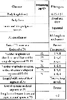 Issued from : S. Y. Moon, S.-H. Youn & H.Y. Soh in ZooKeys, 2014, 456. [p.42, Table I]. Female : marphological characters. Compare with other species of Parvocalanus : ( arabiensis, crassirostris, dubia, elegans, latus, serratipes, scotti).
| | | | | NZ: | 1 | | |
|
Carte de distribution de Parvocalanus leei par zones géographiques
|
| | | | | | | Loc: | | | Yellow Sea, Korea;
Type locality: 34°46'10'' N, 126°20'24'' E. | | | | N: | 1 | | | | Lg.: | | | (1315) F: 0,75-0,92; M: 0,49-0,69; {F: 0,75-0,92; M: 0,49-0,69} | | | | Rem.: | For the authors (p.37, 41), the adult female is very similar to P. arabiensis, crassirostris, latus, scotti, but differs by any differences.
Parvocalanus leei is distinguished from the rest of members of its genus based on the characteristics of the female : 4th and 5th pedigerous somites completely separated; large size (more than 0.7 mm), presence of spinules on the distal end of distal segment of P5 (see the comparison of morphological characteristics in Table I, p.42) | | | Dernière mise à jour : 06/04/2018 | |
|
|
 Toute utilisation de ce site pour une publication sera mentionnée avec la référence suivante : Toute utilisation de ce site pour une publication sera mentionnée avec la référence suivante :
Razouls C., Desreumaux N., Kouwenberg J. et de Bovée F., 2005-2024. - Biodiversité des Copépodes planctoniques marins (morphologie, répartition géographique et données biologiques). Sorbonne Université, CNRS. Disponible sur http://copepodes.obs-banyuls.fr [Accédé le 28 avril 2024] © copyright 2005-2024 Sorbonne Université, CNRS
|
|
|
|


;)
;)
;)
;)
;)
;)

