|
|
 |
Fiche d'espèce de Copépode |
|
|
Calanoida ( Ordre ) |
|
|
|
Clausocalanoidea ( Superfamille ) |
|
|
|
Aetideidae ( Famille ) |
|
|
|
Valdiviella ( Genre ) |
|
|
| |
Valdiviella insignis Farran, 1908 (F,M) | |
| | | | | | | Syn.: | Valdiviella gigas : A. Scott, 1909 (p.77, figs.M, Juv, no adult);
? Valdiviella ignota Sewell, 1929 (p.137, Descr.M, figs.M, );
no V. insignis : Heptner, 1971 (p.113, figs. juv.M, Rem.);
? Valdiviella sp. Vives, 1967 (p.564, tab.II) | | | | Ref.: | | | Farran, 1908 b (p.45, Descr.F, figs.F); Wolfenden, 1911 (p.247, figs.F, Rem.); With, 1915 (p.154, figs.F, juv.M); Lysholm & Nordgaard, 1921 (p.18); Sars, 1925 (p.98, figs.F,M); Sewell, 1929 (p.135, fig.M); Rose, 1933 a (p.125, figs.F,M); Jespersen, 1934 (p.67, fig.16); 1940 (p.28); Lysholm & al., 1945 (p.20); Sewell, 1947 (p.109); Vervoort, 1957 (p.85, Rem.); 1963 b (p.155, Rem.); Owre & Foyo, 1967 (p.50, figs.F,M); Tanaka & Omori,1967 (p.244); 1970 b (p.151, figs.M, Rem.); Razouls, 1972 (p.94, Annexe: p.46, figs.F,M); Zvereva, 1975 (p.1893, figs.F); Park, 1978 (p.197, figs.F); Bradford & Jillett, 1980 (p.83, 84, figs.F,M, fig. 73, distribution chart); Chihara & Murano, 1997 (p.690, Pl.50: F,M); Bradford-Grieve & al., 1999 (p.880, 927, figs.F,M); Vives & Shmeleva, 2007 (p.608, figs.F,M, Rem.) | 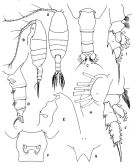 issued from : T. Park in Antarctic Res. Ser. Washington, 1978, 27. [p.198, Fig.62]. Female: A, habitus (dorsal view); B, idem (left lateral side); C, posterior part of metasome and urosome (dorsal); D, idem (lateral); E, forehead (lateral); F, genital segment (ventral); G, rostrum (anterior); H, outer lobe of Mx1; I, P1 (anterior); J, P2 (idem).
|
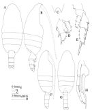 issued from : J.M. Bradford & J.B. Jillett in Mem. N.Z. oceanogr. Inst., 86, 1980. [p.85, Fig.59]. Female: A, habitus (dorsal); B, idem (lateral right side); C, rostrum; D, P1; E, P4. Male: F, habitus (lateral right side); G, idem (dorsal); H, P5.
|
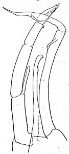
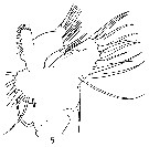
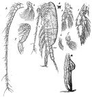 Issued from : G.O. Sars in Résult. Camp. Scient. Prince Albert I, 69, pls.1-127 (1924). [Pl.XXVIII, figs.1-10]. Male: 1, habitus (lateral left side); 2, rostrum (frontal view); 3, A1; 4, Md (mandibular palp); 5, Mx1; 6, Mx2; 7, Mxp; 8, P1; 9, P4; 10, P5.
|
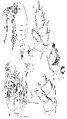
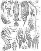 Issued from : G.O. Sars in Résult. Camp. Scient. Prince Albert I, 69, pls.1-127 (1924). [Pl.XXVII, figs.1-16]. Female: 1, habitus (dorsal, with eggs); 2, idem (lateral left side); 3, forehead (lateral); 4, rostrum (frontal view); 5, A1; 6, A2; 7, Md; 8, Mx1; 9, Mx2; 10, Mxp; 11, P1; 12, P2; 13, P4; 14, 1st and 2nd urosomal segments (lateral left side); 15, genital segment (ventral); 16, anal segment and caudal ramus.
|
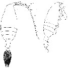
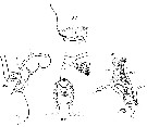
 issued from : R.B.S. Sewell in Mem. Indian Mus., 1929, X. [p.136, Fig.51]. Male (from off SE Sri Lanka): P5.
|
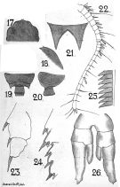 issued from : A. Scott in Siboga-Expedition, 1909, XIX a. [Plate XXII, Figs.17-26]. As Valdiviella gigas. Juvenile non adult. Male (from Banda Sea): 17, forehead (dorsal); 18, idem (lateral); 19, last thoracic and fgenital segments (dorsal); 20, idem (left side); 21, rostrum; 22, A1; 23, P1 (exopodite only); 24, P2 (outer margin of exopodite); 25, part of terminal spine of exopodite of P3; P5.
|
 issued from : R.N. Wolfenden in Die Marinen Copepoden der Deutschen Südpolar-Expedition 1901-1903, 1911. [Pl.XL, Figs.6-7]. Female: 6, habitus (dorsal); 7, idem (lateral).
|
 issued from : O. Tanaka & M. Omori in Publ. Seto mar. Biol. Lab., 1970, XVIII, 3. [p.152, Fig.14]. Male (from Izu Region, Japan): a, forehead (lateral); b, last thoracic segment and genital segment (left latera); c, Mx1; d, Mx2; e, P1; f, P5. Nota: Head and 1st thoracic segment fused, 4th and 5th fused. 5st thoracic segment with a short spine on the lateral distal margin. Urosomal segments and caudal rami in the proportional lengths 19:28:22:16:3:12 = 100. 2nd to 5th urosomal segments striated with fine teeth on the distal margin. A2 exopod 7-segmented, about equal in length of exopod; endopod with 6 setae on the outer lobe, and 6 setae on the inner lobe. Md with cutting blade reduced, 2nd basal segment bears 1 seta, endopod with 1 seta on the 1st segment, and 8 setae on the 2nd segment. Mx1 reduced, the outer lobe lacks setae; exopod with 11 setae, of which the apical one is small; the endopod has 3, and the 2nd basal segmenthas 2 setae; the inner lobes are reduced. Mx2: 1st and 2nd lobes lack setae, 3rd lobe with 1 seta, 4th lobe with 2 setae, 5th lobe with 1 seta and 2 spines; endopod with 5 setae. Mxp: 1st basal segment shorter than the 2nd, and has 1 spine on the anterior distal margin, 2nd basal segment with 3 setae on the anterior margin, and 1 seta on the anterior distal margin; endopod with 3, 3, 2, 2 and 3 setae on the 1st to 5th segments respectively. Caudal ramus about as long as wide P5 agree well with Sewell's (1929) except that the distal segment of the right leg has a small process opposite to the projecting lamella, and that the 1st segment of the exopod of the left leg has a small triangular process near the inner distal margin.
|
 issued from : G.P. Farran in Fish. Ire. Sci. Invest., 1906, II [1908]. [Pl. III, Figs.1-6]. Female (from W Ireland): 1, habitus (lateral); 2, P2; 3, Md (cutting edge); 4, Mx2; 5, P3; 6, P1. Nota: A1 23-segmented (segments 8-9 and 24-25 fused). P2 differs from that of V. oligarthra in having the exopodite imperfectly 3-segmented, the segmentation between the 1st and 2nd segments being faintly indicated though not functional; the endopodite has only a very faint indication of segmentation between the 1st and 2nd segments. Colour, a bright red, deepest on the limbs and mouth parts. Egg sacs orange.
|
 issued from : G.P. Farran in Fish. Ire. Sci. Invest., 1906, II [1908]. [Pl. IV, Fig.5]. Female: 5, Mx1.
|
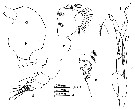
 issued from : C. With in The Danish Ingolf-Expedition, Copepoda I, 1915, III, 4. [p.156, Text-fig. 44]. Female (from 61°30'N, 17°08'W): a, last thoracic segment and urosome with egg-sac (lateral); b, endopodite segment 3 of P3 (anterior view with gland). Nota: Metasome with distinctly rounded lateral cornesrs without tuft of hairs, rather clumsy. Rostrum consists of 2 short, well separated spines. Head and 1st thoracic segment fused: 4th and 5th fused. Prosome 2.5 as long as the abdomen, which has the proportional length 45, 35, 25, 11, 11. Euchaeta; no proximal setae were found in segments 10, 11 and 20-23; rather short triangularly pointed aesthetes were observed in segments 5, 9, 12, 14 and 19.; the segments 8-9 are 1.3 as long as segment 7, and again 1.2 as long as 13; segment 17, which is 3 times as long as the segment 14, is a little longer than the segment 19, and 1.4 as long as 24-25.
A2: exopodite a little longer than the endopodite; 1st segment well developed without any seta, and the 2nd has a short terminal seta.
Md: teeth developed in a curious way (see in Farran).
P1 has the endopodite 1 and 2 completely fused (the endopodite 1 only indicated by a medial incision and a powerful outer spine; ; as shown in fig.2a, a glandular calal and pore was present in the outer margin of the 2nd and 3rd division; the pore in the former was covered by a spine-shaped mass, and both pores were surrounded by numerous hairs. P2 has the distinction between exopodal segments 1-2 indicated by a medial incision and a well developed lateral spine; the articular membrane is anteriorly represented by a faint line, which is not seen posteriorly. Ehe unsegmented endopod has near the tip on the anterior surface a minute pore (text-fig.44b). near the base of outer seta of exopodal segment 2 and outer seta of exopodal segment 3 but not in exopodal segment 1, wide glandular pores are found, in connexion with big sacs with glandular cells, which are placed proximally to the articular membrane between the exopodal segments 1-2 and 2-3 respectively. P3 differs from P2 by the distinct articular line between endopodal segments 2-3; a distinct glandular pore is found at the base of outer seta of endopodal segment 1. P4 is in main features like P3, but the basis has comparatively few hairs along the inner margin.
|
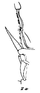 issued from : C. With in The Danish Ingolf-Expedition, Copepoda I, 1915, III, 4. [Pl. VI, Fig.2, a]. Female: left P1 (outer margin of 2nd and 3rd exopodal segment in anterior view).
|
 issued from : C. With in The Danish Ingolf-Expedition, Copepoda I, 1915, III, 4. [Pl. VI, Fig.2, b-e]. Female: b, labrum (anterior surface); c, oral surface of the labrum; d, lamina labialis and serrula 6-dentata; e, labial lobes. Nota : Epistoma represented by a short protuberance, which is placed somewhat behind the insertion of A2 ; it is steep in front, and smoothly sloping behind, and appears quite smooth. Labrum rather prominent ; on the anterior surface, somewhat in front of the free margin, a transverse row of fairly long stiff hairs (fig.2b) ; more laterally, partly covered by this, an oblique row ; anda long the hinder margin the usual row of numerous somewhat curled hairs (fig.2e). Oral surface of the labrum (fig.2c) with the group 1 placed laterally, and consisting of numerous short setae or granules ; the group 2, which is well separated from this, and consists of comparatively few longer setae, is closely followed by groups 3-4 ; the group 5 consists of more numerous and comparatively longer setae. The lamina labialis does not show any features of interest (fig.2d) ; the area in front of it is most like that of Chiridius ; it consists of a lateral, somewhat convex, row of fairly long setae and 2 median, well separated, groups (as shown in fig.). Behind the lamina and between the labial lobes, no setae were observed.
|
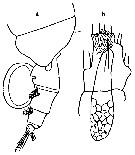
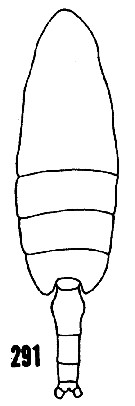 issued from : H.B. Owre & M. Foyo in Fauna Caribaea, 1967, 1, Crustacea, 1: Copepoda. Copepods of the Florida Current, 1967. [p.50, Fig.291]. After Giesbrecht, 1892. Female: 291, habitus (dorsal). Nota: A1 extending posteriorly to the end of the metasome.
|
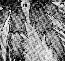 issued from : H.B. Owre & M. Foyo in Fauna Caribaea, 1967, 1, Crustacea, 1967, 1: Copepoda. Copepods of the Florida Current. [p.51, Figs.296, 297]. Female: 296, P2; 297, P4. Nota: Exopodal segments 1 and 2 of P2 and P4 completely fused.
|
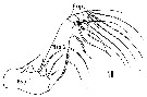 Issued from : H.B. Owre & M. Foyo in Fauna Caribaea, 1, Crustacea, 1: Copepoda. Copepods of the Florida Current, 1967. [p.12, Fig.18]. Female: 18, Mxp. Bsp 1 = Coxa; Bsp 2 = Basis; Enp = endopod.
| | | | | Ref. compl.: | | | Sewell, 1948 (p.384, 500, 521, 524, 546); C.B. Wilson, 1950 (p.350); Grice, 1963 a (p.495); Grice & Hulsemann, 1965 (p.223); 1967 (p.15); 1968 (tab.2); Razouls, 1972 (p.100); Björnberg, 1973 (p.325, 389); Deevey & Brooks, 1977 (p.256, tab.2, Station "S"); Kovalev & Shmeleva, 1982 (p.83); Vives, 1982 (p.291); Lozano Soldevilla & al., 1988 (p.58); Razouls & al., 2000 (p.343, Appendix); Holmes, 2001 (p.51); Park & Ferrari, 2009 (p.143, Table 4, Appendix 1, biogeography); El Arraj & al., 2017 (p.272, table 2, seasonal composition); Belmonte, 2018 (p.273, Table I: Italian zones) | | | | NZ: | 16 | | |
|
Carte de distribution de Valdiviella insignis par zones géographiques
|
| | | | | | | | | | | | | Loc: | | | Antarct. (Indian, SE Pacif.), sub-Antarct. (SW Atlant., SE Pacif.), Atlant. (tropical, temperate and boreal), off da Trindade Is., G. of Guinea, Morocco-Mauritania, Canary Is.- Azores, off Madeira, Barbados Is., Caribbean Sea, off Bermuda: Station "S" (32°10'N, 64°30'W), Sargasso Sea, Davis Strait, Iceland, off W Ireland, off W Scotland, Gibraltar, W Medit. (Banyuls, Tyrrhenian Sea), Arabian Sea, Indian, off E Sri Lanka, G. of Bengal, Indonesia-Malaysia, Philippines, Japan (Izu), New Zealand, S Pacif., SW Galapagos, off Juan Fernandez Is., Chili (N & S, sub-Antarct.) | | | | N: | 33 | | | | Lg.: | | | (1) F: 12; (5) Juv.M: 8; (7) F: 11,75; (11) F: 11,5; (14) F: 11-8,5; M: 8,56-8,02; (20) F: 11,5-10,5; (24) F: 12-11,5 ; (29) M: 8,7; (70) F: 11,3; (112) M: 8,7; (201) F: 11,1-10,6; M: 9,1-9; (866) M: 8,02-10; {F: 8,50-12,00; M: 8,02-10,0} | | | | Rem.: | bathypélagique.
Sampling depth (Antarct., sub-Antarct.): 1000-2000-4000 m.
Voir aussi les remarques en anglais | | | Dernière mise à jour : 25/10/2022 | |
|
|
 Toute utilisation de ce site pour une publication sera mentionnée avec la référence suivante : Toute utilisation de ce site pour une publication sera mentionnée avec la référence suivante :
Razouls C., Desreumaux N., Kouwenberg J. et de Bovée F., 2005-2025. - Biodiversité des Copépodes planctoniques marins (morphologie, répartition géographique et données biologiques). Sorbonne Université, CNRS. Disponible sur http://copepodes.obs-banyuls.fr [Accédé le 26 août 2025] © copyright 2005-2025 Sorbonne Université, CNRS
|
|
|
|


;)
;)
;)
;)
;)
;)
;)
;)
;)
;)
;)
;)
;)
;)
;)
;)

