|
|
 |
Fiche d'espèce de Copépode |
|
|
Calanoida ( Ordre ) |
|
|
|
Arietelloidea ( Superfamille ) |
|
|
|
Arietellidae ( Famille ) |
|
|
|
Paraugaptilus ( Genre ) |
|
|
| |
Paraugaptilus similis A. Scott, 1909 (F,M) | |
| | | | | | | Ref.: | | | A. Scott, 1909 (p.144, figs.F,M); Sewell, 1932 (p.329); Ohtsuka & al., 1994 (p.145, Redescr.F,M, figs.F,M) | 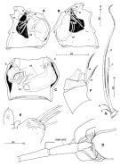 issued from : S. Ohtsuka, G.A. Boxshall & H.S.J. Roe in Bull. nat. Hist. Lond. (Zool.), 1994, 60 (2). [p.146, Fig.27]. Female (central Pacif.): A, genital double-somite (right lateral side); B, idem (left lateral side); C, idem (ventral); D, A1 (segments XXII-XXVIII); E, praecoxal arthrite, coxal endite and inner margin of basis of Mx1; F, 4th and 5th endopod segments of Mxp (innermost seta indicated by arrowhead, mid-margin seta on 4th segment missing); G, 6th endopod segment of Mxp. cd = copulatory duct; cp = copulatory pore; g = gonopore; rd = receptacle duct; o, oviduct; s, spermatothore remnant; sr = seminal receptacle. Scales in mm. Nota: Cephalosome and 1st pedigerous somite separate. A1 20-segmented (segments 7 to 9 and 11 and 12 partly fused near posterior margin, 20 and 21 incompletely fused with suture visible). Praecoxal arthrite of Mx1 with 5 spines (2 of which serrate subterminally, ornamented by minute spinules on both surfaces); coxal endite unarmed; coxal epipodite with 8 setae; basal seta and endopod absent. 4th and 5th endopod segments of Mxp each with unipinnate innermost seta, 6th with reduced setae (a and b), medium-length serrate seta(c) whose tip chitinized, and elongate seta (d) with row of sharp triangular spinules along inner margin. Geniital double-somite asymmetrical, widest at level of paired gonopores, each gonopore covered by operculum, anterior half opening; copulatory pores asymmetrical, right pore located medially on right ventral side, slit-like (approximately 0.043 mm in length), left pore located ventromedially at about 2/3 distance along double-somite, with round opening (about 0.027 mm in diameter); both right and left copulary ducts heavily chitinized, right duct shorter than left (thick); seminal receptacle relatively small, right round in shape, left smaller than right (spindle-shaped).
|
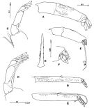 issued from : S. Ohtsuka, G.A. Boxshall & H.S.J. Roe in Bull. nat. Hist. Lond. (Zool.), 1994, 60 (2). [p.148, Fig.29]. Female: A, exopod of A2; B, 2nd endopod segment of A2; C, Md (mandibular exopod); D, basal spine of Mx2. Nota: 1st endopod segment of A2 without inner mid-length seta, 2nd segment about 1.8 times as long as first segment, with 1 inner short seta, and 5 setae and vestigial seta terminally; exopod indistinctly 6-segmented, 6th segment bulbous, unarmed, setal formula 0, 1, 1, 1, 1, 0. Endopod of mandibular palp absent; 1st exopod segment with vestigial seta, 5th segment with 1 normal and 1 reduced seta. Basal spine of Mx2 with 3 rows of spinules. Male: E, exopod of A2; F, detail of exopod of A2 (segmentsI IV to X); G, 2nd endopod segment of A2; Md (mandibular exopod). Nota: 2 nd endopod segment of A2 approximately 1.3 times as long as 1st segment, with 1 long and 1 short seta medially; exopod indistinctly 7-segmented (terminal compound segment bulbous (IX-X, 6th (VIII) with long seta, 7th (IX-X) unarmed. 1st exopod segment of Md palp with long seta. Scales in mm.
|
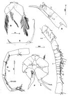 issued from : S. Ohtsuka, G.A. Boxshall & H.S.J. Roe in Bull. nat. Hist. Lond. (Zool.), 1994, 60 (2). [p.149, Fig.30]. Female: A, 1st and 2nd praecoxal endites of Mx2; B, P5 (anterior). Nota: 1st praecoxal endite of Mx2 with 1 serrate seta and 1 vestigial element, 2nd endite endite having single bipinnate seta. P5 with coxae, intercoxal sclerite, basis and endopod fused, endopod represented by plumose seta, exopod absent. Male: C, left A1 (segments I to XVI); D, idem (segments XVII to XXVIII); E, idem (segments XXIV to XXVIII); F, P5 (anterior, minute seta indicated by arrowhead). Nota: Left A1 19-segmented. P5 with coxae and intercoxal sclerite almost completely fused in right leg, but separate in left; right and left endopods consisting of 1 segment; right exopod 2-segmented (ancestral 2nd and 3rd segments almost completely fused), proximal segment triangular, with short seta at outer angle, distal compound segment lamellar, with short outer seta near base, triangular inner process and 2 patches of setules medially; left exopod indistinctly 3-segmented, 1st with short seta at outer angle, 2nd swollen inwards with minute setule subterminally, 3rd incompletely fused with 2nd segment, with 2 processes, outer bifid at tip and minute subterminal outer setule. Remark: The large process on segment XXIV of the left A1 probably represents an extension of the cuticular surface rather than a modified setation element. The presence of 2 aesthetascs located immediately adjacent to each other on the extreme tip is interpreted as an abnormality. Scales in mm.
|
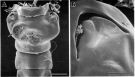 issued from : S. Ohtsuka, G.A. Boxshall & H.S.J. Roe in Bull. nat. Hist. Lond. (Zool.), 1994, 60 (2). [p.147, Fig.28]. Female (SEM micrographs): A, genital double-somite (ventral, copulary pores indicated by arrows); B, left gonopore. Scale bars: 0.100 mm (A); 0.020 mm (B).
|
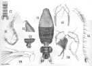 issued from : A. Scott in Siboga-Expedition, 1909, XIX a. [Plate XLIII, Figs.11-19]. Female (from Indonesia-Malaysia): 11, habitus (dorsal); 12, forehead (lateral); 13, last thoracic and genital segments (left side); 14, A1; 15, A2; 16, P5. Male: 17, urosome (dorsal); 18, left A1; 19, P5.
| | | | | NZ: | 2 | | |
|
Carte de distribution de Paraugaptilus similis par zones géographiques
|
| | | | Loc: | | | Indonesia-Malaysia, central equatorial Pacif. | | | | N: | 2 | | | | Lg.: | | | (5) F: 3,75; M: 3,37; (263) F: 3,32; M: 3,03; {F: 3,32-3,75; M: 3,03-3,37} | | | | Rem.: | After Deevey (p.249) , female: A1 extending to urosome; dorsal and lateral thoracic segments covered with hairs; urosome at least 33 % of cephalothorax length. | | | Dernière mise à jour : 28/01/2015 | |
|
|
 Toute utilisation de ce site pour une publication sera mentionnée avec la référence suivante : Toute utilisation de ce site pour une publication sera mentionnée avec la référence suivante :
Razouls C., Desreumaux N., Kouwenberg J. et de Bovée F., 2005-2025. - Biodiversité des Copépodes planctoniques marins (morphologie, répartition géographique et données biologiques). Sorbonne Université, CNRS. Disponible sur http://copepodes.obs-banyuls.fr [Accédé le 26 août 2025] © copyright 2005-2025 Sorbonne Université, CNRS
|
|
|
|


;)
;)
;)
;)
;)
