|
|
 |
Fiche d'espèce de Copépode |
|
|
Calanoida ( Ordre ) |
|
|
|
Arietelloidea ( Superfamille ) |
|
|
|
Arietellidae ( Famille ) |
|
|
|
Scutogerulus ( Genre ) |
|
|
| |
Scutogerulus pelophilus Bradford, 1969 (F) | |
| | | | | | | Syn.: | no Scutogerulus pelophilus (M) Bradford, 1969 b (p.499, figs.M) | | | | Ref.: | | | Bradford, 1969 b (p.495, Descr.F, figs.F); Ohtsuka & al., 1994 (p.120, 152, 161, Rem.F, figs.F); Bradford-Grieve,1999 b (p.39, figs.F, Rem., figs.170, 189) | 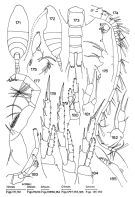 issued from : J.M. Bradford in N.Z. Jl Mar. Freshw. Res., 1969, 3 (4). [p.497, Figs 171-185]. Female (from NE New Zealand: E North Cape): 171, habitus (dorsal); 172, idem (lateral left side); 173, urosome (dorsal); 174, A1; 175, Mx1; 176, Mx2; 177, Mxp; 178, P3; 179, P4; 180, P1; 181-182, P5; 183, A2; 184, P2; 185, Md (mandibular palp). Nota: Head and 1st thoracic segment incompletely separate, 4th and 5th fused. Rostrum large bearing 2 filaments. A1 (23-segmented) almost reaches end of cephalothorax. Md: mandibular palp endopod replaced by 2 setae. Glandular pores open on anterior surfaces of exopods on swimming legs 2-4. P5: endopods unevenly developed in holotype (in paratype, P5 on both sides developped as in fig.181).
|
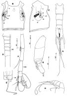 issued from : S. Ohtsuka, G.A. Boxshall & H.S.J. Roe in Bull. nat. Hist. Lond. (Zool.), 1994, 60 (2). [p.153, Fig.33]. Female (paratype, New Zealand): A, urosome (ventral); B, double-somite (ventral); C, genital double-somite (lateral); D, A1 (segments IX to XIV, armature omitted); E, A1 (segments VI and VII (note that aesthetascs on each segment differs in size); F, A1 (segments XXI to XXVIII); G, exopod of A2; H, Md (endopod and exopod). cd = copulatory duct; cp = copulatory pore; g = gonopore; rd = receptacle duct; o, oviduct; s, spermatothore remnant; sr = seminal receptacle. Scales in mm. Nota: Paired gonopores and copulatory pores symmetrically arranged, gonopore sharing common slit-like aperture with copulatory pore; gonopore located anteriorly in slit; genital operculum accompanied by muscles; copulatory pore small, located at innermost corner of slit; copulatory duct swollen anteriorly, relatively short; receptacle seminal simple in shape, pea-like; receptacle duct short, opening beneath gonopore. Left caudal ramus slightly longer than right. 1st endopod segment of A2 without inner seta, 2nd segment with 3 inner setae and 5 terminal setae; exopod indistinctly 8-segmented (setal formula: 0, 1, 1, 1, 1, 1, 0, 3). Mandibular palp with endopod rudimentary, 1-segmented, with 2 setae of unequal lengths; seta on 1st exopod segment not reduced, 5th segment with 2 setae (one of which shorter but not reduced).
|
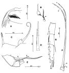 issued from : S. Ohtsuka, G.A. Boxshall & H.S.J. Roe in Bull. nat. Hist. Lond. (Zool.), 1994, 60 (2). [p.154, Fig.34]. Female: A, praecoxal arthrite and coxal endite of Mx1; B, endopod of Mx1 (basal seta indicated by arrowhead); C, 1st and 2nd praecoxal endites of Mx2; D, basal spine of Mx2; E, 4th and 5th endopod segments of Mxp (innermost setae indicated by arrowheads); F, 6th endopod segment of Mxp; G, exopod of P1 (posterior). Scales in mm. Nota: Praecoxal arthrite of Mx1 with 4 spinulose spines and 1 process along inner margin and row of long setules on surface; coxal endite with well-developed spinulose seta; coxal epipodite with 6 setae; basal seta short, endopod rudimentary, 1-segmented, with 1 short seta terminally. 1st praecoxal endite of Mx2 with spinulose seta and 1 vestigial element, 2nd endite with bilaterally spinulose seta. Mxp with innermost seta on 4th and 5th endopod segments not reduced (indicated by arrowhead), 6th endopod segment with stout, elongate setae (c and d) with row of triangular spinules and reduced setae (a and b) (Fig.34 F)
| | | | | Ref. compl.: | | | Bradford-Grieve, 2004 (p.283) | | | | NZ: | 1 | | |
|
Carte de distribution de Scutogerulus pelophilus par zones géographiques
|
| | | | Loc: | | | NE New Zealand (E North Cape) | | | | N: | 2 | | | | Lg.: | | | (230) F: 3,5; 3,60; {F: 3,50-3,60} | | | | Rem.: | bathyal (± 1400 m). | | | Dernière mise à jour : 31/01/2015 | |
|
|
 Toute utilisation de ce site pour une publication sera mentionnée avec la référence suivante : Toute utilisation de ce site pour une publication sera mentionnée avec la référence suivante :
Razouls C., Desreumaux N., Kouwenberg J. et de Bovée F., 2005-2025. - Biodiversité des Copépodes planctoniques marins (morphologie, répartition géographique et données biologiques). Sorbonne Université, CNRS. Disponible sur http://copepodes.obs-banyuls.fr [Accédé le 27 août 2025] © copyright 2005-2025 Sorbonne Université, CNRS
|
|
|
|


;)
;)
;)
