|
|
 |
Fiche d'espèce de Copépode |
|
|
Calanoida ( Ordre ) |
|
|
|
Diaptomoidea ( Superfamille ) |
|
|
|
Candaciidae ( Famille ) |
|
|
|
Candacia ( Genre ) |
|
|
| |
Candacia catula (Giesbrecht, 1889) (F,M) | |
| | | | | | | Syn.: | Candace catula Giesbrecht,1889; 1892 (p.425, 440, 771, figs.F,M);
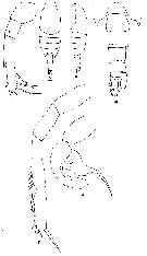
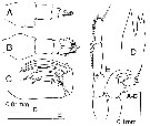
Candacia truncata Brady, 1883 (p.69), fig.) | | | | Ref.: | | | Giesbrecht & Schmeil, 1898 (p.129, Rem. F,M); Thompson & Scott, 1903 (p.235, 250); Carl, 1907 (p.9, Rem.); A. Scott, 1909 (p.152, Rem.); Tanaka, 1935 a (p.213, figs.F,M, juv.); Farran, 1936 a (p.115); Mori, 1937 (1964) (p.81, figs.F,M); Dakin & Colefax, 1940 (p.103, figs.F,M); Pesta, 1941 (p.161, figs.F,M); Chiba, 1957 (p.310); 1957 a (p.11); Tanaka, 1960 (p.55); Brodsky, 1962 c (p.138, figs.F,M); Grice, 1962 (p.234, figs.F,M); 1963 (p.175); Tanaka, 1964 c (p.245); Chen & Zhang, 1965 (p.89, figs.F,M); Kos, 1972 (Vol. I, figs.F,M, Rem.); Lawson, 1977 (p.71, tab.2,3, fig.5); Greenwood, 1978 (p.7, figs.F,M); Marques, 1982 (p.763); van der Spoel & Heyman, 1983 (p.33); Chihara & Murano, 1997 (p.752, Pl.74,76: F,M); Mulyadi, 1997 a (p.82, Redescr.F,M, figs.F,M); Bradford-Grieve & al., 1999 (p.885, 956, figs.F,M); Bradford-Grieve, 1999 b (p.165, figs.F,M, figs.182, 193); Conway & al., 2003 (p.106, figs.F,M, Rem.); Boxshall & Halsey, 2004 (p.85: fig.M); Mulyadi, 2004 (p.82, figs.F,M, Rem.); Phukham, 2008 (p.62, figs.F,M); | 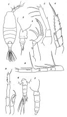 issued from : O. Tanaka in Suisan Gakkai Ho, Tokyo, 1935, 6 (4). [Pl.V, p.225]. Female: 1, habitus (dorsal view); 2, last thoracic segment and urosome (lateral view); 3, Mx2; 4, P3; 5, P5. Male: 6, last thoracic segment and urosome (dorsal view); 7, idem (left lateral view); 8, right A1 (hinge segments); 9, P5 (left and right)
|
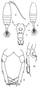 issued from: Q.-c Chen & S.-z. Zhang in Studia Marina Sinica, 1965, 7. [Pl.35, 6-10]. Female (from E China Sea): 6, habitus (dorsal); 7, urosome (ventral); 8, P5 (posterior). Male: 9, habitus (dorsal); 10, P5 (posterior).
|
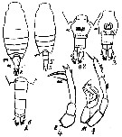
 issued from : J.G. Greenwood in Proc. R. Soc. Qd, 1978, 89. [p.10, Fig.4]. Female (from Moreton Bay, E Australia): a, urosome (lateralleft side); b, idem (dorsal); c, Mx2. Male: d, Md (cutting edge); e, P5.
|
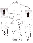
 issued from : T. Mori in The Pelagic copepoda from the neighbouring waters of Japan, 1937 (1964). [Pl. 54, Figs.8-13]. Female: 8, habitus (dorsal); 11, P5; 12, Mx2; 13, P3. Male: 9, habitus (dorsal); 10, P5.
|
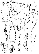
 issued from : G.D. Grice in Fish. Bull. Fish and Wildl. Ser., 1962, 61. [p.233, Pl.31, Figs.17-22]. Female (ffrom equatorial Pacific): 17-18, habitus (dorsal and lateral, respectively); 19, posterior part of thorax and urosome (dorsal); 20, same (lateral, right side); 21, Md (biting edge); 22, Mx2. Nota: Genital segment symmetrical; ventral surface produced into a lobe. The basal tooth of Md ends in 3 cusps, the lowermost one of which is quite small
|
 issued from : G.D. Grice in Fish. Bull. Fish and Wildl. Ser., 1962, 61. [p.236, Pl.32, Figs.1-6]. Female (from equatorial Pacific): 1, P5. Male: 2-3, habitus (dorsal and lateral, respectively); 4, posterior part of thorax and urosome (dorsal); 5, P5; 6, right P5. Nota: Posterior thoracic margins symmetrical. Chela of right P5 small; the thumb of the chela with a long spine protruding from its tip.
|
 issued from : N. Phukham in Species diversity of calanoid copepods in Thai waters, Andaman Sea (Master of Science, Univ. Bangkok). 2008. [p.143, Fig.17]. Female (from W Malay Peninsula): a, habitus (ventral); b, urosome (lateral, right side); c, P5. Male: d, habitus (dorsal); e, P5. Body length after the drawings: F: = 1.636 mm; M = 1.316 mm.
|
 issued from : Mulyadi in Published by Res. Center Biol., Indonesia Inst. Sci. Bogor, 2004. [p.83, Fig.46]. Female (from 03°40'S, 128°10'E): A, habitus (dorsal); B, 5th metasomal somite and urosome (dorsal); C, idem (lateral right side); D, Mx2; E, P3; F, P5; G, right caudal ramus (dorsal). Male: H, habitus (dorsal); I, 5th metasomal somite and urosome (dorsal); J, 5th metasomal somite and urosomal somites 1 and 2 (lateral right side); K, 16th to 19th segments of right A1; L, P5 (posterior view).
|
 Issued from : J.M. Bradford-Grieve, E.L. Markhaseva, C.E.F. Rocha & B. Abiahy in South Atlantic Zooplankton, edit. D. Boltovskoy. 1999, Vol. 2, Copepoda; [p.1064, Fig. 7.363: Candacia catula ]. Ur = urosome. Female characteristics: No spines or spine-like processes present on genital segment; in dorsal view, genital segment with distinctly conxex protrusion on each side; in lateral view, genital segment with ventral, rounded protrusion directed posteriad. Inner edge setae present on terminal segment of P5. - Urosomal segment 2 without lateral,or ventral protrusion and without spine-like process arising from ventral surface. - Posterior prosomal corners pointed. Male characteristics: Left posterior corner of prosome pointed. - In dorsal view, genital segment without process or proyrusion. - Proximal spine on Mx2 lobe 5 considerably thicker that distal spine.
|
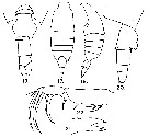
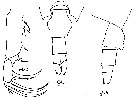
 Issued from : J.M. Bradford-Grieve in NIWA Biodiversity Memoir 111, 1999. [p.167, Fig.117]. After Grenwood, 1978. Female (from SW Pacific): A, urosome (lateral); B, same (dorsal); C, Mx2. male (from SW Pacific): D, end of Md blade; E, P5.
|
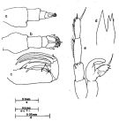
 Issued from : M.S. Kos in Field guide for plankton. Zool Institute USSR Acad., Vol. I, 1972. After Brodsky, 1962. Female: 1, habitus (dorsal); 2 corners of the last thoracic segment and abdomen (dorsal view); 3, abdomen (ventral view); 4, P5. Male: 5, habitus (dorsal); 6, corners of the last thoracic segment and abdomen (dorsal); 7, P5.
| | | | | Ref. compl.: | | | Cleve, 1904 a (p.186); Carl, 1907 (p.17); Sewell, 1912 (p.353, 367); 1914 a (p.229); 1932 (p.335); Rose, 1925 a (p.8); Wilson, 1942 a (p.174); Sewell, 1948 (p.323, 391, 408, 412, 422, 433); Yamazi, 1958 (p.151, Rem.); De Decker, 1964 (p.15, 19, 28); De Decker & Mombeck, 1964 (p.11); Grice & Hulsemann, 1967 (p.19); Fleminger, 1967 a (tabl.1); Itoh, 1970 (tab.1); Timonin, 1971 (p.281, trophic group); Tranter, 1977 (p.596, 600); Carter, 1977 (1978) (p.36); Dessier, 1983 (p.89, Tableau 1, Rem., %); Guangshan & Honglin, 1984 (p.118, tab.); De Decker, 1984 (p.317, 336: chart); Stephen, 1984 (p.161, 169, Distribution vs thermocline & geographic); Brinton & al., 1986 (p.228, Table 1); M. Lefèvre, 1986 (p.33); Madhupratap & Haridas, 1986 (p.105, tab.1); Renon, 1987 (tab.2); Jimenez-Perez & Lara-Lara, 1988; Dessier, 1988 (tabl.1); Hernandez-Trujillo, 1989 a (tab.1); Cervantes-Duarte & Hernandez-Trujillo, 1989 (tab.3); Hirakawa & al., 1990 (tab.3); Othman & al., 1990 (p.561, 564, Table 1);Madhupratap & Haridas, 1990 (p.305, fig.4: vertical distribution night/day; fig.7: cluster); Yoo, 1991 (tab.1); Hernandez-Trujillo, 1991 (1993) (tab.I); Ohtsuka & Kubo, 1991 (p.538); Shih & Young, 1995 (p.69); Padmavati & Goswami, 1996 a (p.85, fig.3, Table 4, vertical distribution); Park & Choi, 1997 (Appendix); Wong & al, 1998 (tab.2); Noda & al., 1998 (p.55, Table 3, occurrence); Padmavati & al., 1998 (p.349); Suarez-Morales & Gasca, 1998 a (p108); Hernandez-Trujillo, 1999 (p.284, tab.1); Lavaniegos & Gonzalez-Navarro, 1999 (p.239, Appx.1); Fernandez-Alamo & al., 2000 (p.1139, Appendix); Suarez-Morales & al., 2000 (p.751, tab.1); Madhupratap & al., 2001 (p. 1345, vertical distribution vs. O2, figs.4, 5: clusters, p.1353); Lo & al., 2001 (1139, tab.I); Uysal & al., 2002 (p.17, tab.1); Hwang & al., 2003 (p.193, tab.2); Hsiao & al., 2004 (p.325, tab.1); Osore & al., 2004 (p.195); Lan & al., 2004 (p.332, tab.1); Lo & al.*, 2004 (p.218, tab.1, fig.6); Gallienne & al., 2004 (p.5, tab.3); Lo & al., 2004 (p.89, tab.1); Zuo & al., 2006 (p.162: tab.1); Lopez-Ibarra & Palomares-Garcia, 2006 (p.63, Tabl. 1, seasonal abundance vs El-Niño); Hwang & al., 2006 (p.943, tabl. I); Hwang & al., 2007 (p.23); Dur & al., 2007 (p.197, Table IV); Jitlang & al., 2008 (p.65, Table 1); Lan Y.C. & al., 2008 (p.61, Table 1, % vs stations); Tseng L.-C. & al., 2008 (p.153, Table 2, fig.5, occurrence vs geographic distribution, indicator species); Tseng L.-C. & al., 2008 (p.46, table 2, abundance vs moonsons); Ayon & al., 2008 (p.238, Table 4: Peruvian samples); Lopez Ibarra, 2008 (p.1, Table 1: abundance, Table 2: Rem.); McKinnon & al., 2008 (p.843: Tab.1); Morales-Ramirez & Suarez-Morales, 2008 (p.518); Fernandes, 2008 (p.465, Tabl.2); C.-Y. Lee & al., 2009 (p.151, Tab.2); Hernandez-Trujillo & al., 2010 (p.913, Table 2); Cornils & al., 2010 (p.2076, Table 3); W.-B. Chang & al., 2010 (p.735, Table 2, abundance); Xu & Gao, 2011 (p.514, figs.3, 4, Table 2: optimal salinity); Hsiao S.H. & al., 2011 (p.475, Appendix I); Kâ & Hwang, 2011 (p.155, Table 3: occurrence %); Maiphae & Sa-ardrit, 2011 (p.641, Table 2); Tutasi & al., 2011 (p.791, Table 2, abundance distribution vs La Niña event); Guo & al., 2011 (p.567, table 2, indicator); Pillai H.U.K. & al., 2011 (p.239, Table 3, vertical distribution); Tseng L.-C. & al., 2011 (p.47, Table 2, occurrences vs mesh sizes); Johan & al., 2012 (2013) (p.1, Table 1); Tseng & al., 2012 (p.621, Table 3: abundance); Jang M.-C & al., 2012 (p.37, abundance and seasonal distribution); Lavaniegos & al., 2012 (p. 11, Appendix); in CalCOFI regional list (MDO, Nov. 2013; M. Ohman, pers. comm.); Tseng & al., 2013 (p.507, seasonal abundance); Hwang & al., 2014 (p.43, Appendix A: seasonal abundance); Lopez-Ibarra & al., 2014 (p.453, Table 2, biogeographical affinity); Nakajima & al., 2015 (p.19, Table 3: abundance); Jerez-Guerrero & al., 2017 (p.1046, Table 1: temporal occurrence); Palomares-Garcia & al., 2018 (p.178, Table 1: occurrence) | | | | NZ: | 11 | | |
|
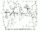
Carte de distribution de Candacia catula par zones géographiques
|
| | | | | | | | | | | |  issued from : T.J. Lawson in Marine Biology, 1977, 43. [Fig.5, p.77]. issued from : T.J. Lawson in Marine Biology, 1977, 43. [Fig.5, p.77].
Distribution map for the Indian Ocean. |
 issued from : Mulyadi in Treubia, 1997, 31 (2). [p.109, Fig.16]. issued from : Mulyadi in Treubia, 1997, 31 (2). [p.109, Fig.16].
Distribution of Candaciidae in Indonesian waters. 3: C. catula. |
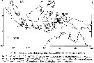
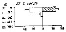 Issued from : M. Madhupratap & P. Haridas in J. Plankton Res., 12 (2). [p.311, Fig.4]. Issued from : M. Madhupratap & P. Haridas in J. Plankton Res., 12 (2). [p.311, Fig.4].
Vertical distribution of calanoid copepod (mean +1 SE), abundance No/100 m3. 27- Candacia catula.
Night: shaded, day: unshaded.
Samples collected from 6 stations located off Cochin (India), SE Arabian Sea, November 1983, with a Multiple Closing Plankton Net (mesh aperture 300 µm), in vertical hauls at 4 depth intervalls (0-200, 200-400, 400-600, 600-1000 m). |
| | | | Loc: | | | South Africa (E & W), Medit. (Monaco, Lebanon Basin), G. of Suez, Red Sea, Arabian Sea, Madagascar (Nosy-Bé), Rodrigues Is. - Seychelles, Monbasa, Sri Lanka, Mascarene Basin, India (Goa - Gujarat), Natal, Indian, Bay of Bengal, Andaman Sea (Barren Island), W Malay Peninsula (Andaman Sea), Ambon Bay, W Australia, Thailand, Indonesia-Malaysia,Ambon Vay, Bintulu coast, Java (Jakarta Bay-Seribu Islands), Lombok Sea Flores Sea, Tioman Is., N Celebes, China Seas (Yellow Sea, East China Sea, South China Sea), Taiwan (S, E, SW, W, NW, N, NE), S Korea, Korea Strait, Japan Sea, Japan, Kuchinoerabu Is., Tanabe Bay, W Pacif. (equatorial), Australia (Great Barrier, Moreton Bay, Shark Bay, North West Cape, G. of Carpentaria), New Caledonia, California, Baja California (Bahia Magdalena, W), G. of California, La Paz, W Mexico, G. of Tehuantepec, W Costa Rica, Clipperton Is., Panama, Bahia Cupica (Colombia), Bikini, Pacif. (equatorial), Moorea Is., Galapagos-Ecuador, Peru | | | | N: | 127 (Medit.: 1, Red Sea: 2; Indian: 40; Indo-Malaysia: 8; Pacif.: 73) | | | | Lg.: | | | (34) F: 1,32; (46) F: 1,6-1,45; M: 1,55-1,4; (91) F: 1,65-1,4; M: 1,6-1,3; (101) F: 1,67-1,53; M: 1,62-1,43; (104) F: 1,7; M: 1,3; (120) F: 1,48-1,35; M: 1,48-1,32; (150) F: 1,67-1,55; M: 1,55-1,45; (290) F: 1,65-1,5; M: 1,5-1,48; (778) F: 1,6-1,55; M: 1,38-1,32; (786) F: 1,46; (991) F: 1,4-1,67; M: 1,3-1,62; (1122) F: 1,6; M: 1,35; {F: 1,32-1,70; M: 1,30-1,62}
The mean female size is 1.540 mm (n = 20; SD = 0.1159), and the mean male size is 1.442 mm (n = 18; SD = 0.1144). The size ratio (male : female) is 0.92 (n = 10; SD = 0.0702). | | | | Rem.: | épipélagique.
Rose (1925 a) et Uysal & al. (2002) signalent cette espèce pour la première fois en Méditerranée (Monaco et bassin Levantin NW). Lessepsian migration possible.
Voir aussi les remarques en anglais | | | Dernière mise à jour : 01/12/2020 | |
|
|
 Toute utilisation de ce site pour une publication sera mentionnée avec la référence suivante : Toute utilisation de ce site pour une publication sera mentionnée avec la référence suivante :
Razouls C., Desreumaux N., Kouwenberg J. et de Bovée F., 2005-2025. - Biodiversité des Copépodes planctoniques marins (morphologie, répartition géographique et données biologiques). Sorbonne Université, CNRS. Disponible sur http://copepodes.obs-banyuls.fr [Accédé le 03 janvier 2026] © copyright 2005-2025 Sorbonne Université, CNRS
|
|
|
|


;)
;)
;)
;)
;)
;)
;)
;)
;)
;)
;)

;)
;)
