|
|
 |
Fiche d'espèce de Copépode |
|
|
Calanoida ( Ordre ) |
|
|
|
Clausocalanoidea ( Superfamille ) |
|
|
|
Euchaetidae ( Famille ) |
|
|
|
Paraeuchaeta ( Genre ) |
|
|
| |
Paraeuchaeta calva Tanaka, 1958 (F,M) | |
| | | | | | | Syn.: | Pareuchaeta calva Tanaka, 1958 (p.347, Descr.F, figs.F);
Pareuchaeta comosa (M) Tanaka, 1958 (p.363: M, no F);
Euchaeta dubia : Vervoort, 1963 b (p.176, figs.M, Rem.);
Pareuchaeta dubia (M) : Tanaka & Omori, 1967 (p.245: M, non F); 1968 (p.136, M, no F;
Pareuchaeta californica : Tanaka & Omori, 1968 (p.231, figs.F, Rem.F);
Pareuchaeta polita Tanaka & Omori, 1968 (p.246, figs.F);
Pareuchaeta simulantis Tanaka & Omori, 1968 (p.253, figs.F);
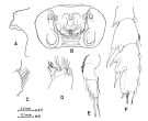
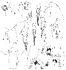
Euchaeta calva : Grice & Hulsemann, 1967 (p.15); Park, 1975 c (p.10, 12, figs.F,M, Rem.) | | | | Ref.: | | | Bradford & al., 1983 (p.20); Park,1994 (p.322); 1995 (p.49, Rem.F, M, figs.F,M); Bradford-Grieve & al., 1999 (p.880, 925, figs.F,M) | 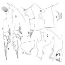 issued from : T. Park in Bull. Scripps Inst. Oceanogr. Univ. California, San Diego, 1995, 29. [p.149, Fig.39]. Female: a, forehead (left side); b, urosome (left); c, genital somite (left); d, outer lobe of Mx1; e, P1 (anterior); f, P2 (anterior). Nota: Similar in habitus to P. sarsi but distinguishable from it. Laterally, rostrum more elongated and pointing more forward. Urosome also more elongated. Ventral margin of genital flange strongly emarginate and posterior lobe of flange produced into a large toothlike process pointing in a posteroventral direction. Posterior edge of genital field scarcely produced beyond margin of genital prominence. Genital prominence is defined posteriorly by a nearly straight margin extending from genital flange to ventral wall of somite. Posterior section of genital somite as measured from distal end of genital flange relatively long, about 1.4-1.8 times length of genital flange, whereas it is about 1.0-1.2 times length of flange in P. sarsi. Cephalosomal appendages similar to those of P. sarsi except that outer lobe of Mx1 with 5 long setae and 3 minute setae proximally. Swimming legs practically identical to those of P. sarsi. Male: g, forehead (left); h, last pedigerous and genital somites (left); i, distal exopodal segments of left 5th leg (lateral, tilted clockwise); j, serrated lamella of left 5th leg (lateral).
|
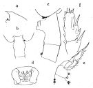 issued from : O. Tanaka in Publs Seto Mar. Biol. Lab., 1958, VI (3). [p.347, Fig.71]. As Pareuchaeta calva. Female: a, forehead (lateral left side); b, last thoracic segment and genital somite (dorsal); c, corner of the last thoracic segment andgenital somite and 2nd urosome segment (lateral left side); d, genital complex (ventral); e, P1; f, exopod of P2. Nota: The first three urosomal segments have the proportional lengths 19:10:9.
|
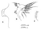 issued from : O. Tanaka & M. Omori in Publs Seto Mar. Biol. Lab., 1968, XVI (4). [p.231, Fig.7]. As Pareuchaeta californica. Female: A, genital somite (lateral left side); B, Mx1; C, outer lobe of Mx1 (right side). Nota: The urosome segments and furca are in the proportional lengths as 46:23:18:2:11 = 100.
|
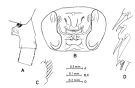 issued from : O. Tanaka & M. Omori in Publs Seto Mar. Biol. Lab., 1968, XVI (4). [p.246, Fig.15]. As Pareuchaeta polita. Female: A, last thoracic segment and urosome (part.) (lateral left side); B, genital complex (ventral); C, outer lobe of Mx1; D, distal part of exopod of P2. Nota: The urosome segments and furca are in the proportional lengths as 42:24:20:2:12 = 100. Prosome and urosome are in the proportional lengths as 72 to 28.
|
 issued from : O. Tanaka & M. Omori in Publs Seto Mar. Biol. Lab., 1968, XVI (4). [p.254, Fig.19]. As Pareuchaeta simulantis. Female: A, genital somite (part.) (lateral left side); B, genital complex (ventral); C, outer lobe of Mx1; D, endopod and 2nd basal segment of Mx1; E, exopod of P1; F, exopod of P2. Nota: The urosome segments and furca are in the proportional lengths as 46:22:19:2:11 = 100.Prosome and urosome are in the proportional lengths as 71 to 29.
|
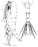 issued from : W. Vervoort in Atlantide Report., 1963, 7. [p.177, Fig.21]. As Euchaeta dubia. Male (from off Ghana): a, habitus (lateral); b, posterior part cephalothorax and urosome). (a: x20; b,: x28). Nota: The length of urosome contained 2 times in that of the cephalothorx.Forehead triangularly produced frontally.base of rostrum just visible. In lateral aspect the cephalosome shows a slight depression in the mid-dorsal line just opposite the insertion of Mxp. On both sides of the laterodorsal corners of the posterior part of prosome there is a small spine . The end of the fused 4th and 5th thoracic somites is slightly asymmetrical, as the left side is slightly more produced than the right side. the genital somite is asymmetrically developped, the genital aperture is situated on the left side. The 2nd and 3rd urosomal somites have a completely closed row of flat, obtuse spines along the distal end; on the 4th somite the row of distal spines is interrupted dorsally and laterally. The anal plate is small but distinct. Each caudal ramus has 4 setae, in addition there is a small seta at the external margin and a geniculate appendicular seta at the internal corner. A1 22-sgmented (8th to 10th and 24th-25th segments fused), reach the end of the genital somite
|
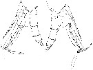 issued from : W. Vervoort in Atlantide Report., 1963, 7. [p.179, Fig.22]. As Euchaeta dubia. Male: a, posterior part cephalothorax, P5 with spermatophore and urosome (lateral view from left side); B, idem (lateral view from right side). (x28)
|
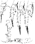 issued from : W. Vervoort in Atlantide Report., 1963, 7. [p.180, Fig.23]. As Euchaeta dubia. Male: a-d, P1 to P4; e, P5 (r = left leg, r = right leg); f, extremity of exopodite of left P5. (a: x55; b-d: x44; e: x28; f: x88).
|
 issued from : T. Park in Smiths. Contr. Zool., 1975, 196. [p.11, Fig.9]. As Euchaeta calva. Female (G. of Mexico): a, forehead (lateral); b, genital segment (lateral); c, same (dorsal); d, genital field (ventral); e, outer lobe of Mx1; f, exopod of P1 (anterior); g, exopod of P2 (anterior). Male: h, forehead (lateral); i, exopod of P1 (anterior); j, exopod of P2 (anterior); k, P5; l, exopod of left P5 (anterior); m, same (lateral); n, same (medial). fl = length of genital flange; sl = length of genital segment posterior to genital flange. Nota: This species constitute the first record in the Gulf of Mexico and Caribbean Sea, most of which were found in tows to depths exceeding 1000 m.
|
 Paraeuchaeta calva Paraeuchaeta calva Female: 1 - See key to species Groups and independent species of Paraeuchaeta (p.30): malayensis species Group. 2 - Outer spine of 2nd exopodal segment (or the 2nd of the first 2 exopodal segments forming a proximal, compound segment) of P1 normally developed (Fig.39-e). 3 - Outer lobe of Mx1 with 5 long setae in addition to some very small setae proximally (Fig.39-d). 4 - Laterally, genital prominence high with emarginate genital flanges (Fig.39-c). 5 - Laterally, genital somite with long posterior ventral wall; genital flange strongly emarginate (Fig.39-c). 6 - Laterally, posterior lobe of genital flange directed posteroventrad (Fig.39-c).
| | | | | Ref. compl.: | | | Suarez-Morales & Gasca, 1998 a (p.109); Razouls & al., 2000 (p.343, Appendix); Park & Ferrari, 2009 (p.143, fig.1, biogeography) | | | | NZ: | 8 | | |
|
Carte de distribution de Paraeuchaeta calva par zones géographiques
|
| | | | | | | | | | | | 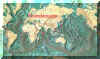 Carte de 1996 Carte de 1996 | |
| | | | Loc: | | | Sub-Antarct. (N Peninsula) (in Yamanaka, 1976), S Atlant (central), G. of Guinea, W Caribbean Sea , G. of Mexico, NW Atlant., W Indian, Indonesia-Malaysia, China Seas (South China Sea, East China Sea), Japan (Suruga, Sagami Bay, SW) | | | | N: | 9 | | | | Lg.: | | | (3) F: 8,4-7,4; M: 7,9-6,8; (14) M: 7,1-6,8; (19) F: 7,91-7,25; M: 7,25-6,66; (63) F: 8,29-8,03; M: ± 7,5; (99) F: 7,44; {F: 7,25-8,40; M: 6,66-7,90} | | | | Rem.: | Sampling depth (Antarct.) : 2000-3000 m.
Voir aussi les remarques en anglais | | | Dernière mise à jour : 27/01/2015 | |
|
|
 Toute utilisation de ce site pour une publication sera mentionnée avec la référence suivante : Toute utilisation de ce site pour une publication sera mentionnée avec la référence suivante :
Razouls C., Desreumaux N., Kouwenberg J. et de Bovée F., 2005-2026. - Biodiversité des Copépodes planctoniques marins (morphologie, répartition géographique et données biologiques). Sorbonne Université, CNRS. Disponible sur http://copepodes.obs-banyuls.fr [Accédé le 05 mars 2026] © copyright 2005-2026 Sorbonne Université, CNRS
|
|
|
|


;)
;)
;)
;)
;)
;)
;)
;)
;)

;)
