|
|
 |
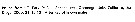
Fiche d'espèce de Copépode |
|
|
Calanoida ( Ordre ) |
|
|
|
Arietelloidea ( Superfamille ) |
|
|
|
Heterorhabdidae ( Famille ) |
|
|
|
Disseta ( Genre ) |
|
|
| |
Disseta palumbii Giesbrecht, 1889 (F,M) | |
| | | | | | | Syn.: | Disseta palumboi: Giesbrecht & Schmeil, 1898 (p.112, Rem.); Farran, 1908 b (p.67); A. Scott, 1909 (p.133, figs.F,M); Sars, 1925 (p.221, figs.F,M); Farran, 1926 (p.279, Rem.); Rose, 1929 (p.34, figs.M, Rem.); Sewell, 1932 (p.309, figs.F,M, Rem.); Rose, 1933 a (p.199, figs.F,M); Hardy & Gunther, 1935 (1936) (p.180, Rem.); Sewell, 1947 (p.185, fig.44, figs.F,M, Rem.); Lysholm & al., 1945 (p.35); Farran, 1948 d (n°15, p.3, figs.F,M); C.B. Wilson, 1950 (p.198, fig.F, Rem.); Brodsky, 1950 (1967) (p.342, figs.F,M, Rem.); Paiva, 1963 (p.58); Grice, 1963 a (p.496); Tanaka, 1964 a (p.32, figs.F); Vervoort, 1965 (p.116, Rem.); Owre & Foyo, 1967 (p.77, figs.F,M); Omori, 1969 (p.5, Table 1); Harding, 1974 (p.141, tab.2, gut contents); Frontier, 1977 a (p.15); Deevey & Brooks, 1977 (p.256, tab.2, Station "S"); Guangshan & Honglin, 1984 (p.118, tab.); Lozano Soldevilla & al., 1988 (p.59); Bradford-Grieve & al., 1999 (p.883, 943, figs.F,M); Chihara & Murano, 1997 (p.816, Pl.117: F,M); Holmes, 2001 (p.15); Hsiao & al., 2004 (p.326, tab.1); Vives & Shmeleva, 2007 (p.294, figs.F,M, Rem.); Gaard & al., 2008 (p.59, Table 1, N Mid-Atlantic Ridge);
Heterorharbdus grandis Wolfenden, 1904 (p.120, fig.F); 1905 a (p.8, Descr.F,M, fig.M); Pearson, 1906 (p.26); van Breemen, 1908 a (p.126); Ohtsuka & al., 1997 (p.579, figs.F); Galbraith, 2009 (pers. comm.);
Disseta grandis Esterly, 1906 a (p.72, figs.F,M); Galbraith, 2009 (pers. comm.);
Disseta sp. Esterly, 1911 (p.331, Descr.M, figs.M);
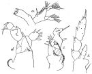
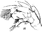
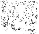
Disseta atlantica Wolfenden, 1911 (p.313, Rem.F,M) | | | | Ref.: | | | Giesbrecht,1889; 1892 (p.369, 772, Descr.F, figs.F); Bradford-Grieve & al., 1999 (p.883, 943, figs.F,M); Bradford-Grieve,1999 b (p.75, Rem., figs.173, 191); Park, 2000 (p.14, figs.F,M, Rem.); Machida & Nishida, 2010 (p.2130, genetic differentiation) | 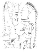 issued from : T. Park in Bull. Scripps Inst. Oceanogr. Univ. California, San Diego, 2000, 31. [p.153, Fig.1]. Female: a, habitus (left side); b, urosome (dorsal); c, d, e, genital somite (dorsal, left, ventral, respectively); f, forehead (ventral); g, left A1 (ventral); h, left A2 (posterior); i, left Md (posterior); j, masticatory edge of left Md; k masticatory edge of right Md. atp = midanterior tubercular process. Nota: Nota: Dorsally, genital somite relatively long, with tubercular lateral swellings. Laterally, posterior end of prosome extending posteriad into rather broadly rounded lappet; dorsally, posterolateral corners more or less asymmetrical, with left side slightly longer than right and each a rather obtuse posterior end.
|
 issued from : T. Park in Bull. Scripps Inst. Oceanogr. Univ. California, San Diego, 2000, 31. [p.154, Fig.2]. Female: a, left Mx1 (posterior); b, left Mx2 (posterior); c, right Mxp (anterior); d, P1 (anterior); e, P1 (with distal part omitted), posterior; f, P2 (anterior); g, P3 (anterior); h, P4 (anterior; i, P5 (anterior). Nota: 2nd exopodal segment of P5 with a short outer spine.
|
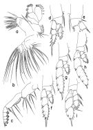
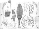
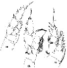
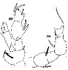
 issued from : T. Park in Bull. Scripps Inst. Oceanogr. Univ. California, San Diego, 2000, 31. [p.155, Fig.3]. Male: a, habitus (left side); b, urosome (dorsal); c, left A1 (ventral); d, right P5 (with 2 distal exopodal segments omitted), anterior; e, left P5 (with 2 distal exopodal segments omitted), anterior; f, basipod and 1st exopodal segment of left P5 (posterior); g, P5 (posterior); h, i, exopod of left P5 (posterior, anterior, respectively); j, exopod of right P5 (tilted clockwise), posterior ; k, idem (posterior). mpr = medial projection.
|
 issued from : J.M. Bradford-Grieve in The Marine Fauna of New Zealand: Pelagic Calanoid Copepoda. National Institute of Water and Atmospheric Research (NIWA). NIWA Biodiversity Memoir, 111, 1999. [p.75, Fig.42]. Female: A, urosome (dorsal); B, idem (left lateral side); C, Md; D, P5; E, spine from inner distal corner of exopod segment 2 of P5. Nota: It should be noted that the female genital somite of Southwest Pacific specimens, if not viewed directly from the dorsal surface, can appear to be quite asymmetrical because the lateral swelling is more dorsally placed on the left side (which may account for some of the variability between descriptions). The right posterior metasomal border extends further posteriorly than that on the left side. Male: F, P5.
|
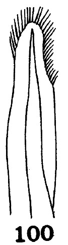
 issued from : O. Tanaka in Publs Seto Mar. Biol. Lab., 1964, XII (1). [p.33, Fig.190]. As Disseta palumboi. Female: a, urosome (dorsal); b, last thoracic segment and genital somite (dorsolateral right side): c, P5. Nota: The urosome segments and furca are in the proportional lengths as 37:16:13:8:26 = 100. The orosome segments and furca are covered with fine short hairs.. A1 exceeds the end of the furca by distal 3 segments Male (immature): d, P5.
|
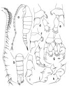
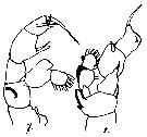
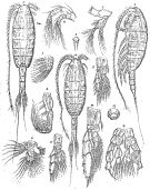 Issued from : G.O. Sars in Résult. Camp. Scient. Prince Albert I, 69, pls.1-127 (1924). [Pl.LX, figs.1-14]. As Disseta palumboi. Female: 1, habitus (dorsal); 2, idem (lateral left side); 3, rostrum; 4, A2; 5, Md; 6, Mx1; 7, Mx2; 8, Mxp; 9, P1; 10, P3; 11, P5; 12, genital complex (ventral). Male: 13, habitus (dorsal); 14, P5. Nota after Brodsky (1950 [1967]): Diagnostic characters female: Ventral process of genital segment situated nearer to proximal part of segment. Posterior thoracic corners of equal length.
|
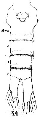
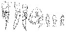
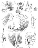 issued from : R.B.S. Sewell in The John Murray Expedition, 1933-34, Scientific Reports, VIII (1), 1947. [p.187, Fig.48]. As Disseta palumboi. Female (from Arabian Sea): A, posterior thoracic margin and genital segment (lateral right side); B, proximal 3 segments of A1; C, Mx1; E, Mxp; F, Mx2; G, exopod of P5. Nota: The proportional lengths of the cephalothorax and abdomen as 67 to 33. The proportional lengths of the various segments of the body (cephalon to caudal rami) in the first specimen as 275:129:84:92:88:111:63:52:33:73 (67) = 1000. In the second specimen: 269:136:78:95:85:111:66:58:29:82 (66). The inequality of the two caudal rami was considerably more marked in the second specimen. A gland composed of granular protoplasm is situated in the postero-lateral region of the combined 4th-5th thoracic segment. The genital segment is covered with numerous minute needle-like spinules in the posterior 2/3 of its length on the dorsal and lateral aspects. The posterior borders of the abdominal segments are fringed with needle-like spines, those on the 4th segment being sowewhat more coarse than the others. A1 25-segmented, overeaches the tip of the caudal rami by 5 segments. In A2 the endopod is slightly longer than the exopod. In Md the endopod is shorter than the exopod or of the same length. In Mx1 the various lobes bear the following number of setae: inner lobe 1 (14 spines or setae); inner lobe 2 (1), inner lobe 3 (3); basal segment 2 (4); endopod (3, 4 and 5 setae on the three segments); exopod (6 large and 5 smaller setae); outer lobe (3 small and 6 large setae). Male: D, Mx1; H, P1; I, P5. Nota: The proportional lengths of the cephalothorax and abdomen as 65 to 35. The proportional lengths of the various segments of the body (cephalon to caudal rami) in the first specimen as 258:128:81:97:89:52:60:65:58:34:78 (58) = 1000. Head and 1st pediger segment separate, 4th and 5th fused. A1 about the length of the body, right 25-segmented, left 22-segmented ( segments 19-20 and 21-23 fused)
|
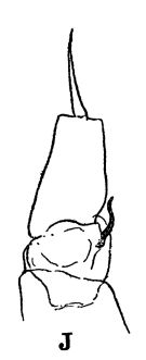 issued from : R.B.S. Sewell in The John Murray Expedition, 1933-34, Scientific Reports, VIII (1), 1947. [p.166, Fig.44, J]. Process on the posterior aspect of the 2nd basal segment of P1. Remarks: The shape of the hook-like or spine-like process shows some variation in different genera, but it is undoubtedly homologous throughout the whole series. The function of this organ is unknown.
|
 Issued from : R.B.S. Sewell in Mem. Indian Mus., 1932, X (continued). [p.311, Fig.103]. As Disseta palumboi. Female (from off Sri Lanka): arrangment of the cutaneous pores (dorsal aspect).
|
 Issued from : R.B.S. Sewell in Mem. Indian Mus., 1932, X (continued). [p.311, Fig.103]. Female: a, Md; b, P3. Male: c, right P5; d, left P5.
|
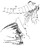
 issued from : A. Scott in Siboga-Expedition, 1909, XIX a. [Plate XLI, Figs.11-21]. Female (from Molucca Passage, Banda Sea): 11, habitus (dorsal); 12, forehead (lateral); 13, last thoracic and genital segments (left side); 14, rostrum; 15, A1; 16, Md (masticatory edge); 17, Mxp; 18, P1; 19, P5. Male: 20, left A1; 21, P5.
|
 issued from : S. Ohtsuka, H.Y. Soh & S. Nishida in J. Crustacean Biol., 1997, 17 (4). [p.580, Fig.1, A-C]. Female (from S Japan): A, right mandibular cutting edge; B, ventralmost tooth; C, dorsal teeth. v = ventarl tooth (or teeth); c = central teeth; d = dorsal teeth.
|
 issued from : S. Ohtsuka, H.Y. Soh & S. Nishida in J. Crustacean Biol., 1997, 17 (4). [p.583, Fig.4, A-B]. Female (from S. Japan): A, right side of labrum (posterior surface; arrowed = type-2 gland opening, small arrow = type-3 gland openings); B, idem, medial pair of gland openings (type-3) .. Scale bars : 0.100 mm (A); 0.010 mm (B).
|
 Issued from : W. Giesbrecht in Systematik und Faunistik der Pelagischen Copepoden des Golfes von Neapel und der angrenzenden Meeres-Abschnitte. Fauna Flora Golf. Neapel, 1892. Atlas von 54 Tafeln. [Taf.38, Fig.44]. Female: 44, urosome (ventral).
|
 Issued from : W. Giesbrecht in Systematik und Faunistik der Pelagischen Copepoden des Golfes von Neapel und der angrenzenden Meeres-Abschnitte. Fauna Flora Golf. Neapel, 1892. Atlas von 54 Tafeln. [Taf.29, Figs.2, 8]. Female: 2, A1 (poximal segments); 8, Mxp (middle segments; anterior view).
|
 Issued from : W. Giesbrecht in Systematik und Faunistik der Pelagischen Copepoden des Golfes von Neapel und der angrenzenden Meeres-Abschnitte. Fauna Flora Golf. Neapel, 1892. Atlas von 54 Tafeln. [Taf.29, Fig.14]. Female: 14, Md (mandibular palp; anterior view).
|
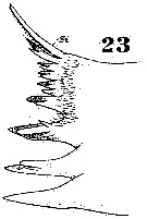 Issued from : W. Giesbrecht in Systematik und Faunistik der Pelagischen Copepoden des Golfes von Neapel und der angrenzenden Meeres-Abschnitte. Fauna Flora Golf. Neapel, 1892. Atlas von 54 Tafeln. [Taf.29, Fig.23] Female: 23, Md (cutting edge)
|
 Issued from : W. Giesbrecht in Systematik und Faunistik der Pelagischen Copepoden des Golfes von Neapel und der angrenzenden Meeres-Abschnitte. Fauna Flora Golf. Neapel, 1892. Atlas von 54 Tafeln. [Taf.29, Fig.27]. Female: 27, A2 (anterior view).
|
 Issued from : W. Giesbrecht in Systematik und Faunistik der Pelagischen Copepoden des Golfes von Neapel und der angrenzenden Meeres-Abschnitte. Fauna Flora Golf. Neapel, 1892. Atlas von 54 Tafeln. [Taf.29, Figs.19, 24, 25]. Female: 19, P2 (posterior view); 24, P5 (anterior view); 25, P1 (anterior view).
|
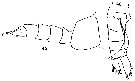
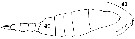 issued from : C.O. Esterly in Univ. Calif. Publs Zool., 1911, 6 (14). [Pl.28, Figs.40, 41]. As Disseta sp. Male (from San Diego Region): 40, habitus (lateral); 41, forehead (lateral). Nota: Rostral filaments are stiff and placed so far beneath the head that they are invisible except from directly below; the sides of the cephalothorax in front seem to be prolonged so as to cover the rostrum. Cephalothorax is twice the length of the urosome. Head not fused with the thorax, the last two thoracic segments fused. The middle segment of the abdomen is the longest one; the 1st and 2nd are of equal lengths and about four-fifths as long as the 3rd; the 4th is three-fourths as long as the 3rd segment; and the 5th is about half as long as the 3rd. left caudal ramus longer than the right. A1 22-segmented and a little longer than the body; the grasping A1 is on the left side and the portion distal to geniculation is 4-segmented
|
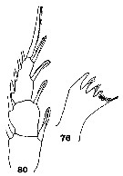 issued from : C.O. Esterly in Univ. Calif. Publs Zool., 1911, 6 (14). [Pl.30, Figs.76, 80]. As Disseta sp. Male: 76, Md (cutting edge); 80, exopod of P1. Nota: The terminal segment of endopod of P2 and P3 has 8 bristles.
|
 issued from : C.O. Esterly in Univ. Calif. Publs Zool., 1911, 6 (14). [Pl.31, Fig.100]. As Disseta sp. Male: 100, distal half of the second outer marginal bristle of the exopod of P1.
|
 issued from : C.O. Esterly in Univ. Calif. Publs Zool., 1911, 6 (14). [Pl.32, Figs.107, 108]. As Disseta sp. Male: 107-108, right and left P5 respectively.
|
 issued from : C.O. Esterly in Univ. Calif. Publs Zool., 1906, 3 (5). [Pl.9, Fig.21]. As Disseta grandis. Female (from San Diego, California): 21, forehead (lateral). Nota: Female rather closely resembling Disseta palumboi Giesbrecht, but much lerger. There are also differences in the structure of P5 and mandibular blade, and the last thoracic segment is asymmetrical, being longer on the left side. The genital segment has a rather ore rounded outline than in D. palumboi, the orifice is of a different shape, and the segment is asymmetrical.
|
 issued from : C.O. Esterly in Univ. Calif. Publs Zool., 1906, 3 (5). [Pl.11, Figs.45, 46]. As Disseta grandis. Female: 46, urosome (ventral). Male: 45, last thoracic segment and urosome (lateral).
|
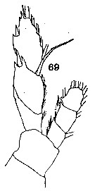 issued from : C.O. Esterly in Univ. Calif. Publs Zool., 1906, 3 (5). [Pl.13, Fig.69]. As Disseta grandis. Female: 69, P5.
|
 issued from : C.O. Esterly in Univ. Calif. Publs Zool., 1906, 3 (5). [Pl.14, Figs.88, 94]. As Disseta grandis. Male: 88, left P5; 94, right P5.
|
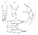
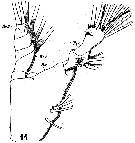
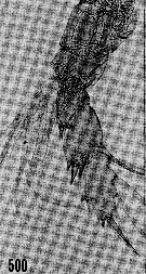
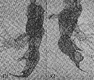
 issued from : M. Rose in Result. Camp. Scient. Prince Albert Ier, Monaco, 1929, 78. [Pl.II, Figs.4]. As Disseta palumboi. Male (from ? 35°30'N, 22°02'W): D, habitus (dorsal); L, idem (lateral); Ab, urosome (dorsal); R, rostrum (lateral view); A1 prehensile; G, detail of geniculation of A1; A2; Md; Mx1 (as Mx); Mx2 (as Mxp1); Mxp (as Mxp2); b1, basipod segment 1 of Mxp; b2, basipod 2nd segment of Mxp (with acinetians: ac); Ac, acinetians epibionts (kystes); Ac l: acinetian free form).
|
 issued from : M. Rose in Result. Camp. Scient. Prince Albert Ier, Monaco, 1929, 78. [Pl.II, Figs.4]. As Disseta palumboi. Male: P1 to P4; End dr, endopod of the right P5; Enp g, endopod of the left P5; Enp g', idem (another view).
|
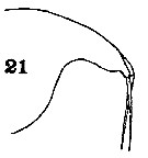
 issued from : R.N. Wolfenden in Plankton Studies Part I. Copepoda. 1905 [Pl. IV, 7-8]. As Heterorhabdus grandis. Male: 7, left P5; 8, right P5.
|
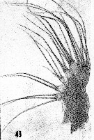
 issued from : H.B. Owre & M. Foyo in Fauna Caribaea, 1, Crustacea, 1: Copepoda. Copepods of the Florida Current. 1967. [p.77, Fig.500]. As Disseta palumboi. Female: 500, P5.
|
 issued from : H.B. Owre & M. Foyo in Fauna Caribaea, 1, Crustacea, 1: Copepoda. Copepods of the Florida Current. 1967. [p.77, Figs.501, 502]. As Disseta palumboi. Male: 501, left P5; 502, tight P5.
|
 issued from : H.B. Owre & M. Foyo in Fauna Caribaea, 1, Crustacea, 1: Copepoda. Copepods of the Florida Current. 1967. [p.17, Fig.49]. As Disseta palumboi. Female: 49, Mx2.
|
 Disseta palumbii Disseta palumbii female: 1 - Dorsally, genital somite (Fig.1-b) relativelylong, with tubercular lateral swellings (Fig.1-c); 2 - P5, 2nd exopodal segment with a short outer spine (Fig.2-i).
|
 Disseta palumbii Disseta palumbii male: 1 - Left P5 (Fig.3-i), 2nd exopodal segment with a large lateral spiniform process.
| | | | | Ref. compl.: | | | Sewell, 1948 (p.371, 503, 514, 525, 530, 534, 538, 539, 547, 549, 558); De Decker & Mombeck, 1964 (p.12); Grice & Hulsemann, 1965 (p.224); 1967 (p.18); Park, 1970 (p.477); Roe, 1972 (p.277, tabl.1, tabl.2); Björnberg, 1973 (p.345, 385); Vives & al., 1975 (p.49, tab.II, XII); Vives, 1982 (p.293); Kovalev & Shmeleva, 1982 (p.84); Shih & Young, 1995 (p.70); Hure & Krsinic, 1998 (p.66, 102); Padmavati & al., 1998 (p.349); Lapernat, 2000 (tab. 3, 4); Razouls & al., 2000 (p.343, Appendix); Park & Ferrari, 2009 (p.143, Table 5, Appendix 1, biogeography); Medellin-Mora & Navas S., 2010 (p.265, Tab. 2); in CalCOFI regional list (MDO, Nov. 2013; M. Ohman, comm. pers.); Peijnenburg & Goetze, 2013 (p.2765, genetic data); Bode & al., 2015 (p.268, Table 1, chemical components, trophic level, geographic zone); Belmonte, 2018 (p.273, Table I: Italian zones) | | | | NZ: | 19 | | |
|
Carte de distribution de Disseta palumbii par zones géographiques
|
| | | | | | | | | | | | | | | | Loc: | | | sub-Antarct. (South Georgia), South Africa (E), off E Tristan da Cunha, Patagonia, off Ghana, G. of Guinea, off E St. Paul Is., off Cape Verde Is. (E, NE & NW), off Morocco-Mauritania, Canary Is., off W Canary Is., Caribbean Sea, Caribbean Colombia, G. of Mexico, Florida, off Bermuda: Station S (32°10N, 64°30W), Sargasso Sea, off E Cape Cod, off S Iceland, Portugal, off Madeira, Azores, Bay of Biscay, off Ireland (S & W), Ibero-moroccan Bay, Medit. (S Adriatic Sea), G. of Oman, Arabian Sea, Laccadive Is., Madagascar (Nosy Bé), Indian, Bay of Bengal, Indonesia-Malaysia, Celebes Sea, Sulu Sea, Philippines, China Seas, Taiwan E, Japan, SW Bering Is., Pacif. (tropical), off Hawaii S, off British Columbia, California, off Ecuador, off Peru, NE Easter Is., Galapagos, E Tuamotu , Pacif. (W equatorial), N Tasman Sea, New Zealand (North Island SW, N), Chile | | | | N: | 53 | | | | Lg.: | | | (1) F: 7; (5) F: 8; M: 7,75; (10) F: 7; (11) F: 7,45-7,33; M: 7-6,667; (16) F: 7,5-6,59; M: 7,5-6,45; (17) F: 8,3; M: 7,6; (22) F: 7-5,7; M: 7,3-6,8; (38) F: 6,95-6,36; M: 6,32; (44) F: 7; (46) F: 5,7; (73) F: 7,74; M: 7,02-6,55; (121) F: 6,57; (143) M: 7,3; (199) F: 7,84-6,88; M: 7,04-5,76; (229) F: 6,6; (238) F: 7; (824) F: 7,75-6,66; M: 7,08-6,35; (866) M: 5,76-7,04; (909) F: 6,5-7,2; M: 5,8-6,2; {F: 5,70-8,30; M: 5,76-7,75} | | | | Rem.: | bathypélagique & abyssopélagique.
Sampling depth (sub-Antarct.) : 500-1000 m.
Pour Rose (1929, p.35) le male dessiné présente des différences avec T. Scott, et pense qu'il pourrait s'agir d'une autre espèce.
Voir aussi les remarques en anglais | | | Dernière mise à jour : 17/08/2021 | |
|
|
 Toute utilisation de ce site pour une publication sera mentionnée avec la référence suivante : Toute utilisation de ce site pour une publication sera mentionnée avec la référence suivante :
Razouls C., Desreumaux N., Kouwenberg J. et de Bovée F., 2005-2024. - Biodiversité des Copépodes planctoniques marins (morphologie, répartition géographique et données biologiques). Sorbonne Université, CNRS. Disponible sur http://copepodes.obs-banyuls.fr [Accédé le 01 mai 2024] © copyright 2005-2024 Sorbonne Université, CNRS
|
|
|
|


;)
;)
;)
;)
;)
;)
;)
;)
;)
;)
;)
;)
;)
;)
;)
;)
;)
;)
;)
;)
;)
;)
;)
;)
;)
;)
;)
;)
;)
;)

