|
|
 |
Fiche d'espèce de Copépode |
|
|
Calanoida ( Ordre ) |
|
|
|
Calanoidea ( Superfamille ) |
|
|
|
Megacalanidae ( Famille ) |
|
|
|
Bradycalanus ( Genre ) |
|
|
| |
Bradycalanus typicus A. Scott, 1909 (F, M) | |
| | | | | | | Syn.: | ? Bradycalanus sp. juv. Sewell, 1947 (p.30);
Megacalanus princeps : Wilson, 1932 a (p.26, figs.F);
Megacalanus typicus : Vervoort, 1946 (p.58, figs.F, juv., Rem.); Owre & Foyo, 1964 (p.343, 344, figs.F, Rem.);
? Bradycalanus pseudotypicus Björnberg, 1968 (p.82, figs.F, juv.5);
Non Bradycalanus pseudotypicus enormis Björnberg, 1968 (p.85, figs.F,M)
Synonymies after Bradford-Grieve & ak. (2017, p.59):
Bradycalanus typicus A. Scott, 1909 (p.14-16, pl.1, figs 1-11)
Megacalanus princeps (Brady, 1883) : Sars, 1924/25 (pp.14-15, Pl. III, figs 1-12).
Megacalanus sarsi Farran, 1939 (pp 355-356).
Megacalanus typicusBradycalanus pseudotypicus Björnberg, 1968 (pp 82-85, figs 55-63).
Bradycalanus sarsi : Brodsky & al. (pp199, fig.88: F).
Bradycalanus typicus : Michel, 1994 (pp 180-181).
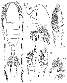
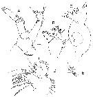
Non Megacalanus princeps Wolfenden, 1904 | | | | Ref.: | | | A. Scott, 1909 (p.14, Descr.F, figs.F); Vervoort, 1946 (p.49); Owre & Foyo, 1967 (p.34, figs. Juv.4F); Michel, 1994 (p.180, Rem.); Bradford-Grieve & al., 1999 (p.877, 906, figs.F); Bradford-Grieve & al., 2017 (p.59, Descr., figs. F, M, phylogeny); ? Fornshell & Ferrari, 2014 (p.114, fig.11: Von Vaupel klein's organ). | 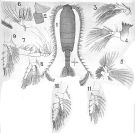 issued frm : A. Scott in Siboga-Expedition, 1909, XIX a. [Plate I, Figs.1-11]. Female (from Halmahera Sea): 1, habitus (dorsal); 2, last thoracic and genital segments (left side); 3, A2; 4, Md; 5, Mx1; 6, Mx2; 7, Mxp; 8, P1; 9, P2; 10, P4; 11, P5. Bar: Natural size. Nota: Head and 1st thoracic segment separated, 4th and 5th completely separated. 5th thoracic segment is produced posteriorly in distint points. Abdomen 4-segmented, symmetrical, equal to one-third of the length of the cephalothorax. Genital segment longer than wide, nearly equal to the combined lengths of 2nd and 3rd segments.. The 2nd segment is about half the length of the genital segment. The 3rd segment is equal to two-thirds of the lonength of the 2nd. The 4th segment is about half as long as the 3rd. Caudal rami short and slightly longer than broad but rather longer than the anal segment. A1 25-segmentd, extend beyond the end of the urosome by at least 4 segments.
|
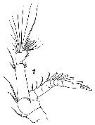
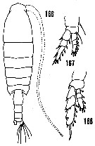 issued from : H.B. Owre & M. Foyo in Fauna Caribaea, 1, Crustacea, 1: Copepoda. Copepods of the Florida Current. 1967. [p.34, Figs.166-168]. Redrawings after A. Scott, 1909. Female (from 23°38'N, 83°06'W): 166, habitus (dorsal); 167, P1; 168, P5.
|
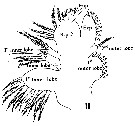 Issued from : H.B. Owre & M. Foyo in Fauna Caribaea, 1, Crustacea, 1: Copepoda. Copepods of the Florida Current, 1967. [p.12, Fig.116]. Female: 16, Mx1 1st inner lobe = arthrite on the praecoxa; 2nd inner lobe: on the coxal endite and outer epipodite or 2nd outer lobe; Bsp 2 = Basis, 3rd lobe = basal endite, and basal exite; Enp = endopod with segments more or less fused; Exp = exopod.
|
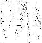
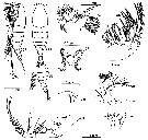
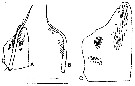
 Issued from : J.M. Bradford-Grieve, L. Blanco-Bercial & G.A. Boxshall in Zootaxa, 2017, 4229 (1). [p.60, Fig. 30]. Female: A-B, habitus (dorsal and lateral, respectively); C, anterior head (lateral); D, exopod of A2. Scale bars: 1.0 mm (A, B); 0.1 mm (C, D) D: specimens from 25.082°S, 09.584°E; A-C: specimens from 20.933°N, 138.533°E. Nota: - Forehead rounded in dorsal view, without projection. - A1 extending about 7 segments beyond caudal rami. - Genital double-somite, in dorsal view, widest at anterior 1/3. - A2 exopod segments I-III each with short but well-developed seta, segment IV with well-developed seta extending as far as distal border of segment VIII. - Mx1 with coxal endite with 5setae, one of them short; endopodal segment 2 with 1 long and 1 vestigial seta. - P1 exopodal segment 3, about 2.7 times as long as maximum width; distal border of endopodal segment 1 not extending beyond exopodal segment 1; exopod outer spines: on segment 1 rxtends less than half way along segment 2; on segment 2 extends half distance to 1st spine on segment 3; segment 3 proximal spine extends almost base of distal spine.
|
 Issued from : J.M. Bradford-Grieve, L. Blanco-Bercial & G.A. Boxshall in Zootaxa, 2017, 4229 (1). [p.61, Fig. 31]. Male: A-B, habitus (dorsal and lateral, respectively); C, caudal ramus (dorsal); D, P1. Scale bars: 1.0 mm (A, B); 0.1 mm C, D. Nota: - Forehead rounded in dorsal view. - Pediger 5 rounded in lateral view. - P1 outer border spines on exopodal segments 1 and 2 extend half way to base of following spine; 1st outer spine inserted at 2/3 distance along outer border of exopodal segment 3 and extending beyond base of outer distal spine; distal border of endopodal segment 1 level with distal border of exopôdal segment 1.
|
 Issued from : K.A. Brodsky , N.V. Vyshkvartzeva, M.S. Kos & E.L. Markhaseva in Opred. Fauna SSSR, Isdav. Inst. Zool., Acad. Sci. SSSR, 1983, 135 (1). [p.198, Fig.88]. As Bradycalanus sarsi. Female (from N Pacif.): habbitus (dorsal); R, rostrum; Ce, anterior head (lateral), Mx1; Mx2; P1, P3, P5. Nota: A1 25-segmented.
|
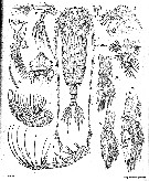
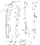
 Issued from : T.K.S. Björnberg in Antarct. Res. Ser., 1968, 11. [p.84, Figs 55-63]. As Bradycalanus pseudotypicus. Female (from SW Valdivia, Chile): 55 A B, habitus (lateral and dorsal, respectively); 56, last thoracic segments and urosome (lateral), another specimen; 57, Mx1; 583, Mx2; 59, mouth plate; 60, rostrum (ventral); 61, A2; 62, Md blade; 63, Mxp. Nota: A1 25-segmented.
|
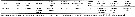
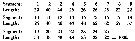 Issued from : T.K.S. Björnberg in Antarct. Res. Ser., 1968, 11. [p.84, Figs 55-63]. As Bradycalanus pseudotypicus. Female A1: Proportional lengths of the segments
|
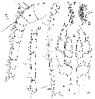
 Issued from: G.O. Sars in Résult. Camp. Scient. Prince Albert I, 69, pls.1-127 (1924). [Pl.III, fig. 1]. As Megacalanus princeps ( Brady, 1883). Female: 1, habitus (dorsal); 2, forehead (lateral); 3, rostrum (fontal view); 4, A2; 5, labrum and labium (ventral view); 6, Md; 7, Mx1; 10, P1; 11, P3; 12, P5.8, Mx2; 8a, distal portion of a seta of Mx2 (enlarged); 9, Mxp.
|
 Issued from : V.N. Andronov in Russian Acad. Sci. P.P. Shirshov Inst. Oceanol. Atlantic Branch, Kaliningrad, 2014. [p.36, Fig.9, 1]. As Bradycalanus sarsi Farran, 1939 (after Sars, 1924 as Megacalanus princeps Pl.III, fig.4). 1, A2.
|
 Issued from : J.M. Bradford-Grieve, L. Blanco-Bercial & G.A. Boxshall in Zootaxa, 2017, 4229 (1). [p.66, Table 8]. Morphological characters after identification key of Bradycalanus females and males. Compare to other species of genus. Main characters identification after species key : 1 - Head rounded; posterior pediger 5 corners triangular in lateral view; Mx1 coxal endite with 5 setae. 2 - Female A1 long extending about 6-7 segments beyond caudal rami; P1 exopod segment 1 and 2 with outer border spines short extending well short of base of following spine. 3 - Female total body length < 13 mm; male right P5 exopod segment 3 inner border spine inserted opposite outer border spine with notch.
|
 Issued from : J.M. Bradford-Grieve, L. Blanco-Bercial & G.A. Boxshall in Zootaxa, 2017, 4229 (1). [p.62, Fig. 32]. Male right A1 ancestral segments: A, segments I-XIV; B, segments XV-XIX; C, segments XX-XXVIII; D, detail segments XIX-XX; E, detail segments XXI-XXIII; F, detail segment XXVIII. Scale bars: 1,0 mm (A-C); 0.1 mm (D, E, F). Illustrated specimen from 38.033°N, 124.183°W. Nota: - A1 extending about 2-3 segments beyond caudal rami; ancestral segments II-IV, Ix-XI and XXI-XXIII fused, double aesthetascs on ancestral segments I, III, V, VII, IX, XI-XIV. Right antennule geniculate between ancestral segments XX-XXI, setation of gripping elements: XIX- 1 fused gripping element extending as far as base of aesthetasc, 1ms, 1a; XX-1 gripping element, 1ms, 1z; XXI-XXIII- 2 short gripping elements, 2nd extending slightly beyond base of aesthetasc, 1a, 1 unknown element, 1ms.
|
 Issued from : J.M. Bradford-Grieve, L. Blanco-Bercial & G.A. Boxshall in Zootaxa, 2017, 4229 (1). [p.63, Fig. 33]. Male left A1 ancsetral segments: A, segments I-XIII; B, hair sensilla of dorsal surface of ancestral segment V; segments XIV-XVIII; D, segmentsXIX-XXVIII; E, P5 male ( arrow indicates notch); F, distal inner corner of left P5 male exopod segment 2; G, distal inner corner of left P5 male exopod segment 2 (another specimen from USNM1208043). Scale bars: 1.0 mm (A, C, D); 0.1 mm (E, F, G). Nota: - P5 left exopodal segment 2 specialised seta tapering, extending short of distal border of endopodal segment 3, with small proximal tapering projection; inner border of left exopodal segment 3 setulose with inner articulated spine inserted just proximal to 2nd outer (terminal) spine; inner border of right exopodal segment 3 naked, inner articulated spine inserted opposite 1st outer border spine, small notch in inner border level with distal border of right endopod.
|
 Issued from : J.M. Bradford-Grieve, L. Blanco-Bercial & G.A. Boxshall in Zootaxa, 2017, 4229 (1). [p.64, Fig. 34]. Male: A, A2; B, Md palp; C, Md gnathobase; D, Mx1; R, details of basal endites 1 and 2 (B1, B2) of mx1. Scale bars: 0.1 mm on all figures. Illustrated specimens from 38.033°N, 124.183°W. Nota: - A2 exopod segments I-IV each withy less well-developed seta than in female. - Mx1 endopodal segment 1 with 1 large and 1 vestigial seta; segment 2 with 1 seta.
|
 Issued from : J.A. Fornshell & F.D. Ferrari in Crustaceana, 2014, 87 (1). [p.105, Fig.1, A-C]. As Megacalanus longicornis Von Vaupel Klein's organ on right endopodal segment 1 and left basal seta of P1. A, anterior, distal up; B, right basal seta; C, proximal endopodal segment of right P1, denticles and pores showed (ventral seta not drawn) . Long scale line; 175 µm; short scale line 290 µm. The Von Vaupel Klein organ is an association (srtucture) of the distal seta from basis and the proximal endopodal segment of P1. This structure shows significant variability among many gymnoplean copepods, in the shape of the distodorsal corner of the proximal endopodal segment, presence and location of denticles on the anterior face of the segment, presence and size of denticles along the distal margin of the segment , number of pores on the segment, shape of the seta that originates on the basis, and the morphology of the basis at the origin of the seta. The function of this complex structure is not known.
| | | | | Ref. compl.: | | | Grice & Hulsemann, 1967 (p.13); 1968 (tab.2); Razouls & al., 2000 (p.343, Appendix) | | | | NZ: | 13 | | |
|
Carte de distribution de Bradycalanus typicus par zones géographiques
|
| | | | | | | | | | Loc: | | | Antarct. (SW Atlant.), Drake Passage, off Namibia, Barbada Is., Caribbean, Florida, G. of Aden, Arabian Sea, NW Indian, off E Réunion Is., Indonesia-Malaysia ( Halmahera Sea, Flores Sea), SE Pacif. (off Juan Fernandez Is.), SW Valdiviella (Chile), off California, off SE Japan | | | | N: | 8 | | | | Lg.: | | | (5) F: 9; (28) M: 11; (85) F: 15,5; (131) F: 11-12; (406) F: 12,5-13,75; (1316) F: 11,5-13,9; M: 9,4-11,3; {F: 9,00-15,50; M: 6,4-11,3} | | | | Rem.: | Bathypélagique.
Sampling depth (Antarct.) : 1000-2000 m.
Sars (1925, p.14) considère cette espèce comme un synonyme de B. sarsi (comme Calanus princeps Brady, 1883, puis Megacalanus princeps (Brady), no Wolfenden,1904. Il est suivi par Jespersen (1934, p.45). Les répartitions géographiques des deux espèces semblent confirmer ce point de vue, bien que les références soient rares. Cependant la présence de pointes postérieures au dernier segment thoracique permet de distinguer les deux espèces.
Voir aussi les remarques en anglais | | | Dernière mise à jour : 14/09/2018 | |
|
|
 Toute utilisation de ce site pour une publication sera mentionnée avec la référence suivante : Toute utilisation de ce site pour une publication sera mentionnée avec la référence suivante :
Razouls C., Desreumaux N., Kouwenberg J. et de Bovée F., 2005-2025. - Biodiversité des Copépodes planctoniques marins (morphologie, répartition géographique et données biologiques). Sorbonne Université, CNRS. Disponible sur http://copepodes.obs-banyuls.fr [Accédé le 27 août 2025] © copyright 2005-2025 Sorbonne Université, CNRS
|
|
|
|


;)
;)
;)
;)
;)
;)
;)
;)
;)
;)
;)
;)
;)

