|
|
 |
Fiche d'espèce de Copépode |
|
|
Calanoida ( Ordre ) |
|
|
|
Metridinidae ( Famille ) |
|
|
|
Gaussia ( Genre ) |
|
|
| |
Gaussia intermedia Defaye, 1998 (F,M) | |
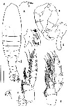
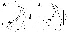
| | | | | | | Syn.: | Gaussia sp.: Cuoc & al., 1997 (p.652, figs.F: genital complex) | | | | Ref.: | | | Defaye, 1998 (p.82, figs.F,M); Alves-Jùnior & al., 2018 (p.501, Table I, Figs. 1, 2, 3, Rem.) | 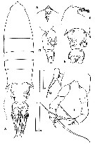 issued from : D. Defaye in Crustaceana, 1998, 71 (1). [p.83, Fig.1]. Female (from S California): A, habitus (dorsal); B, rostrum (ventral); C, forehead (lateral); D-E, urosome (lateral and ventral, respecyively); F, P5; G, apical spine of P5. Scale bars: 1 = 2 mm for A, D, E; 2 = 0.5 mm for B, C; 3 = 0.5 mm for 4; 4 = 0.5 mm for G. Nota: Urosome 3-segmented, 2nd segment very short, widening posteriorly. Genital somite almost symmetrical in dorsal view, except anterior right-lateral edge which is elongated into a spiniform process; ventrally, proximal half of somite with paired , crescent-shaped gonopores,, distal half with 2 ventrolateral, brown, rounded masses corresponding to spermatophores. Anal somite 3 times broader than long at its greatest width, bearing a lateral line of long hairs, and an oblique posterior extension on each side. Caudal rami dorso-ventrally flattened and more or less rectangular in dorsal view, both ramui with 6 setae, outer seta close to lateral expansion of anal somite, the innermost thin, and shorter that the others; 5 main caiudal setae plumose, hairs also present on each side of the ramus, near the basal insertion. Pores of putative luminous glands present on anal segment, particularly at the end of the postero-lateral expansions and on the caudal rami, occuring in the same pattern as described for G. asymmetrica. Rostrum with rostral filaments relatively long, close together and bearing relatively dense hairs.
|
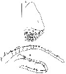
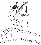 issued from : D. Defaye in Crustaceana, 1998, 71 (1). [p.85, Fig.2]. Female: A, A1; B, A2; C, Md (gnathobasis). Scale bars: 0.5 mm. Nota: A1 23-segmented, longer than body (without caudal rami); 8th segment twice as long as the adjacent segments, with remains of a suture line (indicating the fusion of 2 segments); pores present from 1st to 11th (10 ?) segments, which can be attributed to luminous glands (see in Barnes & Case, 1972). A2 with endopod 2-segmented; exopod 8-segmented.
|
 issued from : D. Defaye in Crustaceana, 1998, 71 (1). [p.86, Fig.3]. Female: A, Mx1; B, Mxp; C, Mx2; D, Md (mandibular palp). Nota: Mx1 with the same morphology and chaetotaxy as G. asymmetrica: 9 setae on exite 1;1 on exite 2; 10 on exopodite; 11 + 6 on endopodite; 4 on endite 2, 16 on endite 1, 5 on endite 3; and 5 on basipodite; (endite 3 and basipodite figured as dotted lines). Mx2 and Mxp as in G. asymmetrica.
|
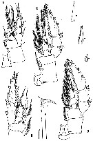 issued from : D. Defaye in Crustaceana, 1998, 71 (1). [p.87, Fig.4]. Female: A-D, P1 to P4; square: detail of hook of endopod 1 of P2 (scale bar: 0.5 mm). Scale bars: 1 = 0.5 mm for detail of setae; 2 = 1 mm for A, B, C, D.
|
 issued from : D. Defaye in Crustaceana, 1998, 71 (1). [p.89, Fig.5]. Male: A, habitus (dorsal); B, rostrum (lateral); C, P2; D, P4; E, P5 (frontal view, excluding last segments of left P5; star: detail of exopod 2. Scale bars: 1 = 1 mm for B, C, D, E; 2 = 1 mm for A. Nota: Buccal appendages as in female.
|
 issued from : D. Defaye in Crustaceana, 1998, 71 (1). [p.85, Fig.2]. Male: D, A1; E, oral field, left part of labrum. Scale bar : 0.5 mm for E. A1 as long as body, the left one 23-segmented (as in female), right A1 geniculated between 17th and 18th segment, rhe five segments before geniculation elongated, the three following segments almost fused into one, these together being as long as the penultimate segment; no gland pore observed on A1.
|
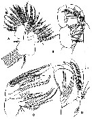
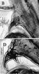
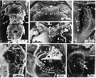 issued from : C. Cuoc, D. Defaye, M. Brunet, R. Notonier & J. Mazza in Mar. Biol., 1997, 129. [p.653, Fig.1]. As Gaussia sp. Scanning electron micrographs of female genital area. 1, outer ventral face of urosome; note position of closed gonopores ( cg) and seminal receptacles ( sr), right receptacle being empty and left containing spermatophoral mass ( sm), thoracic expansions ( stars) of last prosomite segment, and strong hook ( h) and lateral protuberances ( p) of genital double-somite; 2-3, comparative outer ventral (2) and inner dorsal (3) views of anterior part of genital double-somite. 4, insemination site or copulatory pore of noninseminated right receptacle; note copulatory pore closed by a fine membrane, cracked on its external side ( );
5, inner dorsal view of right part of genital area;
6, detail of inner-right gonoporal zone; note semicircular shape of egg-laying duct (d3) with double wall (), and insertion (arrowhead) of muscles (m) of egg-laying duct;
7, inner view of right seminal receptacle (sr) and right seminal duct (d1); note multilobed proximal end (arrow) of seminal duct.
|
 Issued from : F.A. Alves-Jùnior, A. Bertrand, P.A.M.C. Melo, E.P. Correia, L.G.P. Figueiredo & S. Neumann-Lettao in Crustaceana, 2018, 91 (4). [p.506, Fig.2, E, F]. Gaussia intermedia from Rocas Atoll: E, female P5, F, male P5.
|
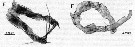
 Issued from : F.A. Alves-Jùnior, A. Bertrand, P.A.M.C. Melo, E.P. Correia, L.G.P. Figueiredo & S. Neumann-Lettao in Crustaceana, 2018, 91 (4). [p.507, Fig.3]. (from Rocas Atoll): A, bidentate hook of 1st segment of endopod of P2 female; B, bidentate hook of 1st segment of endopod of P2 male.
|
 Issued from : F.A. Alves-Jùnior, A. Bertrand, P.A.M.C. Melo, E.P. Correia, L.G.P. Figueiredo & S. Neumann-Lettao in Crustaceana, 2018, 91 (4). [p.506, Fig.2, B, D]. (from Rocas Atoll): B, bidentate hook on the 1st segment of endopod of P2 female; D, bidentate hook on the 1st segment of endopod of P2 male.
| | | | | Ref. compl.: | | | Galbraith, 2009 (pers. comm.) | | | | NZ: | 3 | | |
|
Carte de distribution de Gaussia intermedia par zones géographiques
|
| | | | Loc: | | | S California (33°20'N, 118°43'W), off British Columbia, Rocas Atoll (N Brazil) | | | | N: | 3 | | | | Lg.: | | | (160) F: 10,4; M: 9,5; (1216) F: 10,7-10,5; M: 10,3-10,5; {F: 10,40-10,70; M: 9,50-10,50} | | | | Rem.: | sampling depth 1262-1271 m.Br>For Alves-Jùnior & al. ( 2018) this deep-sea species is found for the first time in western tropical Atlantic (3°51'S, 33°49'W),, collected through oblique hauls from 525 m to the surface, probably associated with transport in upwelling at Rocas Atoll. | | | Dernière mise à jour : 15/03/2018 | |
|
|
 Toute utilisation de ce site pour une publication sera mentionnée avec la référence suivante : Toute utilisation de ce site pour une publication sera mentionnée avec la référence suivante :
Razouls C., Desreumaux N., Kouwenberg J. et de Bovée F., 2005-2025. - Biodiversité des Copépodes planctoniques marins (morphologie, répartition géographique et données biologiques). Sorbonne Université, CNRS. Disponible sur http://copepodes.obs-banyuls.fr [Accédé le 30 août 2025] © copyright 2005-2025 Sorbonne Université, CNRS
|
|
|
|


;)
;)
;)
;)
;)
;)
;)
;)
