|
|
 |
|
Calanoida ( Order ) |
|
|
|
Diaptomoidea ( Superfamily ) |
|
|
| |
| | | |
| Fosshageniidae Suarez-Morales & Iliffe, 1996 ( Diaptomoidea ) | | Ref.: | Suarez-Morales & Iliffe, 1996 (p.755); Boxshall & Halsey, 2004 (p.14; 49; 121: Def.; p.122: Genera & species key); Vives & Shmeleva, 2007 (p.528, spp. Key); Blanco-Bercial & al., 2011 (p.103, Table 1, Fig.2, 3, 4, Biol. mol, phylogeny) ; Laakmann & al., 2019 (p.330, fig. 2, 3, phylogenetic relationships)
Bradford-Grieve J.M., (2002 onwards). Key to calanoid copepod families. Version 1 : 2 oct 2002. http://www.crustacea.net/crustace/calanoida/index.htm  | | Rem.: | Type-genus: Fosshagenia Suarez-Morales & Iliffe, 1996. Total: 2 G.
Suarez-Morales & Iliffe (1996, p.760) create a new superfamily. Fosshagen & Iliffe, 2004 (p.353) deem that the genus Temoropia should be included in this family belonging to the superfamily of the Diaptomoidea. 2 G: Fosshagenia, Temoropia.
Vives & Shmeleva, 2007 (p.521, 528) maintain the genus Temoropia in the family of the Temoridae.
Key to genera and species after Boxshall & Halsey (2004, p.122) :
1 Urosome 3-segmented in female, with 2 free abdominal somites ; exopod of right male P5 3-segmented, lacking terminal claw
. Fosshagenia ferrarii.
1 Urosome 4-segmented in female, with 3 free abdominal somites ; exopod of right male P5 2-segmented, terminating in curved claw
2.
2 Rostrum very thin filament-like in both sexes ; genital double-somite of female distinctly asymmetrical in dorsal view, with short process on left side ; male P5 with protopod much more powerfully developed on right leg than on left
.. Temoropia mayumbaensis.
2 Rostrum strong in both sexes ; genital double-somite of female symmetrical, without process on left side ; more or less equally developed
.. 3.
3 Female P5 with 2nd segment 2 to 2.5 times longer than wide ; 2nd segment of male right P5 about 3 times longer than wide and lacking ornamentation of spinules on anterior surface of segment
.. Temoropia minor.
3 Female P5 with 2nd segment about 1.5 times longer than wide ; 2nd segment of male right P5 about 2 times longer than wide and ornamented with several rows of fine spinules on anterior surface of segment
Temoropia setosa.
|  Issued from : G.A. Boxshall & S.H. Halsey in An Introduction to Copepod Diversity. The Ray Society, 2004, No 166, Part. I. [p.121]. Armature formula of swimming legs P1 to P4. Nota: Female P5 with separate coxae and intercoxal sclerite; basis distinct, without outer seta. Endopod represented by bifid process, as in Temoropia, or by single spine, as in Fosshagenia. Free exopod unsegmented, armed with strong spine on inner margin; distal margin of exopodal segment bearing 5 spinous processes; - Male P5 asymmetrical; carried on bilobed trnsverse plate formed by fusion of coxae and intercoxal sclerite or on distinct coxae; right leg comprising unarmed basis and 2 or 3-segmented exopod; distal exopodal segment forming long curved subchela in Temoropia. Endopod comprising single segment plus apical digitiform process, or absent. Left leg indistinctly subchelate, with exopod opposing curved digitiform process (possibly representing endopod) on medial margin of basis. Bais distinct, exopod 1 or 2-segmented. - Eggs probably released into water. |
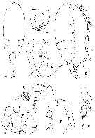 Issued from : G.A. Boxshall & S.H. Halsey in An Introduction to Copepod Diversity. The Ray Society, 2004, No 166, Part. I. [p.123, Fig.24]. Fosshageniidae. A, Fosshagenia ferrarii habitus female (dorsal view); B, habitus male (lateral view); C, Temoropia setosa female A2; D, female P1; E, Fosshagenia ferrarii female P5; F, Temoropia setosa female P5; G, Fosshagenia ferrarii male P5; H, Temoropia minor male P5. [Suarez-Morales & Iliffe, 1996: A-B, E, G; Schulz, 1986: C-D, F, H]. | | | | | (1) Fosshagenia Suarez-Morales & Iliffe, 1996 | |
| | Ref.: | Suarez & Iliffe, 1996 (p.755); Mauchline, 1998 (p.77); Fosshagen & Iliffe, 2004 a (p.346: emend, Rem.); Boxshall & Halsey, 2004 (p.122) | | Rem.: | Type: Fosshagenia ferrarii Suerez-Morales & Iliffe, 1996. Total: 2 spp.
For Fosshagen & Iliffe (2004, p.353) the appearance of this genus, with a transparent body, long A1, unmodified mouthparts, and not particularly strong outer spines on swimming legs, suggests a planktonic existence in caves. For the time being it seems most relevant to keep this genus as a cave-adapted genus belonging to the superfamily Diaptomoidea. It does not seem unlikely that some ancestral deep-water species closely related to Temoropia have invaded caves quite recently in geological terms and become established in an environment also characterized by darkness and scarcity of food. The close resemblance between Fosshagenia and Temoropia was pointed out by Boxshall & Halsey (2004, p.122). | | Remarks on dimensions and sex ratio: | | The mean female size is 0.650 mm (n = 4; SD = 0.0528) and the mean male size is 0.665 mm (n = 4; SD = 0.0443). The size ratio (male : female) is 1.023. The sex ratio (female : male) is 1. | | | | (2) Temoropia T. Scott, 1894 | |
| | Syn.: | Temeropia : Rose, 1933 a (p.174, lapsus calami); Marques, 1953 (p.109) | | Ref.: | T. Scott, 1894 b (p.79); Giesbrecht & Schmeil, 1898 (p.96); A. Scott, 1909 (p.119); Sewell, 1932 (p.246); Tanaka, 1963 (p.14); Razouls, 1982 (p.403); Schulz, 1986 (p.140, clé spp.); Razouls, 1993 (p.308); Mauchline, 1998 (p.77); Bradford-Grieve,1999 b (p.159, Déf.); Boxshall & Halsey, 2004 (p.122, clé spp.); Vives & Shmeleva, 2007 (p.527, spp. Key) | | Rem.: | type: Temoropia mayumbaensis T. Scott, 1894. Total: 3 spp.
Diagnosis after Bradford-Grieve (1999 b, p.112) :
- As in family definition.
- Head and pedigerous segment 1 separate, pedigerous segments 4 and 5 separate.
- Posterior corners of prosome rounded.
- Rostral filaments paired, of variable thickness.
- Urosme female 4-segmented.
- Urosome male 5-segmented.
- Female genital segment large and protruding ventrally by about the same amount as depth of urosome itself.
- Caudal rami small with 3 terminal setae (2 for minor).
- A1 24-segmented (segments 24 and 25 fused).
- Male right A1 geniculate, joint between free segments 16 and 17.
- A2 with both rami of about equal length ; exopod 7-segmented.
- Mx1 inne lobe 1 with 15 spines and setae, inner lobes 2 and 3 with 2 and 4 setae respectively, basis with 2 setae, endopod with 2, 2, 5 setae, exopod with 9 setae, outer lobe 1 with 9 setae.
- Mx2 with lobes 1-6 with 5, 3, 3, 3, 4, 3 setae respectively.
- Mxp basis distally broaded.
- P1 endopod 2-segmented ; P2-P4 3-segmented.
- Exopod segment 3 of P1 with 2 outer edge spines, of P2-P4 with 3 outer edge spines.
- Posterior surfaces of some of the segments of P1-P4 bearing patches of spinules.
- P5 female 3-segmented with a seta-like endopod ; exopod with inner and distal tooth-like processes.
- Male P5 2-segmented on right, 3-segmented on left ; right segment 2 has a long, strongterminal spine ; left segment 1 has a long distal spine and a smaller inner bulb tapering to a point, segment 2 with an inner distal, hairy flap. | | Remarks on dimensions and sex ratio: | | The mean female size is 0.767 mm (n = 6; SD = 0.2124) and the mean male size is 0.723 mm (n = 6; SD = 0.1883). The size ratio (Male : Female) is 0.940. The sex ratio (Female : Male) is 1. | | |
|
|
 Any use of this site for a publication will be mentioned with the following reference : Any use of this site for a publication will be mentioned with the following reference :
Razouls C., Desreumaux N., Kouwenberg J. and de Bovée F., 2005-2026. - Biodiversity of Marine Planktonic Copepods (morphology, geographical distribution and biological data). Sorbonne University, CNRS. Available at http://copepodes.obs-banyuls.fr/en [Accessed June 07, 2026] © copyright 2005-2026 Sorbonne University, CNRS
|
|
|
|


;)
