|
|
 |
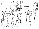
| Lubbockiidae Huys & Böttger-Schnack, 1996/97 ( Cyclopoida ) | | Syn.: | Oncäidae part. Giesbrecht,1892 (p.81) | | Ref.: | Huys & Böttger-Schnack, 1996/97 (p.243, 247: Redef.; p.260: Genera key); Boxshall & Halsey, 2004 (p.17, 575, Def., spp. Key; Rem. p.1: For the authors the order Poecilostomatoida is questionable); Vives & Shmeleva, 2010 (p.245, Rem., Genera key) | | Rem.: | The family comprises 6 frequently encountered pelagic genera: Atrophia, Haplopodia, Homeognathia, Lubbockia, Pseudolubbockia, Rhamphochela, and one, the genus Laitmatobius Humes,1987, very rare, only known by numerous males in the Guaymas Basin (Gulf of California) from depth 2,004 m. | 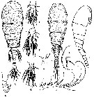 Issued from : F. Vives & A.A. Shmeleva in Fauna Iberica, 2010, 33. [p.253, Fig.118]. After Heron & Damkaer, 169. As Pseudolubbockia dilatata type species. Female: A, habitus (dorsal); B, Md; C, Mx1; D, Mxp; E, P1; F, P2; G, P4. Male: H-I, habitus (dorsal and lateral, respectively); J, A1. |
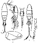 Issued from : F. Vives & A.A. Shmeleva in Fauna Iberica, 2010, 33. [p.251, Fig.117]. After Giesbrecht, 1893 [1892]. As Lubbockia squillimana type species. Female: A-B, habitus (dorsal and lateral with eggs, respectively); C, Mxp; D, urosome. Male: E, habitus (dorsal); F, anal segment and furca. |
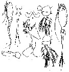 Issued from : F. Vives & A.A. Shmeleva in Fauna Iberica, 2010, 33. [p.255, Fig.119]. After Heron & Damkaer, 1978. As Atrophia minuta type species. Female: A, habitus (lateral); B, labrum (ventral); C, right Md; D, Mx1; E, Mxp; F, urosome (dorsal); G, P1; H, P3. Male: I, habitus (ventral). |
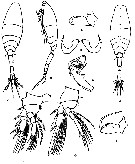 Issued from : F. Vives & A.A. Shmeleva in Fauna Iberica, 2010, 33. [p.257, Fig.120]. After Heron & Damkaer, 1978. As Haplopodia petersoni type species. Female: A-B, habitus (dorsal and lateral, respectively); C, Mxp; D, labrum; E, right Md; F, P1; G, P3. Male: H, habitus (dorsal); I, Mxp. |
 Issued from : F. Vives & A.A. Shmeleva in Fauna Iberica, 2010, 33. [p.247, Fig.115]. After Farran, 1908; Heron & al., 1984. As Homeognathia brevis. Female: A, E, habitus (dorsal and lateral, respectively); B, A2; C, Mxp; D, urosome (ventral); F, P1; G, P4. Male: H, J, habitus (dorsal and lateral, respectively); I, urosome (ventral). |
 Issued from : Boxshall & Halsey in The Ray Society, 2004, Part II. [p.575]. Spine and setal formula of swimming legs P1 to P4. Spines: Roman numerals; setae Arabic numerals. The element or elements on the outer margin of any segment are given in first, separated by a hyphen from the inner margin element or elements. Nota: Swimming legs 1 to 4 biramous, with 3-segmented rami. Intercoxal sclerites present in legs 1 to 4. Inner seta on basis of P1 present or secondarily absent. Inner coxal seta present. - P5 laterally located on somite; protopod incorporated into somite, represented by seta on surface of somite; exopod 1-segmented typically with 2 setae; exopod armed with 4 setation elements in Pseudolubbockia; absent in Haplopodia. - P6 represented by genital opercula; unarmed or armed with 1 or 2 setae in female, unarmed in male. Egg sacs paires, multiseriate. |
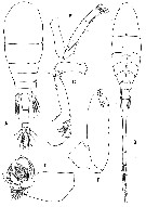 Issued from : Boxshall & Halsey in The Ray Society, 2004, Part II. [p.576, Fig.186]. A, Pseudolubbockia dilatata female; B, Lubbockia wilsonae male; C, Lubbockia glacialis female A2; D, Md; E, Laitmatobius crinitus male A2; F, Rhamphochela forcipula female Mxp. Heron & Damkaer, 1969: A; Heron & Damkaer, 1978: B; Heron & al., 1984: C-D; Huys & Böttger-Schnack, 1997: E-F. | | | | | (1) Atrophia Huys & Böttger-Schnack, 1996/97 | |
| | Ref.: | Huys & Böttger-Schnack, 1996/97 (p.259: Def.); Boxshall & Halsey, 2004 (p.577); Vives & Shmeleva, 2010 (p.254, Rem..) | | Rem.: | Type: Lubbockia minuta Wolfenden, 1905. Total: 2 spp.
Diagnosis after Boxshall & Halsey (2004, p.577):
1 - P1 and P2 with 3 outer spines on 3rd exopodal segment.
2 - 3rd endopodal segment of P3 and P4 with 2 and 1 setae/spines respectively; A2 seually dimorphic; male mouthparts atrophied. | | | | (2) Haplopodia Huys & Böttger-Schnack, 1996/97 | |
| | Ref.: | Huys & Böttger-Schnack, 1996/97 (p.258: Def.); Boxshall & Halsey, 2004 (p.577); Vives & Shmeleva, 2010 (p.256, Rem.) | | Rem.: | Type: Lubbockia petersoni Heron & Damkaer, 1978. Total: 1 sp.
Diagnosis after Boxshall & Halsey (2004, p.577):
1 - P1 and P2 with 3 outer spines on 3rd exopodal segment.
2 - 3rd endopodal segment of P3 and P4 with 3 and 2 setae/spines respectively; A2 not sexually dimorphic; male mouthparts not atrophied.
3 - Mxp sexually dimorphic; 3rd endopodal segment of P3 and P4 with armature formula I, II, 2 + I and I, II, 1 + I respectively; P5 of both sexes represented by single seta, with no free segment. | | | | (3) Homeognathia Huys & Bottger-Schnack, 1996/97 | |
| | Ref.: | Huys & Bottger-Schnack,1996/97 (p.259: Def.); Boxshall & Halsey, 2004 (p.577: spp. key); Vives & Shmeleva, 2010 (p.246, Rem..) | | Rem.: | Type: Lubbockia flemingi Heron & Damkaer, 1978. Total: 2 spp.
Diagnosis after Boxshall & Halsey (2004, p.577):
1 - P1 and P2 with 3 outer spines on 3rd exopodal segment.
2 - 3rd endopodal segment of P3 and P4 with 3 and 2 setae/spines respectively; A2 not sexually dimorphic; male mouthparts not atrophied.
3 - Mxp not sually dimorphic; 3rd endopodal segment of P3 and P4 with armature formula I, II, 3 and I, II, 2 respectively; P5 of both sexes represented by cylindrical exopod with 2 setal elements. | | Remarks on dimensions and sex ratio: | | The mean female size is 0.9075 mm. (n = 4, SD = 0.095), and the mean male size is 0.985 mm. (n = 4, SD = 0.297). The size ratio male: female is 1.085. Sex ratio Female: Male = 1. | | | | | (4) Laitmatobius Humes, 1987 | |
| | Ref.: | Humes, 1987 (p.645; p. : Def.); Huys & Böttger-Schnack, 1996/97 (p.257: Def.); Boxshall & Halsey, 2004 (p.577) | | Rem.: | Type: Laitmatobius crinitus Humes, 1987. 1 sp.
Diagnosis Male after Humes (1987, p.663):
Segment bearing P1 separated from cephalosome.
- Urosome 6-segmented.
- Caudal ramus with 6 setae.
A1 5-segmented, with aesthete on last segment.
A2 4-segmented, with 4 setiform terminal claws.
- Labrum indented medially.
- Md with 2 spines and 1 seta followed by long lash.
- Mx1 bilobed, lobes with 2 and 3 setae, respectively.
- Mx2 2-segmented, 2nd segment with 3 elements.
- Mxp 4-segmented (assuming proximal part of claw to represent 4th segment), 3rd segment showing subdivision; long slender claw.
P1-P4 biramous with 3-segmented rami. 3rd segment of endopod in legs P2-P4 armed as I, II, 3; I, III, 2; and I, III, 1 , respectively. Exopod of P4 with 3rd segment II, I, 5.
P5 with small free segment bearing 2 setae.
Diagnosis after Boxshall & Halsey (2004, p.577):
1 - P1 and P2 with outer spines on 3rd exopodal segment.
2 - A2 endopod 3-segmented. | | Remarks on dimensions and sex ratio: | | The mean male size is 1.31 mm (n = 10; 1.29 mm - 1.38 mm) | | | | (5) Lubbockia Claus, 1862 | |
| | Ref.: | Claus, 1863 (p.147, 149, 163); Giesbrecht, 1892 (p.82, 606); Sars, 1900 (p.114); van Breemen, 1908 a (p.192); A. Scott, 1909 (p.244); Rose, 1933 a (p.304); Mori, 1937 (1964) (p.121); Boxshall, 1977 a (p.109, spp. key); Heron & Damkaer,1978 (p.3, 4: clé spp.); Razouls, 1982 (p.679); Gardner & Szabo, 1982 (p.125); Heron & al.,1984 (p.449); Zheng Zhong & al., 1984 (1989) (p.263, Rem.); Heron & Bradford-Grieve, 1995 (p.12, Déf.); Huys & Böttger-Schnack, 1996/97 (p.245, 246, 256: Def., clé spp.); Chihara & Murano, 1997 (p.978); Bradford-Grieve & al., 1999 (p.887, 970, spp. key); Boxshall & Halsey, 2004 (p.577); Vives & Shmeleva, 2010 (p.248, Rem., spp. key) | | Rem.: | Type: Lubbockia squillimana Claus,1863. Total: 5 spp. (1 doubtful).
Diagnosis after Heron & Bradford-Grieve (1995, p.12):
Prosome elongate.
Urosome slender.
A1 7-segmented, with 1 or more incomplete sutures on female, 3rd segment with 2 spines; at least 3 terminal segments always fused on male.
A2 with elongate 3rd segment; female armature formula: 0, 1, 3 (sub-apical) + 3 (terminal) +3 or 4 terminal claws.
Md blade with scale-like denticles on median edge and terminating in a lash; 1 inner and 2 dorsal elements.
Mx2 with short setule and 3 (1 bifurcate) or 4 ornamented elements.
Oral appendages of male degenerate in some species; Mxp usually dimorphic.
All species, except L. flemingeri, with a hyaline papilla partially covering vent on outer edge of P1 coxa.
Species usually with small or conspicuous conical projections between 2 terminal spines on the endopods of P2 and P3, and sometimes of P4; these terminal projections appear to support a vent, which is present even when the projection is lacking. Often a vent is also on the inner distal corner of basis of each swimming leg.
P5 a free segment bearing 2 terminal elements, or P5 absent and represented by 1 lateral seta.
Diagnosis after Boxshall & Halsey (2004, p.577):
1 - P1 and P2 with 2 outer spines on 3rd exopodal segment.
2 - A2 endopod 2-segmented.
3 - P5 with cylindrical free exopodal segment bearing 2 setae/spines.
4 - 3rd exopodal segment of P3 and P4 with armature formula I, II, 2 and I, II, 1 respectively; Mxp basis without anterior keel-like process in female; male mouthparts atrophied. | | | | | (6) Pseudolubbockia Sars, 1909 | |
| | Ref.: | Sars, 1909 (p.4); Heron & Damkaer, 1978 (p.3); Razouls, 1982 (p.682); Gardner & Szabo, 1982 (p.131); Heron & Bradford-Grieve, 1995 (p.11, Def.); Huys & Böttger-Schnack, 1996/97 (p.245, 246, 257: Def.); Boxshall & Halsey, 2004 (p.577); Vives & Shmeleva, 2010 (p.252, Rem..) | | Rem.: | Type: Pseudolubbockia dilatata Sars, 1909. Total: 1 sp.
Diagnosis after Boxshall & Halsey (2004, p.577):
1 - P1 and P2 with 2 outer spines on 3ed exopodal segment.
2 - A2 endopod 2-segmented.
3 - P5 of both sexes with flattened free exopodal segment bearing 4 serrate spines. | | | | (7) Rhamphochela Huys & Böttger-Schnack, 1996/97 | |
| | Ref.: | Huys & Böttger-Schnack, 1996/97 p.258: Def.); Boxshall & Halsey, 2004 (p.577: spp. Key) | | Rem.: | Type: Lubbockia carinata Heron & Damkaer, 1978. Total: 2 spp.
Diagnosis after Boxshall & Halsey (2004, p.577):
1 - P1 and P2 with 2 outer spines on 3rd exopodal segment.
2 - A2 endopod 2-segmented.
3 - P5 with cylindrical free exopodal segment bearing 2 setae/spines .
4 - 3rd endopodal segment of P3 and P4 with armature formula I, II, 2 + I and I, II, 1 + I respectuvely; Mxp basis with anterior keel-like process in female. Male mouthparts atrophied. | | | | | |
|
|
 Any use of this site for a publication will be mentioned with the following reference : Any use of this site for a publication will be mentioned with the following reference :
Razouls C., Desreumaux N., Kouwenberg J. and de Bovée F., 2005-2026. - Biodiversity of Marine Planktonic Copepods (morphology, geographical distribution and biological data). Sorbonne University, CNRS. Available at http://copepodes.obs-banyuls.fr/en [Accessed April 07, 2026] © copyright 2005-2026 Sorbonne University, CNRS
|
|
|
|


;)
;)
;)
;)
;)
;)
