|
|
 |
|
Calanoida ( Order ) |
|
|
|
Calanoidea ( Superfamily ) |
|
|
| |
| | | |
| Mecynoceridae Andronov, 1973 ( Calanoidea ) | | Syn.: | Mecynoceridae : Andronov, 1970 (p.985, Rem.); 1973 b (p.1721); 1974 a (p.1005); Razouls, 1982 (p.101); Bowman & Abele, 1982 (p.10); Brodsky & al., 1983 (p.142, 147, 216); Mauchline, 1988 (p.722: pores cuticulaires); Hiromi, 1987 (p.147, Rem.); Nishida, 1989 (p.174); Bradford-Grieve, 1994 (p.71, Rem.); Chihara & Murano, 1997 (p.833); Barthélémy, 1999 a (p.30); Bradford-Grieve & al., 1999 (p.878, 902, 905, 911); Ohtsuka & Huys, 2001 (p.461); Boxshall & Halsey, 2004 (p.14; 49; 136: Def.); Vives & Shmeleva, 2007 (p.907); Cornils & Blanco-Bercial, 2013, p.861, figs.1, 3, 5, phylogeny, molecular analysis); Ohtsuka & Nishida, 2017 (p.568, Rem. concerning the 3 families Calocalanidae, Mecynoceridae and Paracalanidae).
Bradford-Grieve J.M., (2002 onwards). Key to calanoid copepod families. Version 1 : 2 oct 2002. http://www.crustacea.net/crustace/calanoida/index.htm
Rem.: 1 G.: Mecynocera.
| | Ref.: | Bradford-Grieve, 2008 (p.59, Rem.).
Bradford-Grieve J.M., (2002 onwards). Key to calanoid copepod families. Version 1 : 2 oct 2002. http://www.crustacea.net/crustace/calanoida/index.htm  | | Rem.: | 1 G.: Mecynocera.
Previously placed in the subfamily of the Eucalaninae by Giesbrecht, 1892 (p.45, 47), then in the Eucalanidae (Sars, 1902, p.14), next in the Calocalanidae Bernard, 1958 a (p.199). Placed in the Paracalanidae family by Bradford-Grieve 2008 (p.59) and Cornils & Blanco-Bercial, 2013 (p.861, 870).
Taxonomic notes after Boxshall & Halsey (2004, p.136) (summary) :
- Body slender.
- Cephalosome and 1st pedigerous somite separate or no.
- Male with dorsal cephalic hump on cephalothorax.
- Posterolateral angles of prosome rounded.
- Urosome female 3-segmented.
- Genital apparatus female comprising common genital aperture located medially on ventral surface of genital double-somite ; copulatory pore contained within median genital aperture.
- Urosome male apparently 5-segmented in male ; comprising genital somite and 4 free abdominal somites ; single genital aperture located ventrolaterally on right side at posterior rim of genital somite.
- Caudal rami short, separate at base, with up to 6 setae.
- Rostrum divided into paired rostral filaments.
- Nauplius eye present.
- A1 much longer than body, 23-segmented in female with considérable fusion of proximal segments ; segmental homologies : segment ½ compound (I-IV), segment 3 (V) to 7 (IX) separate, segments 8 (X) and 9 (XI) fused, segments 10 (XII) to 21 (XXVI) separate, apical (23rd) seggment double (XXVII-XXVIII) ; proximal segments with non-functional sutures making plane of fusion between segments 1 (I-IV) and 7 (XI). Aesthetascs present on segments III, V, VII, XI, XXI and XXVI-XXVIII. Many setae unusually long. Mid-length segments especially elongate, ornamented with rows of spinules along posterior margins.
- A1 non-geniculate in males, shorter than in female ; 20-segmented (according to Andronov, 1973) with additional segmental fusions, 1 and 2 (I-IV) and 3 to 6 (V-VIII), and thickenings proximally.
- A2 biramous, with separate coxa and basis ; coxa with 1 seta ; basis with 2 setae ; endopod 2-segmented ; compound distal segment bilobed ; setation formula 2, 9+7 ; exopod 8-segmented, shorter than endopod, terminal segment short ; segmental fusions : I-II, III-IV, V, VI, VII, VIII, IX, X ; setation formula : 2, 2, 1, 1, 1, 1, 1, 3.
- Md with well developed coxal gnathobase ; palp consisting of basis, armed with 4 setae, 2-segmented endopod and 5-segmented exopod ; endopodal segments 1 and 2 with 4 and 10 setae ; exopodal setation formula 1, 1, 1, 1, 2.
- Mx1 with well developed praecoxal arthrite bearing about 12 elements, including 4 setae on posterior surface ; coxa with endite bearing 3 setae and 9 setae on epipodite ; basis without outer seta, with proximal endite bearing 2/3 setae and distal group of 4 setae representing distal endite ; endopod distinctly 3-segmented, setation formula 4, 4, 6/7 ; exopod 1-segmented, armed with 11 setae.
- Mx2 7-segmented ; praecoxa and coxa separate, setation formula of endites 4/5, 3, 3, 3 ; allobasdis with ¾ setae on basal endite and 2 on first endopodal endite, free endopod indistinctly segmented, with total og 6 setae.
- Mxp 7-segmented ; syncoxa with endite setation formula 1, 2, 3, 4 ; basis armed with 3 setae, plus 2 setae on incorporated first endopodal segment, ornamented with medial spinule row proximal to basal setae, free endopod 5-segmented, segmental setation formula 4, 4, 3, 2+1, 4.
- Swimming legs 1 (P1) to 4 (P4) biramous with 3-segmented rami, except for 1-segmented or indistinctly 2-segmented endopod of leg . P1 with group tiny spinules at outer distal angle. Second endopodal segment of legs 2 to 4 ornamented with conspicuous spinule row, smaller spinules present on exopodal and distal endopodal segments. |  Issued from : G.A. Boxshall & S.H. Halsey in An Introduction to Copepod Diversity. The Ray Society, 2004, No 166, Part. I. [p.136]. Armature formula of swimming legs P1 to P4. Nota: Female P5 comprising 2-segmented protopod and 3-segmented exopod; 2nd exopodal segment armed with inner seta; distal exopodal segment bearing 1 to 6 setation elements around apex and inner margin (according to different descriptions). -Male P5 asymmetrical, both uniramous and 5-segmented; coxa fused to intercoxal sclerite; basis free, unarmed; exopod 3-segmented, distal segment armed with 2 apical spines, 1 large and 1 small; legt leg longer than right. - Eggs released into water. |
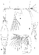 Issued from : G.A. Boxshall & S.H. Halsey in An Introduction to Copepod Diversity. The Ray Society, 2004, No 166, Part. I. [p.137, Fig.28]. Mecynoceridae. A, Mecynocera clausii habitus female; B, female P5; C, Mxp; D, habitus male; E, male P5. [Giesbrecht, 1893a (1892): A-C; Brodsky & al., 1983: D-E]. | | | | |
|
|
 Any use of this site for a publication will be mentioned with the following reference : Any use of this site for a publication will be mentioned with the following reference :
Razouls C., Desreumaux N., Kouwenberg J. and de Bovée F., 2005-2026. - Biodiversity of Marine Planktonic Copepods (morphology, geographical distribution and biological data). Sorbonne University, CNRS. Available at http://copepodes.obs-banyuls.fr/en [Accessed May 15, 2026] © copyright 2005-2026 Sorbonne University, CNRS
|
|
|
|


;)
