|
|
 |
|
Calanoida ( Order ) |
|
|
|
Diaptomoidea ( Superfamily ) |
|
|
| |
| | | |
| Temoridae Giesbrecht, 1893 ( Diaptomoidea ) | | Syn.: | Temorina Giesbrecht, 1892 (p.60); Temorinae Esterly, 1905 (p.173) | | Ref.: | Sars, 1902 (1903) (p.95, Rev.); Gurney, 1931 a (p.84); Wilson, 1932 a (p.103); Rose, 1933 a (p.169); Brodsky, 1950 (1967) (p.83, 279); Gonzalez & Bowman, 1965 (p.248); Andronov, 1974 a (p.1005); Razouls, 1982 (p.395); 1993 (p.308); Bowman & Abele, 1982 (p.9); Dussart & Defaye, 1983 (p.47); Brodsky & al., 1983 (p.142, 146); Zheng Zhong & al., 1984 (1989) (p.241, Genera Key); Sazhina, 1985 (p.50); Mauchline, 1988 (p.712, 740: cuticular pores); Huys & Boxshall, 1991 (p.467); Razouls, 1993 (p.308); Madhupratap & al., 1996 (p.863, Table 5: %/copepods); Chihara & Murano, 1997 (p.916); Barthélémy, 1999 a (p.24); Bradford-Grieve & al., 1999 (p.884, 902, 904, 953, 954: Genera Key); Bradford-Grieve,1999 b (p.155, Def., Rem.); Ohtsuka & Huys, 2001 (p.461); Boxshall & Halsey, 2004 (p.12; 49; 205: Def., Genera Key); Vives & Shmeleva, 2007 (p.521); Blanco-Bercial & al., 2011 (p.103, Table 1, Fig.2, 3, 4, molecular biology, phylogeny); Kos, 2016 (p.39, Def., Gennera Key); Laakmann & al., 2019 (p.330, fig. 2, 3, phylogenetic relationships)
Bradford-Grieve J.M., (2002 onwards). Key to calanoid copepod families. Version 1 : 2 oct 2002. http://www.crustacea.net/crustace/calanoida/index.htm  | | Rem.: | This family encloses only one marine genus: Temora Baird,1850; and three brackish- and freshwater genera: Eurytemora Giesbrecht,1881; Heterocope Sars,1863; Epischura Forbes, 1882; plus maybe Lamellipodia Schmeil,1897 . Oliveira, 1946 (1947) (p.463 & foll.) describes three genera: Lahmeyeria, Ganchosia, Manaia, of which the type species are juvenile forms belonging to other families.
Vives & Shmeleva, 2007 (p.521, 528) maintain the genus Temoropia in the Temoridae family.
Key to genera after Boxshall & Halsey (2004, p.207) :
1 - Female P5 with proximal exopodal segment produced medially into spinous process, distal segment armed with 2 setae; male P5 without inner process on basis of left leg .......... Eurytemora (in estuarine and brackish waters).
1' - Female P5 without spinous inner process on proximal exopodal segment; male P5 typically with inner process on basis og left leg ...... 2.
2 - P1 to P4 with 2-segmented endopods ........... Temora (in coastal waters).
2' - P1 to P4 with 1-segmented endopods ........ Heterocope and Epischura (in fresh waters).
Definition from J.M. Bradford-Grieve (1999 b, p.155) :
Female :
- Cephalosome and pediger segment 1 separate, pediger segments 4 and 5 fused, partly fused or separate
Forehead unarmed or provided with 2 solt rostral filaments or 1-2-pointed (Temoropia).
- Urosome 3-4 segmented.
- Genital segment without seminal receptacles.
- Caudal rami of different structure in different genera, sometimes elongate, with 6 setae.
- A1 24- or 25-segmented.
- A2 endopod more or less equal in length to exopod which is 6-7-segmented.
Md with a broad blade and 1 large tooth set slightly apart from remaining teeth ; endopod segment 1 often fused to basis.
- Mx1 inner lobe 1 with 12-15 spines and setae, inner lobes 2 and 3 usually with 4 and 3 setae respectively (with 2 and 4 setae respectively in Temoropia setosa), basis and proximal endopod segment often fused, exopod with 8-10 setae, outer lobe 2 with 1 or no setaa, outer lobe 1 with 8-9 setae.
- Mx2 with lobes 1-5 with 4-5, 2-3, 3, 3, 2-4 setae respectively.
- Mxp of moderate size, may be slighly modified with recurved endopod or with fusion between endopod segments ; coxa with 2, 2-3, 2-3, setae, basis with 0-3+2 setae, endopod with 2-4, 1-4, 1-3,0-3+1,4 setae respectively.
- Simming legs 1-4 with the endopods 1-, 2- or 3-segmented ; exopod segments 1 and 2 may be fused.
Male :
- Urosome 5-segmented.
- A1 distinctly geniculate on the right, 20-23-segmented.
- Segmentation of the body and the form of the mouthparts and swimming legs similar to those of the female. |  Issued from : G.A. Boxshall & S.H. Halsey in An Introduction to Copepod Diversity. The Ray Society, 2004, No 166,
Part. I. [p.205]. Armature formula of swimming legs P1 to P4. Nota: Setation sometimes reduced, often by loss of outer margin spine from 3rd exopodal segments, and by loss of outer margin element from distal endopodal segment. - Female P5 with coxae and intercoxal sclerite forming transverse plate; basis distinct, with outer seta. Endopod absent. Free exopod 2-segmented; 1st segment double (formed by fusion of 1st and 2nd exopodal segments) and armed with 1 or 2 outer spines, inner margin produced into spinous process in some genera; distal exopodal segment armed with up to 2 outer margin spines and distal element, plus, in some genera, several spinous processes along inner margin. - Male P5 asymmetrical; carried on bilobed trnsverse plate formed by fusion of coxae and intercoxal sclerite; right leg comprising basis with outer seta and 2 or 3-segmented exopod. Endopod absent. Exopod sometimes short, sometimes forming long curved subchela; segments typically with smallmarginal spines. Left leg either subchelate or chelate, with exopod opposing curved digitiform process (possibly representing endopod) on medial margin of basis. basis didtinct. Exopod 2-segmented, distal segment often withexpanded apex bearing spinous processes (reduced setation elements usually present. - Eggs released into water, retained in mass on ventral side of urosome, or contained in paired, multiseriate sacs. |
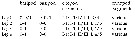 Issued from : J.M. Bradford-Grieve in NIWA Biodiversity Memoir 111, 1999. [p.155]. Spine and setal formula of swimming legs P1 to P4. Female P5 simple, not natatory, usually without an endopod; 2-3-segmented with common basal segment. Ovisac present in some cases. Male P5 simple, not natatory, usually without an endopod; larger than those of the female and prehensile, often pincer-like on one side, 2-4-segmented with common basal segment. | | | | | (0) Eurytemora Giesbrecht, 1881 | |
| | Syn.: | Temorella Claus, 1881; Canu, 1892 (p.173) | | Ref.: | Giesbrecht & Schmeil, 1898 (p.102, clé spp. Key); Sars, 1902 (1903) (p.97, 99); van Breemen, 1908 a (p.98, spp. Key); Esterly, 1924 (p.93); Gurney, 1931 a (p.182, spp. Key); Wilson, 1932 a (p.107, spp. Key); Rose, 1933 a (p.171, spp. Key); Mori, 1937 (1964) (p.66); Brodsky, 1950 (1967) (p.279, spp. Key); Jeffries, 1962 (p.291, Salinity vs. distribution); Tanaka, 1963 (p.15); Dussart, 1967 (p.69, clé spp.); Heron & Damkaer, 1976 (p.127, Rem.); Kos, 1977 a (p.20, Rev.); Razouls, 1982 (p.398); Gardner & Szabo, 1982 (p.313); Dussart & Defaye, 1983 (p.47); Zheng Zhong & al., 1984 (1989) (p.242, Rem.); Ferrari, 1992 (p.392, tab.3); Razouls, 1993 (p.308); Kos, 1993 (p.30, spp. Key); Chihara & Murano, 1997 (p.917); Mauchline, 1998 (p.93: F,M); Bradford-Grieve,1999 b (p.155, Def., Rem.); Boxshall & Halsey, 2004 (p.207); Dodson & al., 2010 (p.653, table 1, 2, fig.1, 2, 4, 5, 6); Kos, 2016 (p.52, Species Key: p.53-55); Sukhikh & Alekseev, 2013 (p.85, Remark concerning the species type E. affinis. | | Rem.: | Freshwater species, some forms are brackish. Analysis of this genus provisional, partially included in the matrix and in the treatment of geographic data (for example for E. caspica from the Caspian Sea..
Type: Temora affinis Poppe, 1880. Total: 24 spp.(noted up to 2019)
After Heron & Damkaer (1976, p.127), Gurney (1931) suggested that Eurytemora may have evolved from the marine genus Temora in the ''arctic sea of glacial times''.
Definition from Bradford-Grieve (1999 b, p.155) :
- As for the family definition.
- Head with a posterodorsal prominence similar to that in Temora, anterior head only slightly prominent and with 2 soft rostral filaments.
- Pedigerous segments 4 and 5 separated by a distinct suture, sometimes greatly expanded laterally.
- Urosome slender with female genital segment somewhat protuberant below..
- Caudal rami elongated and slightly divergent, setae normal in number.
- Eye of moderate size.
- A1 comparatively short, scarcely exceeding the prosome in length, 24-segmented in the female. Right A1 in male distinctly geniculate.
- A2 exopod 7-segmented, longer than the endopod.
- Anterior lip rather prominent ventrally.
- Md, Mx1 and Mx2 similar to Temora.
- Mxp shorter and stouter than in Temora, basipod 2 dilated and the endopod is recurved and carries delicate plumose setae.
- Endopod of P1 1-segmented, of P2-P4 2-segmented; exopodal segment 3 of P2-P4 with 2 outer edge spines, terminal spine finely toothed on the outer edge.
- P5 female 4-segmented, penultimate segment with the inner edge produced into a strong, pointed process; last segment small with 2 unequal spines terminally.
- Male P5 large and less asymmetrical than in Temora, both legs nearly the same size, 4-segmented, more or less incurved, right terminal segment claw-shaped, that of the left spatulate terminally.
- Ovisac present in the female. | | Remarks on dimensions and sex ratio: | | The mean female size is 1.488 mm (n = 42; SD = 0.3550), and for male 1.324 mm (n = 39; SD = 0.3230). The size ratio (male: female) is 0.89. The sex ratio is 1.1. | | | | | | Syn.: | Halitemora Giesbrecht,1881 (p.257)
Cyclops (part.): Müller, 1785 (p.115).
Calanus (part.): Dana, 1848 (p.12).Diaptomus (part.): Lubbock, 1856 (p.10). | | Ref.: | Brady, 1878 (part., p.53); Giesbrecht, 1892 (p.60, 328); Giesbrecht & Schmeil, 1898 (p.100, clé spp.); Wheeler, 1901 (p.174); Sars,1902 (1903) (p.96); A. Scott, 1909 (p.118); Sewell, 1932 (p.244); Wilson, 1932 a (p.103, spp. Key); Rose,1933 a (p.169); Mori, 1937 (1964) (p. 64); Dakin & Colefax, 1940 (p.92, clé spp.); Carvalho, 1952 a (p.147); Tanaka, 1963 (p.13); Gonzalez & Bowman, 1965 (p.248); Ramirez, 1966 (p.13); Razouls,1982 (p.395); Zheng Zhong & al.,1984 (1989) (p.241, clé spp.); Mauchline, 1988 (p.712, cuticular pores); Ferrari, 1992 (p.392, tab.3); Razouls,1993 (p.308); Chihara & Murano, 1997 (p.916); Mauchline, 1998 (p.76; p.95);Bradford-Grieve & al., 1999 (p.954: clé spp.); Bradford-Grieve,1999 b (p.157, Def.); Boxshall & Halsey, 2004 (p.207); Vives & Shmeleva, 2007 (p.521, spp. Key); Kos, 2016 (p.42, Def., p.43: species Key). | | Rem.: | Type: Cyclops longicornis Müller, 1785. Total: 5 spp. (of which 1 doubtful).
Definition from Bradford-Grieve (1999 b, p.157) :
- As in the family definition.
- Body short and compact.
- Head vaulted dorsally; remarkably dilated with a posterodorsal prominence.
- Head with 2 slender rostral filaments.
- Pedigerous segments 4 abd 5 fused.
- Female genital segment comparatively short and hardly protuberant ventrally.
- Caudal ramui narrow and elongate, sometimes asymmetrical, setae comparatively short and of the usual number, one is on the outer border some distance from others.
- Eye small.
- A1 slender and elongate, 24-segmented in female, last 2 segments fused, geniculate on right in male.
- A2 exopod 7-segmented, scarcely longer than endopod.
- Anterior lip not prominent.
- Mouthparts of a normal structure.
- Swimming legs with endoipods small and 2-segmented; exopodal segments 1 and 2 of P2-P4 partly fused in female; exopodal segment 3 with 3 outer edge spines and 1 terminal coarsely toothed spine.
- Female P5 small, , 3-segmented, first two simple, last segment dentate terminally.
- Male P5 asymmetrical, left leg much larger, 4-segmented, segment 2 produced on inner edge into a long curved thumb-like process, which opposes the 2 terminal segments; right leg 3-segmented, terminal segment incurved, claw-like.
- No ovisac present in the female. | | Remarks on dimensions and sex ratio: | | The mean female size is 1.418 mm (n = 8; SD = 0.5205), and the mean male size is 1.471 mm (n = 9; SD = 0.5054). The size ratio (Male: Female) is 0.989 (n = 4; SD = 0.0848). The sex-ratio (Female: Male) is 1. | | | |
|
|
 Any use of this site for a publication will be mentioned with the following reference : Any use of this site for a publication will be mentioned with the following reference :
Razouls C., Desreumaux N., Kouwenberg J. and de Bovée F., 2005-2026. - Biodiversity of Marine Planktonic Copepods (morphology, geographical distribution and biological data). Sorbonne University, CNRS. Available at http://copepodes.obs-banyuls.fr/en [Accessed June 06, 2026] © copyright 2005-2026 Sorbonne University, CNRS
|
|
|
|



;)
;)