|
|
 |
|
Calanoida ( Order ) |
|
|
|
Clausocalanoidea ( Superfamily ) |
|
|
|
Scolecitrichidae ( Family ) |
|
|
|
Parkius ( Genus ) |
|
|
| |
Parkius karenwishnerae Ferrari & Markhaseva, 1996 (F, Juv.M) | |
| | | | | | | Ref.: | | | Ferrari & Markhaseva, 1996 (p.266, Descr.F, figs. F, juv. F,M); Boxshall & Halsey, 2004 (p.194, figs.F); Ferrari & Dahms, 2007 (p.66, 68, Rem., fig.23, Table IV) | 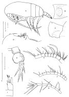 issued from : F.D. Ferrari & E.L. Markhaseva in Proc. Biol. Soc. Washington, 1996, 109 (2). [p.268, Fig.1]. Female (from E tropical Pacif. : Volcano 7):A, habitus (lateral left side) (firca cross hatched); B, genital complex (ventral); C, idem (lateral right side); D, rostrum, labrum and labium (lateral right side); E, A1 (segments 1-8); F, idem (segments 9-17); G, idem (segments 18-24); H, P5; I, caudal ramus (setae cutoff). Bars: 1 = 0.1 mm (A); 2 = 0.1 mm (D-G); 3 = 0.1 mm (I); 4 = 0.1 mm (B-C); 5 = 0.1 mm (H).
|
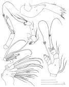 issued from : F.D. Ferrari & E.L. Markhaseva in Proc. Biol. Soc. Washington, 1996, 109 (2). [p.269, Fig.2]. Female: A, A2 (exopod); B, idem (coxa, basis and endopod); C, Md (gnathobase (anterior); D, Md (mandibular palp, anterior); E, Mx2 (inner lobes 1-4 on coxa and medial lobe on basis, posterior); F, Mx2 (inner lobe 5 on basis, without setae, and inner lobe 6 + endopod, posterior);. Bars: 1 = 0.1 mm (A); 2 = 0.1 mm (E); 3 = 0.1 mm (C, D).
|
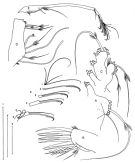 issued from : F.D. Ferrari & E.L. Markhaseva in Proc. Biol. Soc. Washington, 1996, 109 (2). [p.270, Fig.3]. Female: A, Mx1 (syncoxa with inner lobe 1 detached, posterior); B, Mx1 ( (inner lobe 1, posterior); C, Mx1 (palp, posterior); D, Mxp (syncoxa); E, Mxp (basis and endopod); F, Mxp (detached segments of endopod, b = distal tip of basis; numbers to right indicate relative appearence of ramal segments during development). Bars: 1 = 0.1 mm (F); 2 = 0.1 mm (A-C); 3 = 0.1 mm (D-E).
|
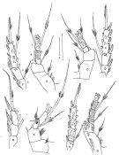 issued from : F.D. Ferrari & E.L. Markhaseva in Proc. Biol. Soc. Washington, 1996, 109 (2). [p.271, Fig.4]. Female: A, P1 (coxa, basis and endopod, anterior, arrow indicates approximate position of presumptive boundary between endopodal segments 1 and 2, between proximal two medial setae); B, P1 (exopod, anterior); C, P2 (coxa, basis and endopod, posterior); D, P2 (exopod, posterior); E, P3 (coxa, basis and endopod, posterior; F, P3 (exopod, posterior); G, P4 (coxa, basis and endopod, posterior); H, P4 (endopod, posterior) Bars: 1 = 0.1 mm (A-B); 2 = 0.1 mm (C-H). Numbers indicate relative appearence of ramal segments during development).
| | | | | Compl. Ref.: | | | Bradford-Grieve, 2004 (p.285) | | | | NZ: | 1 | | |
|
Distribution map of Parkius karenwishnerae by geographical zones
|
| | | | Loc: | | | NE Pacif. (off E Clipperton Is.) | | | | N: | 1 | | | | Lg.: | | | (440) F: 2,15-1,8; {F: 1,80-2,15} | | | | Rem.: | hyperbenthic (± 3000 m). | | | Last update : 28/01/2015 | |
|
|
 Any use of this site for a publication will be mentioned with the following reference : Any use of this site for a publication will be mentioned with the following reference :
Razouls C., Desreumaux N., Kouwenberg J. and de Bovée F., 2005-2026. - Biodiversity of Marine Planktonic Copepods (morphology, geographical distribution and biological data). Sorbonne University, CNRS. Available at http://copepodes.obs-banyuls.fr/en [Accessed June 15, 2026] © copyright 2005-2026 Sorbonne University, CNRS
|
|
|
|


;)
;)
;)
;)
