|
|
 |
|
Calanoida ( Order ) |
|
|
|
Clausocalanoidea ( Superfamily ) |
|
|
|
Phaennidae ( Family ) |
|
|
|
Xanthocalanus ( Genus ) |
|
|
| |
Xanthocalanus obtusus Farran, 1905 (F) | |
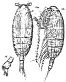
| | | | | | | Ref.: | | | Farran, 1905 (p.40, Descr.F, figs.F); Wolfenden, 1908 (p.35, Rem.); Sars, 1925 (p.131, figs.F); Rose, 1933 a (p.130, figs.F); Bradford & al., 1983 (p.71); Vyshkvartzeva, 2002 (p.94, figs.F); Markhaseva & Schnack-Schiel, 2003 (p.112, Rem.) |  Issued from : G.O. Sars in Résult. Camp. Scient. Prince Albert I, 69, pls.1-127 (1924). [Pl.XXXV, figs.11-13]. Female: 11, habitus (dorsal); 12, idem (lateral left side); 13, P5.
|
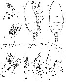
 issued from : G.P. Farran in Ann. Rep. Fish. Brch., Ireland, 1902-1903, II, App. 2, 1905. [Plate IX, Figs.10-19]. Female (from W Ireland): 10-11, habitus (lateral and dorsal, respectively); 12, A1; 13, A2; 14, Mx1; 15, Mx2; 16-19, P1 to P4.
|
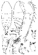
 Issued from : N.V. Vyshkvartzeva in Zoosyst. Rossica , St. Petersburg, 2002, 11. [p.95, Figs.17-26]. Female (from 45°11'N, 152°28'E): 17-18, habitus (dorsal and lateral, respectively); 19, rostrum (ventral view); 20, 5th thoracic segment with P5 and urosome (right side); 21, genital segment (ventral); 22, A1; 23, A2; 24, Md (masticatory edge of gnathobase); 25, Md palp; 26, Mxp. Nota: - Cephalosome and 1er pedigerous segment, 4th and 5th thoracic segments separated by fine sutures. - Rostrum as a short plate with 2 fine filaments. - Posterolateral corners of last thoracic segment slightly produced, broadly rounded in lateral view, triangular in dorsal view. - Urosome as long as 1/4 of prosome. - Genital segment as long as wide, laterally with a small genital swelling in distal half of segment. Transverse common genital opening (fig.21) located ventrally just before midlength of genital segment; lateral triangular skeletal plates well developed; spermatheca elongate, its distal part curved forward. - Urosomal segments 2 and 3 wider than long. - Posterior margin of Urosomal segments 1-3 striated. - Caudal rami longer than wide, with 4 long apical and short inner and outer setae. - A1 24-segmented, reaching posterior margin of urosomal segment 1. - Coxa of A2 with 1, basis and endopodal segment 1 with 2 setae; 2nd endopodal segment with 6+7 setae; endopod as long as 2/3 of exopod; exopodal segments 1-6 with 0, 1, 1, 1, 1, 0+3 setae, respectively. - Md gnathobase with 10 teeth and 1 dorsal, slender, spinulose seta. 3 ventral teeth with tall and narrow multicusped crowns; central and dorsal teeth shorter, without crowns; one tip of dorsalmost tooth remarkably elongate, as long as dorsal seta; basis with 2 strong inner setae; endopodal segment 1 without setae; 2nd endopodal segment with 8 setae; endopod as long as exopod.
|
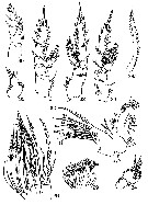
 Issued from : N.V. Vyshkvartzeva in Zoosyst. Rossica , St. Petersburg, 2002, 11. [p.96, Figs.27-35]. Female: 27, Mx1; 28, Mx2 (inner lobes 1 to 5); 28a, Mx2 claw-like seta of inner lobe 5 in another position; 29, Mx2 (sensory setae of distal endopodal complex); 30, P1; 31, P2; 32, P3; 33, P4; 34, P5 (dorsal side); 35, P5 (two distal segments, ventral side). Nota: - Mx1: inner lobe 1 (= arthrite) with 9 marginal, 4 tapering, with short spinules posterior (S11-S14), and 1 short, slender anterior setae (S6). Marginal setae longer than inner lobe 1, and their lengths remarkably increase distally; S1 and S2 tapering towards the tip, with long staff spinules; S3-S5 and S10 strongly sclerotized, with scythe-like tip, with setules along their length and sharp spinules along proximal margin in distal half. inner lobe 2 with 2 long and 1 short setae; inner lobe 3 with 2 long and 2 shorter setae; inner lobe 4 with 3 long and 2 shorter setae; endopod partly fused with basis; endopodal segment 2 fused with the 1st, each with 2 very long and 1 short setae; endopodal segment 3 with 4 setae; exopod with 10 and outer lobe 1 with 9 setae. - Mx2 compact. Inner lobes 1-3 short, subequal; inner lobe 1 with 5 setae (3 long plumose and 2 naked and shorter); inner lobe 2 with 2 long plumose and 1 shorter but more coarsely plumose setae; inner lobe 3 with 1 long plumose and 1 shorter plumose seta and short worm-like sensory seta; inner lobe 4 with 1 long plumose seta, 1 shorter and more coarsely plumose seta and 1 more strongly sclerotized long seta with long, strong, dense spines; inner lobe 5 with 1 strong claw-like seta furnished with 31 strong widely spaced spines, 1 slightly longer plumose seta and 2 worm-like sensory setae; distal endopodal complex (fig.29) indistinctly 4-segmented, with 7 brush-like and 1 worm-like sensory setae; brush-like setae slender, slightly differing in length. - P5 uniramous, 3-segmented; proximal segment slightly longer than wide, with patch of short, thickened spinules along inner margin and on outer distal margin; 2nd segment about as long as wide, globular, slightly longer than distal segment, with patches of spinules along inner margin and distal half of lateral margin and with a row of long, lancet-like spinules on posterior surface; distal segment the shortest, separated from preceding only on one side; posterior surface covered woth spinules; segment bearing 4 serrate spines (1, the longest, inner; one, outer, 293 times as long as inner, situated opposite to the inner, and 2 apical, inner apical as long as outer, outer apical slightly shorter).
| | | | | Compl. Ref.: | | | Pearson, 1906 (p.20); Sewell, 1948 (p.501); Grice & Hulsemann, 1967 (p.16); Campaner, 1978 a (p.968, Rem.); Bradford-Grieve, 2004 (p.284) ; Salah S. & al., 2012 (p.155, Tableau 1); El Arraj & al., 2017 (p.272, table 2); | | | | NZ: | 5 | | |
|
Distribution map of Xanthocalanus obtusus by geographical zones
|
| | | | | | | Loc: | | | Morocco (Cap Ghir), Madeira, Azores, off S Cape Cod, W Ireland, W Indian, NW Pacif. (off E Kuril Is.) | | | | N: | 9 | | | | Lg.: | | | (1) F: 3,3; (32) F: 2,4; (58) F: 2,4; (878) F: 3,12; {F: 2,40-3,30} | | | | Rem.: | bathypelagic (1465-1500 m), hyperbenthic.
For Vyshkvartzeva (2002, p.97) the specimen from the NW Pacific is similar in the body shape and size and in the length of spines of P5 distal segment to that described by Sars rather than to that of Farran. The armament of P1-P4 seems quite well coincide with Farran's description. P5 of the examined Pacific specimen differs slightly from that described by Farran and Sars in the presence of spinules on outer distal corner of 1st segment, more globular 2nd segment partly separated from the 3rd one, presence on posterior surface of 2nd segment of spinules much longer than the other spinules | | | Last update : 25/10/2022 | |
|
|
 Any use of this site for a publication will be mentioned with the following reference : Any use of this site for a publication will be mentioned with the following reference :
Razouls C., Desreumaux N., Kouwenberg J. and de Bovée F., 2005-2026. - Biodiversity of Marine Planktonic Copepods (morphology, geographical distribution and biological data). Sorbonne University, CNRS. Available at http://copepodes.obs-banyuls.fr/en [Accessed February 08, 2026] © copyright 2005-2026 Sorbonne University, CNRS
|
|
 |
 |


