|
|
 |
|
Calanoida ( Order ) |
|
|
|
Diaptomoidea ( Superfamily ) |
|
|
|
Pontellidae ( Family ) |
|
|
|
Labidocera ( Genus ) |
|
|
| |
Labidocera acutifrons (Dana, 1849) (F,M) | |
| | | | | | | Syn.: | Pontella acutifrons Dana,1849 (p.30, type locality: Kingsmill, W. Pacif., after Jeong & al., 2009, p.509); Brady,1883 (p.91, figs.F,M);
Pontella agilis Dana, 1849;
Pontella bairdii Lubbock, 1853 b (p.117, 159);
Pontia Edwardsii Kröyer, 1849 (in Damkaer & Damkaer, 1979, p.31);
L.abidocera agilis : Giesbrecht & Schmeil, 1898 (p.138); C.B. Wilson, 1950 (p.242, figs.F); Weikert, 1975 (p.141); Lakkis, 1984 (p.294, figs.M); Mauchline, 1998 (p.506); Zenetos & al., 2005 (p.63, Rem.: p.82, casual occurrence); Kovalev, 2006 (p.67: Lessepsian migration); Zenetos & al., 2010 (p.397);
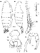
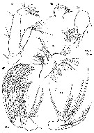
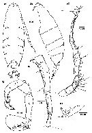
L. albatrossi C.B. Wilson, 1950 (p.243, figs.juv.F) | | | | Ref.: | | | Giesbrecht, 1892 (p.445, 458, 773, figs.F,M); Giesbrecht & Schmeil, 1898 (p.134, Rem. F,M); Wolfenden, 1911 (p.361); Früchtl, 1924 b (p.55); Sars, 1925 (p.354); Farran, 1929 (p.210, 274); Wilson, 1932 a (p.145, figs.F,M); Dakin & Colefax, 1933 (p.206); Rose, 1933 a (p.262, figs.F,M); Farran, 1936 a (p.116); Dakin & Colefax, 1940 (p.101, figs.F,M); Wilson, 1942 a (p.191, fig.F); Sewell, 1947 (p.249); Heinrich, 1960 a (p.43, fig.1); Fish, 1962 (p.18); Paiva, 1963 (p.72, figs.F); Tanaka, 1964 c (p.253, figs.F,M); Fleminger, 1965 (p.126, Rem.); Vervoort, 1965 (p.187, Rem.); Owre & Foyo, 1967 (p.97, figs.F,M); Vidal, 1968 (p.43, figs.M); Björnberg, 1972 (p.59, Rem.N, figs.); Silas & Pillai, 1973 (1976) (p.793, figs.F,M); Goswami & Goswami, 1979 a (p.259, figs. caryotypes); Greenwood, 1979 (p.97, Rem.); Björnberg & al., 1981 (p.659, figs.F,M); Sazhina, 1982 (p.1160, Rem.N, fig.); Chahsavar-Archad & Razouls, 1982 (p.32, figs.F, M); Zheng & al., 1982 (p.73, figs.F,M); Sazhina, 1985 (p.67, figs.N); Baessa de Aguiar, 1986 (1989) (p.61, figs.F,M, Rem.); Land, 1988 (p.381, fig.M); Ohtsuka & Onbé, 1991 (p.214); Chihara & Murano, 1997 (p.866, Pl.147,149: F,M); Bradford-Grieve & al., 1999 (p.885, 959, figs.F,M); Bradford-Grieve, 1999 b (p.188, figs.F,M, Rem., figs.184, 194); Conway & al., 2003 (p.126, figs.F,M, Rem.); Avancini & al., 2006 (p.105, Pl. 74, figs.F,M, Rem.);Jeong & al., 2009 (p.509, Rev.F,M, figs.F,M, distribution chart) | 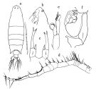 issued from : O. Tanaka in Publs Seto Mar. Biol. Lab., 1964, XII (3). [p.253, Fig.231]. Female: a, habitus (dorsal); b, forehead (left lateral side); c, P5. Nota: The head has a median crest. Male: d, right A1; e, left P5; f, right P5. Nota: The urosome segments and furca are in the proportional lengths as 19:18:22:9:6:26 = 100.
|
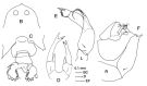 issued from : J.M. Bradford-Grieve in The Marine Fauna of New Zealand: Pelagic Calanoid Copepoda. National Institute of Water and Atmospheric Research (NIWA). NIWA Biodiversity Memoir, 111, 1999. [p.188, Fig.136]. Female: B, forehead (dorsal); C, urosome (dorsal); D, P5. Male: E, left P5; R, right P5.
|
 issued from : E.G. Silas & P.P. Pillai in J. mar. biol. Ass. India, 1973 (1976), 15 (2). [p.794, Fig.8]. Female (from Andaman Islands): a, urosome (dorsal); b, rostrum (anteror view); c, P5. Male: d, right A1 (geniculate and distal part); e, P5. Scale as in Calanopia minor.
|
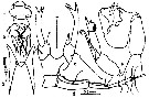
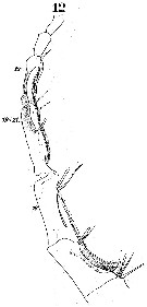
 Issued from : W. Giesbrecht in Systematik und Faunistik der Pelagischen Copepoden des Golfes von Neapel und der angrenzenden Meeres-Abschnitte. Fauna Flora Golf. Neapel, 1892. Atlas von 54 Tafeln. [Taf.41, Fig.3]. Female: 3, habitus (dorsal);
|
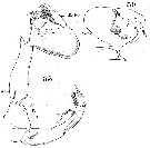
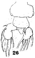 Issued from : W. Giesbrecht in Systematik und Faunistik der Pelagischen Copepoden des Golfes von Neapel und der angrenzenden Meeres-Abschnitte. Fauna Flora Golf. Neapel, 1892. Atlas von 54 Tafeln. [Taf.41, Fig.26]. Female: 26, urosome (dorsal).
|
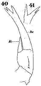 Issued from : W. Giesbrecht in Systematik und Faunistik der Pelagischen Copepoden des Golfes von Neapel und der angrenzenden Meeres-Abschnitte. Fauna Flora Golf. Neapel, 1892. Atlas von 54 Tafeln. [Taf.23, Figs.40, 41]. Female: 40, P5 (posterior view); 41, P5 (exopod, distal part, another view).
|
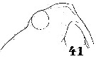 Issued from : W. Giesbrecht in Systematik und Faunistik der Pelagischen Copepoden des Golfes von Neapel und der angrenzenden Meeres-Abschnitte. Fauna Flora Golf. Neapel, 1892. Atlas von 54 Tafeln. [Taf.41, Fig.41]. Male: 41, forehead (lateral).
|
 Issued from : W. Giesbrecht in Systematik und Faunistik der Pelagischen Copepoden des Golfes von Neapel und der angrenzenden Meeres-Abschnitte. Fauna Flora Golf. Neapel, 1892. Atlas von 54 Tafeln. [Taf.23, Fig.2] Male: 2, left A1 (anterior view, ventral surface, setae missing).
|
 Issued from : W. Giesbrecht in Systematik und Faunistik der Pelagischen Copepoden des Golfes von Neapel und der angrenzenden Meeres-Abschnitte. Fauna Flora Golf. Neapel, 1892. Atlas von 54 Tafeln. [Taf.23, Fig.12]. Male: right A1 (distal part, , anterior view, ventral surface).
|
 Issued from : W. Giesbrecht in Systematik und Faunistik der Pelagischen Copepoden des Golfes von Neapel und der angrenzenden Meeres-Abschnitte. Fauna Flora Golf. Neapel, 1892. Atlas von 54 Tafeln. [Taf.23, Figs.30, 33]. Male: 30, right P5 (distal part, anterior surface); 33, P5 (postrior view).
|
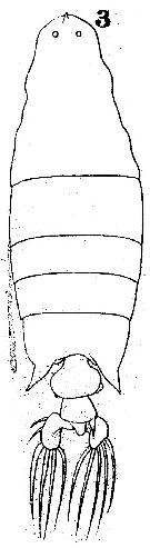
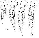
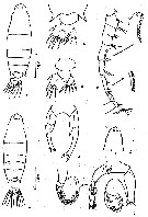 issued from : Z. Zheng, S. Li, S.J. Li & B. Chen in Marine planktonic copepods in Chinese waters. Shanghai Sc. Techn. Press, 1982 [p.74, Fig.43]. Female: a, habitus (dorsal); b, urosome (dorsal); c, idem (another specimen); d, P5. Male: e, habitus (dorsal); f, forehead (another specimen); g, A1; h, P5. Scale bars in mm.
|
 Issued from : H.G. Jeong, H.-L. Suh, S.B. Jeong, Y.H. Yoon & H.Y. Soh in Zoological Studies, 48 (4). [p.509, Fig.1]. Female (from 34°26'N, 127°34'E): A-B, habitus (dorsal and lateral, respectively); ; C, A1: D, urosome (ventral); E, P5. Nota: A1 symmetrical, 23-segmented; posterior margin of 2nd-12th segments fringed with fine hairs; ancestral segments II_IV and XXVII-XXVIII completely fused while VII-IX incompletely fused. Anal somite covered to 2nd urosomite, anal operculum extend posteriad and slightly to the right. Caudal rami strongly asymmetrical, left ramus larger than right, left ramus bearing 7 setae: 2 inner setae (I and II), 3 terminal setae (III-V) proximally thickened, 2 outer setae (VI and VII); right ramus with 6 setae except for dorsal seta (VII).
|
 Issued from : H.G. Jeong, H.-L. Suh, S.B. Jeong, Y.H. Yoon & H.Y. Soh in Zoological Studies, 48 (4). [p.510, Fig.2]. Female: A, A2; B, Md (cutting edge of mandibular gnathobase); C, Md (mandibular palp); D, Mx1; E, Mx2; F, Mxp. Nota: Md with large coxal gnathobase bearing 5 teeth along its distomedial margin and dorsal seta, 3rd and 4th dorsalmost teeth bicuspidate; patch of dagger-like spinules ornamented on base of 3rd-5th dorsalmost teeth; basis of mandibular palp bearing 4 elongated setae; endopod 2-segmented (setal formula: 4, 8); exopod 5-segmented (setal formula: 1, 1, 1, 1, 2). Mx1 praecoxal arthrite with 16 setae; coxa with 3 setae on endite and 9 setae on epipodite; basis with 4 and 3 setae on proximal and distal endite, respectively, with 1 seta on exite; 1st and 2nd endopod segments, each with 2 setae, incorporated into basis, distal endopod segment with 5 apical setae; exopod 1-segmented, with 10 setae and 1 setule distally.
|
 Issued from : H.G. Jeong, H.-L. Suh, S.B. Jeong, Y.H. Yoon & H.Y. Soh in Zoological Studies, 48 (4). [p.511, Fig.3]. Female: A, right P1; B, left P2; C, left P3; D, left P4.
|
 Issued from : H.G. Jeong, H.-L. Suh, S.B. Jeong, Y.H. Yoon & H.Y. Soh in Zoological Studies, 48 (4). [p.513]. Female: Armature formula of swimming legs P1 to P4. Spines in Roman numerals; setae in Arabic numerals.
|
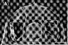 Issued from : H.G. Jeong, H.-L. Suh, S.B. Jeong, Y.H. Yoon & H.Y. Soh in Zoological Studies, 48 (4). [p.519, Fig.9, A]. Female: Scanning electron micrograph of rostrum (ventral view). Scale bar = 100 µm.
|
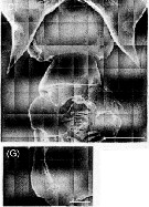 Issued from : H.G. Jeong, H.-L. Suh, S.B. Jeong, Y.H. Yoon & H.Y. Soh in Zoological Studies, 48 (4). [p.519, Fig.9, D-G]. Female: Scanning electon micrographs of genital area. The arrow indicates the genital operculum. Scale bars: 200 mm (D); 50 µm (G).
|
 Issued from : H.G. Jeong, H.-L. Suh, S.B. Jeong, Y.H. Yoon & H.Y. Soh in Zoological Studies, 48 (4). [p.512, Fig.4]. Male: A-B, habitus (dorsal and lateral, respectively); C, right A1; D, ancestral segments XIX-XXIV in right A1; E, P5; F, 2nd and 3rd segments of right P5.
|
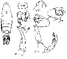 Issued from : J.M. Bradford-Grieve, E.L. Markhaseva, C.E.F. Rocha & B. Abiahy in South Atlantic Zooplankton, edit. D. Boltovskoy. 1999, Vol. 2, Copepoda; [p.1068, Fig. 7.382]. Ce = cephalon; Ur = urosome; l = left leg; r = right leg,. B = basis. Labidocera acutifrons. Female characters (from key, p.959): Posterior prosome corners divergent; urosomal segment 2 with right side, posteriorly directed, posterodorsal spine which covers some of right caudal ramus; anterior cephalosome with crest, visible in dorsal view as an anterior spine; cephalon without lateral hooks. Male characters (p.959): left P5 exopodal segment 1 more than twice as long as exopodal segment2-3; right exopodal segment1-2 with very short 'thumb' and broad rounded projection. Left P5 basis with one of distal corners produced into large process which extends at least beyond distal border of exopodal segment 1. Left P5 basis with the process extending beyond distal border of whole exopod and decorated with spinules.
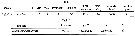
| | | | | Compl. Ref.: | | | Oliveira, 1945 (p.191); Sewell, 1948 (p.322, 451, 512); C.B. Wilson, 1950 (p.242); Yamazi, 1958 (p.151, Rem.); Heinrich, 1960 a (p.43: fig.1); Fagetti, 1962 (p.34); Cervigon, 1962 (p.181, tables: abundance distribution); Ganapati & Shanthakumari, 1962 (p.9, 15); Grice & Hart, 1962 (p.287, table 3); Sherman, 1963 (p.216: fig.2, 3, p.221: fig.10, p. 222: fig.12, p. 224: fig.14); Ahlstrom & Thrailkill, 1963 (p.57, Table 5, abundance); Gaudy, 1963 (p.29, Rem.); Björnberg, 1963 (p.60, Rem.); Heinrich, 1964 (p.86: fig.4, p.91: fig.7); De Decker & Mombeck, 1964 (p.12); Mazza, 1966 (p.72); Mullin, 1966 (p.546, Table I, III, diet); Fleminger, 1967 a (tabl.1); Sherman & Schaner, 1968 (p.583, fig.1); Omori, 1969 (p.5, Table 1); Heinrich, 1969 (p.1457, fig.4); Morris, 1970 (p.2301); Itoh, 1970 (tab.1); Gueredrat, 1970 (p.293, durnal migration); Salah, 1971 (p.319); Morris R.J., 1971 (p.275, Table 1, 2, lipids composition); Bainbridge, 1972 (p.61, Appendix Table I: vertical distribution vs day/night); Zagalsky & Herring, 1972 (p.397, carotenoprotein); Björnberg, 1973 (p.352, 387); Heinrich, 1974 (p.43, fig.1); Weikert, 1975 (p.141, chart); Raymont & al., 1975 (p.261, amino-acids); Carter, 1977 (1978) (p.36); Stephen & Iyer, 1979 (p.228, tab.1, 3, 4, figs.3, 4); Turner & al., 1979 (p.289, 293, Tab.3, Rem.); Dessier, 1979 (p.207); Turner & Collard, 1980 (p.527, Tab.1); Svetlichnyi, 1980 (p.28, Table 1, passive submersion); Vives, 1982 (p.295); Kovalev & Shmeleva, 1982 (p.85); Dessier, 1983 (p.89, Tableau 1, Rem., %); Guangshan & Honglin, 1984 (p.118, tab.); De Decker, 1984 (p.317, 352: chart); Brenning, 1985 a (p.28, Table 2); Brinton & al., 1986 (p.228, fig.11: spatial distribution, Table 1); Madhupratap & Haridas, 1986 (p.105, tab.1); Diouf & Diallo, 1987 (p.260); Brenning, 1987 (p.31, T-S diagram, Rem.); Dessier, 1988 (tabl.1); Heinrich, 1988 (p.89, fig.1); Land, 1988 (p.381, eye movements); Hernandez-Trujillo, 1989 (tab.1); 1989 a (tab.1); Cervantes-Duarte & Hernandez-Trujillo, 1989 (tab.3); Heinrich, 1990 (p.19); Suarez & al., 1990 (tab.2); Suarez & Gasca, 1991 (tab.2); Baessa De Aguiar, 1991 (1993) (p.107); Suarez, 1992 (App.1); Séguin & al., 1993 (p.23); Hernandez-Trujillo, 1994 (tab.1); Shih & Young, 1995 (p.71); Suarez-Morales & Gasca, 1997 (p.1525); Alvarez-Cadena & al., 1998 (tab.4); Mauchline, 1998 (tab.8, 30, 64); Suarez-Morales, 1998 (p.345, Table 1); Suarez-Morales & Gasca, 1998 a (p.110); Hernandez-Trujillo, 1999 (p.284, tab.1); Lavaniegos & Gonzalez-Navarro, 1999 (p.239, Appx.1); Neumann-Leitao & al., 1999 (p.153, tab.2); Razouls & al., 2000 (p.343, tab. 5, Appendix); Alvarez-Silva & Gomez-Aguirre, 2000 (p.163: tab.2); Lopez-Salgado & al., 2000 (tab.1); Hidalgo & Escribano, 2001 (p.159, tab.2); Lopez-Ibarra & Palomares-Garcia, 2006 (p.63, Tabl. 1, seasonal abundance vs El-Niño); Lavaniegos & Jiménez-Pérez, 2006 (p.146, tab.2, 3, Rem.); Neumann-Leitao & al., 2008 (p.799: Tab.II, fig.6); Muelbert & al., 2008 (p.1662, Table 1); Ayon & al., 2008 (p.238, Table 4: Peruvian samples); Hernandez-Trujillo & al., 2010 (p.913, Table 2); Schnack-Schiel & al., 2010 (p.2064, Table 2: E Atlantic subtropical/tropical); Medellin-Mora & Navas S., 2010 (p.265, Tab. 2); Teuber & al., 2013 (p.1, Table 2, 3, fig.5, abundance vs oxygen minimum zone, respiration rates, enzyme activity); in CalCOFI regional list (MDO, Nov. 2013; M. Ohman, comm. pers.); Lidvanov & al., 2013 (p.290, Table 2, % composition); Beltran Castro, 2014 (p.36, Table 1, 2: molecular CO1); Bonecker & a., 2014 (p.445, Table II: frequency, horizontal & vertical distributions); Dias & al., 2015 (p.483, Table 2, abundance, biomass, production); Hernandez-Trujillo & Esqueda Escarcega, 2016 (p.1, egg production); Marques-Rojas & Zoppi de Roa, 2017 (p.495, Table 1); Palomares-Garcia & al., 2018 (p.178, Table 1: occurrence); Belmonte, 2018 (p.273, Table I: Italian zones) | | | | NZ: | 20 | | |
|
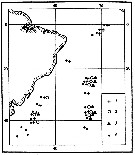
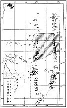
Distribution map of Labidocera acutifrons by geographical zones
|
| | | | | | | | | | | | | | |  Issued from : A.K. Heinrich inTrudy Inst. Okeanol., 1974, 98 [p.44, Fig.1]. Issued from : A.K. Heinrich inTrudy Inst. Okeanol., 1974, 98 [p.44, Fig.1].
Distibution of three species of Pontellidae from SW Atlantic (Cruise of the R/V 'Akademik Kurchatov', 1971 and 1972).
Points = stations; cross = Labidocera acutifrons ; circle= Pontellopsis regalis ; triangle= Pontellopsis villosa . |
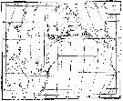
 Issued from : A.K. Heinrich in Trudy Inst. Okeanol., 1960, 41. [p.43, Fig.1]. Issued from : A.K. Heinrich in Trudy Inst. Okeanol., 1960, 41. [p.43, Fig.1].
Distribution in the equatorial and tropical West Pacific of seven species of Pontellidae (Cruise of the R/V 'Vityaz', November 1957 to December 1958.
1: Stations; 2: Pontellopsis villosa; 3: Pontella fera; 4: Labidocera acutifrons; 5: Pontella whiteleggi; 6: Pontella tenuiremis; 7: Pontella chierchiae; 8: Labidocera acuta. |
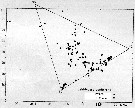
 issued from : A.K. Heinrich in Zool. Zh., 1969, 48 (10) [p.1459, Fig.4]. issued from : A.K. Heinrich in Zool. Zh., 1969, 48 (10) [p.1459, Fig.4].
Distribution of Labidocera acutifrons in the Pacific Ocean.
1: occurrence of L. acutifrons; 2: stations. |
 issued from : U. Brenning in Wiss. Z. Wilhelm-Pieck-Univ. Rostock - 36. Jahrgang 1987. Mat.-nat. wiss. Reihe, 2. [p.31, Fig.4]. issued from : U. Brenning in Wiss. Z. Wilhelm-Pieck-Univ. Rostock - 36. Jahrgang 1987. Mat.-nat. wiss. Reihe, 2. [p.31, Fig.4].
T-S Diagram for Labidocera acutifrons from 8° S - 26° N; 16°- 20° W.
SO: Southern Surface Water (S °/oo: 34,50; T°C: 29,0); ND: Northern Water of the Surface Layer (S °/oo: 37,5; T°C: 21,0); SD: Southern Deep Water of the surface layer (S °/oo: 35,33; T°C: 13,4). See commentary in Temora stylifera and Brenning (1985 a, p.6). |
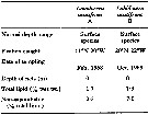 Issued from : R. J. Morris in Comp. Biochem. Physiol., 1971, 40B. [p.277, Table 1]. Issued from : R. J. Morris in Comp. Biochem. Physiol., 1971, 40B. [p.277, Table 1].
Total lipid and saponification for Labidocera acutifrons from Northeastern Atlantic : off Guinea Bissau (A) and off north Cape Verde Is. (B). |
 Issued from : S. Hernandez-Trujillo & G.Ma. Esqueda Escarcega in CICIMAR Oceanides, 2016, 31 (1). [p.3, Table 1 & 2]. Issued from : S. Hernandez-Trujillo & G.Ma. Esqueda Escarcega in CICIMAR Oceanides, 2016, 31 (1). [p.3, Table 1 & 2].
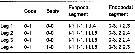
Table 1: Egg production rate (TPH: eggs female-1 day -1 mean during Marca Roja-VII cruise in Abril 2015. n = number of experiments with 5 replicates. Superficial water temperature, salinity and chlorophyll a at 10 m depth. Min: minimum number of eggs; Max: number maximum of eggs.
Table 2: Mean length of prosome (mm), quantity of carbone (µgC), K: enviromental and physiological factor according to Froese (2006), Rennie & Verdon (2008), Blackwell & al. (2000), Trudel & al;, (2005), Hernandez-Trujillo & al. (2008, 2013). |
 Issued from : S. Hernandez-Trujillo & G.Ma. Esqueda Escarcega in CICIMAR Oceanides, 2016, 31 (1). [p.4, Table 3]. Issued from : S. Hernandez-Trujillo & G.Ma. Esqueda Escarcega in CICIMAR Oceanides, 2016, 31 (1). [p.4, Table 3].
Egg production rate (TPH: eggs female-1 day -1). TSM: temperature (°C) of egg incubation. After Ekau & al. (2008). |
| | | | Loc: | | | Antarct. (Indian: continent), sub-Antarct. (SE Pacif.), South Africa (E & W), Namibia, Angola, off S St. Helena Is., Congo, off S Ascension Is., G. of Guinea, Casamance, Dakar, Cape Verde Is., off Morocco-Mauritania, off Canary Is., Azores, Uruguay (continental shelf), Brazil (N & S, off Rio de Janeiro, Campos Basin, off Natal), Venezuela, Bahia de Mochima, Cariaco Basin, Yucatan, Barbada Is., Caribbean Colombia, Caribbean Sea, Yucatan, G. of Mexico, Florida, Cape Hatteras, Woods Hole, Georges Bank, Medit. (Tyrrhenian Sea, Adriatic Sea, Lebanon Basin, Alexandria), Red Sea, Arabian (coast), off SE La Réunion Is., S Indian Ocean, Natal, Madagascar (Nosy Bé), India (off Cochin, Lawson's Bay), Indonesia-Malaysia, Philippines, China Seas (East China Sea, South China Sea), Viet-Nam (Cauda Bay), Japan, Tanabe Bay, off E Mariana Is. Pacif. (W equatorial), Australia (Great Barrier, Moreton Bay, Cape York), New Caledonia, off W Kermadec Is., Pacif. (N central subtropical), off California, W Baja California, Bahia Magdalena, G. of California, Bahia de La Paz, Guerrero coast (W Mexico), Central America, Hawaii, Galapagos, off Peru, Chile, N Chile (Mejillones Peninsula) | | | | N: | 145 | | | | Lg.: | | | (16) F: 4,25-3,41; M: 4,04-3,3; (34) F: 3,55-3,36; M: 3,48-3,3; (45) F: 3,85-3,5; M: 4-3,75; (46) F: 3,85-3,7; M: 3,9-3,8; (73) F: 4,26-3,9; M: 4,16-3,69; (104) F: 3,7; M: 3,8; (120) F: 3,75; M: 3,38; (135) ?F: 4,2; (137) F: 3,15; (187) M: 3,6; (237) F: 4,1; M: 4,0; (256) F: 3,8-3,2; M: 3,28; (458) F: 3,81-3,75; (785) F: 3,97-3,79; M: 4,06-3,68; (909) F: 3,4; (991) F: 3,2-4,15; M: 3,28-4,08; (1023) F: 3,25-3,72; M: 3,4-3,61; (1026) F: 3,49-3,67; M: 3,45-3,53; (1110) F: 4,1-4,7; M: 3,88-4,56; {F: 3,15-4,70; M: 3,28-4,56}
Prosome length: (1254) F: 1.93 ±0.11. | | | | Rem.: | epipelagic-?.
Over Georges Bank with an incursion of the Gulf Stream (Sherman & Schaner, 1968).
For Zagalsky & Herring (1972, p.397) this species is a blue pigmented member of the eastern North Atlantic neuston and is occasionaly encountered in dense swarms. Collected at 10°58'N, 20°00'W). The blue pigment is contained in both the carapace chitin and the underlying hypodermis and is believed to provide the animals with protective coloration.
. The presence and distribution of this species in the Pacific Ocean, where it can be used as an indicator of certain bodies of water, is discussed by Heinrich (1960) and Sherman (1963). The occurrence near the antarctic continent is astonishing by the fact of the thermical preferences in the genus.
For Itoh (1970 a, fig.2, from co-ordonates) the Itoh's index value of the mandibular gnathobase = 945.
For Prusova & Al-Yamani (2014, p.1163) this species belongs ''darwinii'' species group. The species shows resemblance to L. kuwaitiana.
Diagnosis for L. acutifrons in the 'darwinii' species group after Prusova & Al-Yamani (2014, p.1165) : Females:
- 1: Urosme of 4 somites.
- 2: Genital somite asymmetrical, caudal rami approximately equal length.
- 3: Caudal rami asymmetrical, partly fused to anal somite.
- 4: Genital somite with small lateral lump on right side, genital pore lateral; both P5 exopod with 3 terminal points, outermost the largest.
Males:
- 1: Urosome of 5 somites.
- 2: Prosome posterior corners symmetrical.
- 3: Left P5 endopod, if fully extended, extends beyond distal border of exopodal segment 2, and terminates in thin elongate appendix laciniate at its mid-length; right A1 segment's XXI-XXIII toothed process extends to distal 1/3 of segment XXIV.
See in DVP Conway & al., 2003 (version 1) | | | Last update : 13/11/2020 | |
|
|
 Any use of this site for a publication will be mentioned with the following reference : Any use of this site for a publication will be mentioned with the following reference :
Razouls C., Desreumaux N., Kouwenberg J. and de Bovée F., 2005-2026. - Biodiversity of Marine Planktonic Copepods (morphology, geographical distribution and biological data). Sorbonne University, CNRS. Available at http://copepodes.obs-banyuls.fr/en [Accessed June 15, 2026] © copyright 2005-2026 Sorbonne University, CNRS
|
|
|
|


;)
;)
;)
;)
;)
;)
;)
;)
;)
;)
;)
;)
;)
;)
;)
;)
;)

;)
;)
;)
;)
;)
