|
|
 |
|
Calanoida ( Order ) |
|
|
|
Diaptomoidea ( Superfamily ) |
|
|
|
Pontellidae ( Family ) |
|
|
|
Labidocera ( Genus ) |
|
|
| |
Labidocera minuta Giesbrecht, 1889 (F,M) | |
| | | | | | | Syn.: | Labidocera minutum Giesbrecht, 1889; 1892 (p.446, 459, 773, figs.F,M); Dakin & Colefax, 1940 (p.101, figs.F,M); De Decker, 1964 (p.15, 22, 28); De Decker & Mombeck, 1964 (p.13); Carter, 1977 (1978) (p.36); Maiphae & Sa-ardrit, 2011 (p.641, Table 2, 3, Rem.);
? Labidocera chubbi (M) Brady, 1915;
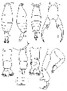
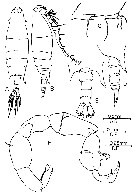
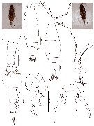
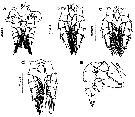
Non Labidocera minuta : Greenwood, 1979 (p.101, figs.F,M, Rem.); | | | | Ref.: | | | Giesbrecht & Schmeil, 1898 (p.137, Rem. F,M); Thompson & Scott, 1903 (p.235, 251); Wolfenden, 1905 (1906) (p.1018, figs.); A. Scott, 1909 (p.234); Pesta, 1912 a (p.53, fig.F); Sewell, 1912 (p.354, 370); 1914 a (p.234); Früchtl, 1924 b (p.56, fig.M); Sewell, 1932 (p.364); Farran, 1936 a (p.116); Sewell,1947 (p.249); C.B. Wilson, 1950 (part., p.247, figs.F,M); Voronina, 1962 a (p.68); Kasturirangan, 1963 (p.50, 51, figs.F,M); Tanaka, 1964 c (p.257, figs.F,M); Chen & Zhang, 1965 (p.99, figs.F,M); Fleminger, 1965 (p.125, Rem.); Saraswathy, 1966 (1967) (p.82); Kos, 1972 (Vol. I, figs.F,M, Rem.); Silas & Pillai, 1973 (1976) (p.800, Rem., figs.F,M); Goswami & Goswami, 1979 a (p.259, figs. caryotypes); Kimmerer & al., 1985 (p.428); Ohtsuka & Onbé, 1991 (p.214); Chihara & Murano, 1997 (p.868, Pl.149,151: F,M); Bradford-Grieve & al., 1999 (p.885, 959, figs.F,M); Bradford-Grieve, 1999 b (p.195, figs.F,M, Rem., figs.185, 194); Mulyadi, 2002 (p.71, figs.F,M, Rem.); Al-Yamani & Prusova, 2003 (p.81, figs.F,M); Conway & al., 2003 (p.132, figs.F,M, Rem.); Othman & Toda, 2006 (p.310, F,M); Phukham, 2008 (p.81, figs.F,M); Jeong & al., 2009 (p.517, Rev.F,M, figs.F,M, distribution chart); Al-Yamani & al., 2011 (p.70, figs.F,M); Lacuna & al., 2013 (p.61, figs.F,M, Rem.); Abo-Taleb, 2019 (p.367, 369: Key F, M, figs.F, M). | 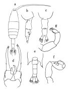 issued from : O. Tanaka in Publs Seto Mar. Biol. Lab., 1964, XII (3). [p.257, Fig.233]. Female: a, habitus (dorsal); b, last thoracic segment and urosome (right lateral side); c, idem (ventral); d, P5. Nota: The head has small side hooks. Male: e, last thoracic segment and urosome (dorsal); f, right P5; g, left P5.
|
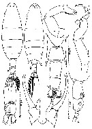
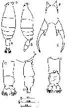
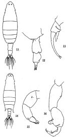 issued from: Q.-c Chen & S.-z. Zhang in Studia Marina Sinica, 1965, 7. [Pl.41, 11-16]. Female (from E China Sea): 11, habitus (dorsal); 12, urosome (lateral right side); 13, left P5 (posterior). Male: 14, habitus (dorsal); 15, left P5 (posterior); 16, right P5 (anterior).
|
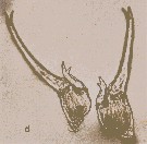
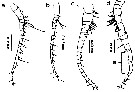
 issued from : E.G. Silas & P.P. Pillai in J. mar. biol. Ass. India, 1973 (1976), 15 (2). [p.801, Fig.12]. Female (from Indian Ocean): a, urosome (lateral right side); b, rostrum (anterior view); c, P5. Nota: Urosome 3-segmented. Urosomal segment 2 as broad as long, ventrally with chitenous tubercles which are spread laterally along iths right margin. Caudal rami longer than broad, asymmetrical, right ramus larger. A1 23-segmented. P5 symmetrical. Male: d, 5th metasomal somite (lateral right side); e, A1 (geniculate part); f, P5. Nota: Dorsal eye lenses conspicuously large and placed close together. 5th metasimal segment asymmetrical.
Scale as in Calanopia minor.
|
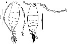
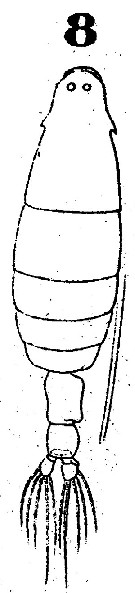 Issued from : W. Giesbrecht in Systematik und Faunistik der Pelagischen Copepoden des Golfes von Neapel und der angrenzenden Meeres-Abschnitte. Fauna Flora Golf. Neapel, 1892. Atlas von 54 Tafeln. [Taf.41, Fig.8]. As Labidocera minutum. Female: 8, habitus (dorsal).
|
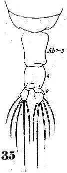 Issued from : W. Giesbrecht in Systematik und Faunistik der Pelagischen Copepoden des Golfes von Neapel und der angrenzenden Meeres-Abschnitte. Fauna Flora Golf. Neapel, 1892. Atlas von 54 Tafeln. [Taf.41, Fig.35]. As Labidocera minutum. Female: Th5 and urosome (dorsal).
|
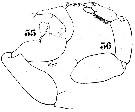
 Issued from : W. Giesbrecht in Systematik und Faunistik der Pelagischen Copepoden des Golfes von Neapel und der angrenzenden Meeres-Abschnitte. Fauna Flora Golf. Neapel, 1892. Atlas von 54 Tafeln. [Taf.25, Fig.32]. As Labidocera minutum. Female: 32, P5.
|
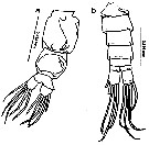
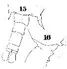 Issued from : W. Giesbrecht in Systematik und Faunistik der Pelagischen Copepoden des Golfes von Neapel und der angrenzenden Meeres-Abschnitte. Fauna Flora Golf. Neapel, 1892. Atlas von 54 Tafeln. [Taf.41, Figs.15, 16]. As Labidocera minutum. Male: 15, Th5 and urosome (dorsal); 16, idem (lateral right side).
|
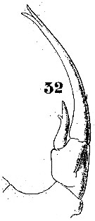
 Issued from : W. Giesbrecht in Systematik und Faunistik der Pelagischen Copepoden des Golfes von Neapel und der angrenzenden Meeres-Abschnitte. Fauna Flora Golf. Neapel, 1892. Atlas von 54 Tafeln. [Taf.23, Figs.35, 36]. As Labidocera minutum. Male: right P5 (posterior view); 36, left P5 (posterior view).
|
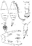
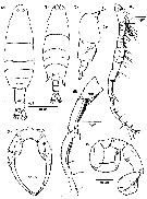
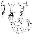
 issued from : F.Y. Al-Yamani & I. Prusova in Common Copepods Northwestern Arabian Gulf : Identification Guide. Kuwait Institute for Scientific Research, 2003. [p.80, Fig.28]. Female: A, habitus (dorsal); B, idem (lateral right side); C, urosome (dorsal); D, idem (lateral right side); E, idem (ventral); F, rostrum; G, P5. Male: H, habitus (dorsal); I, P5. Nota: Right leg of P5 3-segmented, with chela; left leg terminal segment with 2 pairs of subequal stout processes and with inner marginal hairs.
|
 issued from : Mulyadi in Treubia, 2002, 32; [p.72, Fig.22]. Female (from Indonesian waters): a, habitus (dorsal); b-c, urosome (dorsal and ventral, respectively); d, metasomal somite 5 and urosome (ight lateral); e, rostrum (anterior view); f, P5. Male: g, habitus (dorsal); h, right P5; i, left P5.
|
 issued from : Mulyadi in Treubia, 2002, 32; [p.59, Table 2]. Characteristics features of L. minuta in Indo-West Pacific. Comparison with L. detruncata, L. kroyeri and L. pectinata (see at these species).
|
 issued from : B.H.R. Othman & T. Toda in Coastal Mar. Sc., 2006, 30 (1). [p.311, Fig.10]. Female (from Sisters Island, Singapore): A-B, habitus (dorsal and lateral, respectively); C-D, posterior part of prosome and urosome (dorsal and lateral, respectively); E, P5. Nota: Prosome to urosome length ratio 2.78 : 1. - Body similar in shape as L. bengalensis. - Cephalon with lateral hooks. - Dorsal eye lenses small. - Thoracic segment rounded posteriorly; posterolateral ends produced into short spine, right side directed ventrally. - Urosome 3-segmented. - Genital segment elongated, as long as urosomitr 2 and urosomite 3 combined, right posterior margin modified into a short lobular projection, ventrally with chitinous tubercles which are spread laterally along its right margin. - P5 symmetrical and inramous; exopod longer than endopod, all ending bifurcated at apex.
|
 issued from : B.H.R. Othman & T. Toda in Coastal Mar. Sc., 2006, 30 (1). [p.311, Fig.11]. Male (from Sister's Island, Singapore): A-B, habitus (dorsal and lateral, respectively); C, A1 (segments 21-24); D-E, posterior part of prosome and urosome (lateral and dorsal, respectively); F, P5. Nota: Prosome to urosome length ratio 3.33 : 1. - Dorsal eye lenses prominent and configuous. - Posterolateral end of prosome produced into asymmetrical pointed processes, right process slightly longer, brad-like extending to distal end of urosmite 2. - Urosome 5-segmented, naked. - Right A1 geniculate with 1 conspicuous spine on segment 17; anterior margin of segment 18 with villiform denticulate ridge; fused segments 19-21 with blunt denticulate plate, segment 22 with spur-like process distally. - P5: right leg, thumb of chela short, broader toward tip with 1 process and 2 setae, exopodal segment 2 bent inwards at distal half, inner margin with 1 transparent flap, 3 setae along its inner margin and 2 setae at apex; left leg 3-segmented, exopodal segment 1 with 1 distolateral rudimentary spine, exopodal segment 2 with 2 pairs of unequal stout processes, outer one of longer pair pointed, inner margin hirsute.
|
 issued from : Y. Al-Yamani, V. Skryabin, A. Gubanova, S. khvorov & I. Prusova in Marine Zooplankton Practical Guide for the Northwestern Arabian Gulf, 2, 2011. [p.70, Foig.70]. Female (from Kuwait): A-B, habitus (dorsal and lateral, respectively); C-E, urosome (dorsal, lateral and ventral, respectively); F, rostrum; G, P5.
|
 issued from : Y. Al-Yamani, V. Skryabin, A. Gubanova, S. khvorov & I. Prusova in Marine Zooplankton Practical Guide for the Northwestern Arabian Gulf, 2, 2011. [p.71, Fig.200, d]. Female: d, P5.
|
 issued from : Y. Al-Yamani, V. Skryabin, A. Gubanova, S. khvorov & I. Prusova in Marine Zooplankton Practical Guide for the Northwestern Arabian Gulf, 2, 2011. [p.71,Fig.201, c]. Male: c, left P5.
|
 issued from : N. Phukham in Species diversity of calanoid copepods in Thai waters, Andaman Sea (Master of Science, Univ. Bangkok). 2008. [p.162, Fig.36]. Female (from W Malay Peninsula): a, habitus (dorsal); b-c, urosome (dorsal and lateral, respectively); d, P5. Male: e, habitus (dorsal); f, distal part of A1; g, P5. Body length after the drawings: F = 1.800 mm; M = 1.436 mm.
|
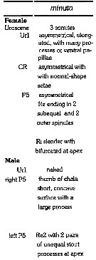
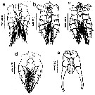
 Issued from : H.G. Jeong, H.-L. Suh, S.B. Jeong, Y.H. Yoon & H.Y. Soh in Zoological Studies, 48 (4). [p.518, Fig.8]. Female (from 34°30'N, 128°E): A, habitus (dorsal); C, urosome (right lateral view); F, P5 (L = left leg; R = right leg). Nota: Cephalosome and 1st pedigerous somite completely separated, 4th and 5th incompletely fused. Posterior corners of prosome rounded in dorsal view, but with short ventrally directed process visible in lateral view. Cephalosome with lateral hooks and small pair of dorsal lenses. Urosome 3-segmented: genital double-somite asymmetrical with small processes (1 anteroventral and 1 posteroventral); anterior and posterior parts of right side swollen; genital operculum located ventroposteriorly of midline; right ventral surface of 2nd urosomite with chitinous tubercles . Caudal rami asymmetrical, right ramus slightly wider than left one. P5 slightly asymmetrical, left leg longer than right: exopod with 2 outer spinules and 2 apical processes; endopod bifurcate, inner process shorter. Male: B, habitus (dorsal); D, right A1; E, ancestral segments XIX-XXIV of right A1; G, P5. Nota: Cephalosome with a pair of large contiguous dorsal lenses.
Cephalosome and 1st pedigeous somite completely separated, 4th and 5th completely fully fused.
Posterior corners of prosome asymmetrical, acutely pointed an left and blade-like on right and longer than left.
Urosome 5-segmented: genital segment wider than long.
Caudal rami symmetrical.
P5 uniramous, asymmetrical; right basis with outer seta and patch of inner spinules; right exopod 2 segmented, 1st segment comprising palm with bilobed process and 2 setae, 2nd segment elongate, finger-like, with 1 transparent flap, 3 setae along inner margin, and 2 apical setae; left exopod segment 2-segmented, 1st segment with small distolateral spine, 2nd segment with 2 stout processes, 2 triangular processes, and hirsute inner margin.
|
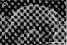 Issued from : H.G. Jeong, H.-L. Suh, S.B. Jeong, Y.H. Yoon & H.Y. Soh in Zoological Studies, 48 (4). [p.519, Fig.9, C]. Female: Scanning electon micrograph of rostrum (ventral view). Scale bar = 50 µm. Rostrum bifid, widely spaced and distant from ventral ocellus.
|
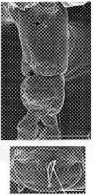 Issued from : H.G. Jeong, H.-L. Suh, S.B. Jeong, Y.H. Yoon & H.Y. Soh in Zoological Studies, 48 (4). [p.519, Fig.9, F, I]. Female: Scanning electon micrograph of the genital area. The arrow indicates the genital operculum Sale bars: 200 µm (F); 20 µm (I).
|
 ssued from : M.L.D.G. Lacuna, D.C. Sagrado, R.O. Mejorada, D.D. Simyunn & M.J.J. Pueblos in ABAH Bioflux, 2013, 5 (1). [p.61, Fig.8]. Female (from Gingoog, Mindanao): a, habitus (dorsal). Male: b, habitus (dorsal). Nota: The dorsal eye lenses in males are large and in contact with each other, contrary for the female with small and sepated eye lenses .
|
 ssued from : M.L.D.G. Lacuna, D.C. Sagrado, R.O. Mejorada, D.D. Simyunn & M.J.J. Pueblos in ABAH Bioflux, 2013, 5 (1). [p.62, Fig.9]. Female: a, urosome (ventral). Male: urosome (dorsal).
|
 ssued from : M.L.D.G. Lacuna, D.C. Sagrado, R.O. Mejorada, D.D. Simyunn & M.J.J. Pueblos in ABAH Bioflux, 2013, 5 (1). [p.62, Fig.10]. Female: a, right A1; b, left A1. Male: c, left A1; d, right A1 (G = geniculation). Nota: Both A1 female 19-segmented, for the male, the left A1 male 21-segmented while the right A1 where geniculation occurs, is 14-segmented.
|
 ssued from : M.L.D.G. Lacuna, D.C. Sagrado, R.O. Mejorada, D.D. Simyunn & M.J.J. Pueblos in ABAH Bioflux, 2013, 5 (1). [p.63, Fig.11]. Female: a, P1; b, P2; c, P3; d, P4; e, P5.
|
 ssued from : M.L.D.G. Lacuna, D.C. Sagrado, R.O. Mejorada, D.D. Simyunn & M.J.J. Pueblos in ABAH Bioflux, 2013, 5 (1). [p.64, Fig.12]. Male: a, P1; b, P2; c, P3; d, P4; e, P5.
|
 Issued from : J.M. Bradford-Grieve, E.L. Markhaseva, C.E.F. Rocha & B. Abiahy in South Atlantic Zooplankton, edit. D. Boltovskoy. 1999, Vol. 2, Copepoda; [p.1069, Fig. 7.386: Labidocera minuta. Ur = urosome; B = basis; r = right leg; l = left leg. Female characters (from key, p.959): - Anterior cephalosome rounded in dorsal view; posterior prosomal corners rounded on left and with an elongate, ventrally directed spine on right. - Cephalosome with lateral hooks. Male characters (from key p.959): - Left P5 terminal segment with 3 distal rounded lobes. - Right P5 basis relatively short, shorter than or only slightly longer than exopodal segment 1.. - Left P5 basis especially produced distally.
|
 Issued from : H.A. Abo-Taleb in Egyptian J. Aqua. Res., 2019, 45. [p.369]. Key to species of Labidocera in the Red Sea. Female: 1 - Anterior end of cephalosome rounded without median crest .... 2. 2 - Lateral hooks on cephalosome present and small. 2' - Other species lateral hooks absent. Male: 1 - Anterior end of cephalosome rounded without a median crest. 2 - Last posterolateral end of metasome asymmetrical. 3 - Cephalosome with lateral hooks.
| | | | | Compl. Ref.: | | | Gurney, 1927 (p.140, 154); Sewell, 1948 (p.325); Krishnaswamy, 1953 (p.134); Anraku & Azeta, 1965 (p.13, Table 2, fish predator); Weikert, 1975 (p.137, chart); Patel, 1975 (p.660); Grice & Gibson, 1978 (p.23, tab.8); Stephen & Iyer, 1979 (p.228, tab.1, 3, 4, figs.3, 4); Chen Q-c, 1980 (p.795); Sreekumaran Nair & al., 1981 (p.493), Guangshan Honglin, 1984 (p.118, tab.); Stephen, 1984 (p.161, Distribution vs thermocline & geographic); De Decker, 1984 (p.317, 353: chart); Binet, 1984 (tab.3); 1985 (p.85, tab.3); Sarkar & al., 1986 (p.178); Madhupratap & Haridas, 1986 (p.105, tab.1); Heinrich, 1988 (p.89, fig.2); Hernandez-Trujillo, 1989 (tab.1); 1989 a (tab.1); Mizushima, 1990 (fig.2, 3); Mitra & al., 1990 (fig.3); Othman & al., 1990 (p.561, 564, Table 1); Shih & Young, 1995 (p.72); Sharaf & Al-Ghais, 1997 (tab.1); Hwang & al., 1998 (tab.II); Wong & al, 1998 (tab.2); Suarez-Morales & Gasca, 1998 a (p.110); Mauchline, 1998 (tab.8); Achuthankutty & al., 1998 (p.1, Table 2, seasonal abundance vs monsoon); El-Serehy, 1999 (p.172, Table 1, occurrence); Madhupratap & al., 2001 (p. 1345, vertical distribution vs. O2, figs.4, 5: clusters, p.1353); El-Serehy & al., 2001 (p.116, Table 1: abundance vs transect in Suez Canal); Lo & al., 2001 (1139, tab.I); Osore & al., 2003 (p.69); Rezai & al., 2004 (p.489, tab.2); Chang & Fang, 2004 (p.456, tab.1); Lan & al., 2004 (p.332, tab.1); Lo & al., 2004 (p.89, tab.1); Kazmi, 2004 (p.228); Hwang & al., 2006 (p.943, tabl. I); Rakhesh & al., 2006 (p.93, Table 2, spatial distribution); Dur & al., 2007 (p.197, Table IV); Rakhesh & al., 2008 (p.154, abundance vs stations); McKinnon & al., 2008 (p.844: Tab.I, p.848: Tab. IV); Ohtsuka & al., 2008 (p.115, Table 5); Tseng L.-C. & al., 2008 (p.153, Table 2, occurrence vs geographic distribution); Tseng L.-C. & al., 2008 (p.46, table 2, abundance vs moonsons, fig.8); Cornils & al., 2010 (p.2076, Table 3); Shanthi & Ramanibai, 2011 (p.132, Table 1); Tseng L.-C. & al., 2011 (p.47, Table 2, occurrences vs mesh sizes); Johan & al., 2012 (2013) (p.1, Table 1); Naz & al., 2012 (p.61, Table 4, relative abundance); Tseng & al., 2012 (p.621, Table 3: abundance); in CalCOFI regional list (MDO, Nov. 2013; M. Ohman, comm. pers.); Tseng & al., 2013 (p.507, seasonal abundance); Rakhesh & al., 2013 (p.7, Table 1, abundance vs stations); Jagadeesan & al., 2013 (p.27, Table 3, seasonal distribution); El-Serehy & al., 2013 (p.2099, Rem.: p.2101); Hwang & al., 2014 (p.43, Appendix A: seasonal abundance); Nakajima & al., 2015 (p.19, Table 3: abundance); Jerez-Guerrero & al., 2017 (p.1046, Table 1: temporal occurrence) | | | | NZ: | 10 | | |
|
Distribution map of Labidocera minuta by geographical zones
|
| | | | | | | | |  issued from : C.T. Achuthankutty, N. Ramaiah & G. Padmavati in Pelagic biogeography ICoPB II. Proc. 2nd Intern. Conf. Final report of SCOR/IOC working group 93, 9-14 July 1995. Workshop Report No. 142, Unesco, 1998. [p.8, Fig.6]. issued from : C.T. Achuthankutty, N. Ramaiah & G. Padmavati in Pelagic biogeography ICoPB II. Proc. 2nd Intern. Conf. Final report of SCOR/IOC working group 93, 9-14 July 1995. Workshop Report No. 142, Unesco, 1998. [p.8, Fig.6].
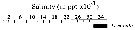
Salinity ranges for L. minuta in coastal and estuarine waters of Goa (India).
Shaded area indicates the range of higher abundance. |
| | | | Loc: | | | South Africa (E & W), N Suez Canal, Bitter Lakes, Kabret, Suez Bay Red Sea, Arabian Gulf, Kuwait, Arabian Sea, Arabia (south coast, UAE coast), Laccadive Is., Maldive Is., Karachi coast, Goa, off Cochin, Sri Lanka, Natal, Madagascar (Nosy Bé), Seychelles, India (Saurashtra coast, Madras, Mandarmani creek, Hooghly estuary, Gulf of Mannar, Palk Bay, Godavari region, Kakinada Bay), Bay of Bengal, Andaman Is., Nicobar Is., W Malay Peninsula, Nankauri Harbour, Strait of Malacca, Singapore, Malaysia (Sarawak: Bintulu coast), G. of Thailand, Indonesia (Sunda Strait, Jakarta Bay, S & N Java, Lombok Is., Ambon Is., Tioman Is., NE & SW Celebes, Philippines (N Mindanao: Gingoog Bay), Viet-Nam (Cauda Bay), Hong Kong, China Seas (East China Sea, South China Sea), Taiwan (SW, Kaohsiung Harbor, N, NE, Mienhua Canyon), S Korea, Nagazaki, Japan, Pacif. (W equatorial), Australia (Great Barrier, Shark Bay, North West Cape, G. of Carpentaria), New Caledonia, Galapagos, Bahia Cupica (Colombia), Pacif. (tropical), W Baja California, G. of California
Type locality: Hong Kong. | | | | N: | 113 | | | | Lg.: | | | (34) F: 2,26-1,76; M: 1,68; (46) F: 2,05-1,95; M: 1,65; (104) F: 2,1; M: 1,75; (120) F: 2,16-1,96; M: 1,66-1,36; (256) F: 2,2-1,96; M: 1,83-1,46; (290) F: 2-2,05; M: 1,45-2; (334) F: 2,1; M: 1,75-1,54; (530) F: 2; M: 1,8; (786) F: 2,04-1,94; (795) F: 2; M: 1,5; (937) F: 1,97-2,07; M: 1,76-1,83; (991) F: 1,76-2,26; M: 1,46-1,83; (1026) F: 1,77-1,92; M: 1,63-1,71; (1086) F: 1,84-1,95; M: 1,55-1,57; (1087) F: 1,95-2,15; M: 1,65-1,8; (1085) F: 1,6-1,9; M: 1,4-1,6; (1130) F: 1,73; M: 1,60; {F: 1,60-2,26; M: 1,36-2,00} | | | | Rem.: | Inshore; Coastal. Epipelagic.
This species belongs to the group.
For Othman & Toda (2006, p.310) this species is often mistaken for L. bengalensis in the general shape and size of both sexes. For Jeong & al. (2009, p.518) this species is closely related to L. bengalensis, but can be distinguished by: In females the genital double-somite is about 1.5 times longer than the 2nd urosomite (2.5 times longer in L. bengalensis), the 2nd urosomite has prominent chitinous tibercles (no tubercles in L. bengalensis), the caudal rami are asymmetrical (symmetrical in L. bengalensis, the P5 have a bifurcate endopod (conical endopod in L. bengalensis).
See in DVP Conway & al., 2003 (version 1) | | | Last update : 22/06/2020 | |
|
|
 Any use of this site for a publication will be mentioned with the following reference : Any use of this site for a publication will be mentioned with the following reference :
Razouls C., Desreumaux N., Kouwenberg J. and de Bovée F., 2005-2026. - Biodiversity of Marine Planktonic Copepods (morphology, geographical distribution and biological data). Sorbonne University, CNRS. Available at http://copepodes.obs-banyuls.fr/en [Accessed March 25, 2026] © copyright 2005-2026 Sorbonne University, CNRS
|
|
|
|


;)
;)
;)
;)
;)
;)
;)
;)
;)
;)
;)
;)
;)
;)
;)
;)
;)
;)
;)
;)
;)
;)
;)
;)
;)
;)

