|
|
 |
|
Calanoida ( Order ) |
|
|
|
Diaptomoidea ( Superfamily ) |
|
|
|
Pontellidae ( Family ) |
|
|
|
Pontellina ( Genus ) |
|
|
| |
Pontellina platychela Fleminger & Hulsemann, 1974 (F,M) | |
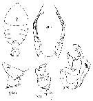
| | | | | | | Ref.: | | | Fleminger & Hulsemann, 1974 (p.75, figs.F,M); Hulsemann & Fleminger, 1975 (p.174, Juv.V F,M; M, Rem.); Björnberg & al., 1981 (p.659, figs.F,M); Hulsemann & Fleminger, 1990 (p.99, figs.F, pores cuticulaires); Bradford-Grieve & al., 1999 (p.885, 960, figs.F,M, p.876: chart) | 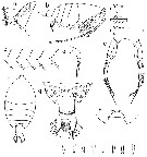 issued from : A. Fleminger & K. Hulsemann in Fishery bulletin, 1974, 72 (1). [p.77, Fig.6]. Female:: a, Th 4-5 and genital segment (lateral view); b, habitus (lateral right side; specimen from another area); c, range variation in Th 4-5 (lateral view; different specimen); d, rostrum (lateral; same as a); e, P5 (anterior view; same as a); f, habitus (dorsal; same as b); g, Th 4-5 and urosome (dorsal; same as a); h, Th4-5 (dorsal; another specimen); i, endopod of P5 (other specimens; first four: right side, five to height: left side). Nota: Genital segment with several isiolated lateral sensory hairs and line of slender hairs along distal margin, lacking lateral clusters of coarse hairs. Right caudal ramus somewhat shorter than in , typically 1.4 times longer than wide (median 1.44:1, range 1.28-1.55:1, lateral process anterior to proximal seta lacking, but interior of ramus with glandlike tissue and dctlike structure leading to lateral margin as in P. plumata. P5 essentially as in P. plumata
|
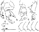 issued from : A. Fleminger & K. Hulsemann in Fishery bulletin, 1974, 72 (1). [p.78, Fig.7]. Male: a, P5 (posterior view); b, chela (lateral view; same as a); c, Th 4-5 and P5 (lateral view (other area); d, range of variation in Th 4-5 (specimens from different area). Nota: Chela of P5 differing markedly from that in P. plumata due to strong antero-posterior expansion of both segments; Left P5 with proximal segment of exopod somewhat longer than that in P. plumata. Length of right caudal ramus exceeds endopod by 1.3-1.5 times; right caudal ramus as in P. olumata, but relatively wider, ratio of length to width typically 2:1 (median 2.06:1, range 1.91-2.34:1.
|
 issued from : A. Fleminger & K. Hulsemann in Fishery bulletin, 1974, 72 (1). [p.101, Fig.33, C-e]. Female: Th 4-5 and urosome with attached spermatophore (dorsal, lateral and ventral, respecticely).
|
 issued from : A. Fleminger & K. Hulsemann in Fishery bulletin, 1974, 72 (1). [p.111, Table 19]. List of identified particles from microscopic analysis of stomach contents in adulte female.
|
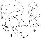 issued from : K. Hulsemann & A. Fleminger in Bull. Mar. Sci., 1975, 25 (2). [p.181, Figs.18-19]. Male: 18, P5 (posterior view); 19, chela in lateral view. Striation on process in fig.18 is assumed to function as friction pad.
|
 issued from : K. Hulsemann & A. Fleminger in Mar. Biol., 1990, 105. [p.101, Fig.1, b]. Female: b, genital segment (dorsal view). Nota: Scanning electron microscope examination revealed on the genital segment many patches of small spinules ornamenting the integument dorsally and laterally. The spinules are flattened, triangular, non-articulated, rigid structures rising from the integument, usually pointing obliquely posteriad.
|
 issued from : K. Hulsemann & A. Fleminger in Mar. Biol., 1990, 105. [p.101, Fig.2, b]. Female: b, genital segment (right lateral view).
|
 issued from : K. Hulsemann & A. Fleminger in Mar. Biol., 1990, 105. [p.104, Fig.5-7]. Female: genital segment (left to right: dorsal view, right latera viewl and ventral view).Generalized distribution of peg sensilla (o) and pores of integumental glands (filled circle); symbols are larger than organs. Organs shown occurred in at least 40 % of specimens examined (n = 25).
|
 Issued from : J.M. Bradford-Grieve, E.L. Markhaseva, C.E.F. Rocha & B. Abiahy in South Atlantic Zooplankton, edit. D. Boltovskoy. 1999, Vol. 2, Copepoda; [p.1072, Fig. 7.398: Pontellina platychela ]. r = right leg; l = left leg; r male P5 = lateral view of male right P5. Female characters (from key, p.960): - Posterior prosome corners very short, extending only about 1/3 of way along genital segment; P5 basis seta extends beyond base of 1st outer edge spine on terminal segment. Male characters (from key, p.960): - Posterolateral corners of prosome, in lateral view, rounded and lacking denticle; right chela with strong distal expansions, in anterior-posterior direction, of both exopodal segment1-2 and exopodal segment 3.
| | | | | Compl. Ref.: | | | Turner & Collard, 1980 (p.527, Tab.1, Rem.: p.528); Suarez-Morales & Gasca, 1998 a (p.111); Schnack-Schiel & al., 2010 (p.2064, Table 2: E Atlantic subtropical/tropical); Tutasi & al., 2011 (p.791, Table 2, abundance distribution vs La Niña event) | | | | NZ: | 4 | | |
|
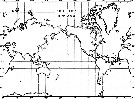
Distribution map of Pontellina platychela by geographical zones
|
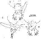
| | |  issued from : A. Fleminger & K. Hulsemann in Fishery bulletin, 1974, 72 (1). [p.79, Fig.8]. issued from : A. Fleminger & K. Hulsemann in Fishery bulletin, 1974, 72 (1). [p.79, Fig.8].
Geographical distribution of captures recorded during the present study. |
| | | | Loc: | | | Atlant. (equatorial), G. of Mexico, Caribbean, Brazil, Galapagos-Ecuador (in Tutasi & al., 2011) | | | | N: | 6 | | | | Lg.: | | | (275) F: 1,96-1,54; M: 1,74-1,41; {F: 1,54-1,96; M: 1,41-1,74} | | | | Rem.: | epiplagic.
Likely, Tutasi & al. (2011) point to this rare species for the first time in the eastern equatorial Pacific. | | | Last update : 20/01/2016 | |
|
|
 Any use of this site for a publication will be mentioned with the following reference : Any use of this site for a publication will be mentioned with the following reference :
Razouls C., Desreumaux N., Kouwenberg J. and de Bovée F., 2005-2026. - Biodiversity of Marine Planktonic Copepods (morphology, geographical distribution and biological data). Sorbonne University, CNRS. Available at http://copepodes.obs-banyuls.fr/en [Accessed June 17, 2026] © copyright 2005-2026 Sorbonne University, CNRS
|
|
|
|


;)
;)
;)
;)
;)
;)
;)
;)
