|
|
 |
|
Calanoida ( Order ) |
|
|
|
Diaptomoidea ( Superfamily ) |
|
|
|
Pontellidae ( Family ) |
|
|
|
Pontellopsis ( Genus ) |
|
|
| |
Pontellopsis laminata C.B. Wilson, 1950 (F,M) | |
| | | | | | | Ref.: | | | C.B. Wilson, 1950 (p.308, Descr.F, figs.F); Silas & Pillai, 1973 (1976) (p.777); Pillai, 1977 (1982) (p.61, figs.F,M, Rem.F,M) | 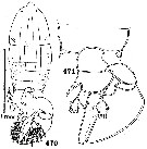 issued from : C.B. Wilson in Bull. U.S. natn Mus., 100, 14 (4), 1950. [Pl.31, Figs.470, 471]. Female (from Palawan Island: Philippines): 470, habitus (dorsal); 471, urosome (dorsal). Nota: Genital segment increasing in width to its posterior margin, where it is as wide as long. 2nd abdominal segment with an acute spine on each lateral margin; the one on the left is long and narrow and points diagonally bacward, while the right one is shorter and wider and extends outward at right angles to the urosome axis; to the dorsal surface of the segment at the right posterior corner are attached two rounded laminae, the smaller anterior one is elliptical in outline and is usually turned down over the ventral surface, the larger posterior one extends backward and inward above the anal segment and caudal rami and reaches the tips of the caudal setae; there is another smaller lamina attached to the posterior margin of the segment and extending back over the the anal segment and beyond its posterior margin. Usually yhese three complete the laminate armature of the urosome, but in one female there was a fourth large lamina attached to the left side and sweeping around backward and overlapping the one from the right. These laminae are chitinous and perfectly transparent but of course brittle and likely to be broken off. They still remained intact in 75 percent of the specimens. The genital protuberance on the ventral surface of the genital segment is at the posterior margin, and in most of the females a single spermatophore was attached to it; the long narrow discharge tube swept around and up over the right side of the urosome, and the body of the spermatophore trailed backward on the top of everything else
|
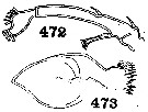 issued from : C.B. Wilson in Bull. U.S. natn Mus., 100, 14 (4), 1950. [Pl.31, Figs.472, 473]. Female: 472, A2; 473, Md (masticatory blade).
|
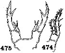 issued from : C.B. Wilson in Bull. U.S. natnl Mus., 100, 14 (4), 1950. [Pl.31, Figs.474, 475]. Female: 474, P1; 475, P5.
|
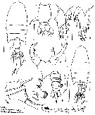 issued from : P.P. Pillai in J. mar. biol. Ass. India, 1977 (1982), 19 (1-2). [p.60, Fig.1]. Female: a, habitus (dorsal); b, urosome (dorsal); c, idem (lateral left side); d, idem (lateral right side); e, P5; f, distal outer marginal spine of P5 (enlarged). Male: g, habitus (dorsal); h, urosome (dorsal); i, right margin of urosomal segments 2 and 3 (enlarged); j, right A1 (distal portion).
|
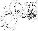 issued from : P.P. Pillai in J. mar. biol. Ass. India, 1977 (1982), 19 (1-2). [p.62, Fig.2, a-b]. Female: a, urosome with spermatophore sac and coupler sheath/ Male: b, P5.
| | | | | NZ: | 2 | | |
|
Distribution map of Pontellopsis laminata by geographical zones
|
| | | | | | | Loc: | | | Philippines (off Palawan Island), China Seas, Hong-Kong | | | | N: | 2 | | | | Lg.: | | | (137) F: 2; (276) F: 2,32-1,912; M: 2,016-1,864; {F: 1,912-2,320; M: 1,860-2,016} | | | | Rem.: | epipelagic. | | | Last update : 28/01/2016 | |
|
|
 Any use of this site for a publication will be mentioned with the following reference : Any use of this site for a publication will be mentioned with the following reference :
Razouls C., Desreumaux N., Kouwenberg J. and de Bovée F., 2005-2026. - Biodiversity of Marine Planktonic Copepods (morphology, geographical distribution and biological data). Sorbonne University, CNRS. Available at http://copepodes.obs-banyuls.fr/en [Accessed January 28, 2026] © copyright 2005-2026 Sorbonne University, CNRS
|
|
 |
 |


