|
|
 |
|
Calanoida ( Order ) |
|
|
|
Diaptomoidea ( Superfamily ) |
|
|
|
Pontellidae ( Family ) |
|
|
|
Pontellopsis ( Genus ) |
|
|
| |
Pontellopsis regalis (Dana, 1849) (F,M) | |
| | | | | | | Syn.: | Pontella regalis Dana,1849;
Pontellina regalis : Dana,1853;
Monops regalis : Giesbrecht, 1892 (part., p.486, 496, 773, figs.F,M); Wheeler, 1901 (p.182, figs.M); Oliveira, 1945 (p.191); Crisafi, 1960 c (p.279, figs.F,M, juv.); Maiphae & Sa-ardrit, 2011 (p.641, Table 2, 3, Rem.)
no Monops grandis Lubbock, 1853 b (p.122);
Pontella strenua (part.) Brady,1883 (p.95, Pl.45, fig.18);
Monachops grandis : Wilson, 1924 (p.16)
Pontellopsis regalis : Tanaka, 1964 c (p.268, figs.240, F) | | | | Ref.: | | | Giesbrecht & Schmeil, 1898 (p.147, Rem. F,M); Thompson & Scott, 1903 (p.236, 253); A. Scott, 1909 (p.171, Rem.); Sharpe, 1910 (p.413); Wolfenden, 1911 (p.362); Pesta, 1920 (p.544); Sars, 1925 (p.354); Farran, 1929 (p.210, 280); Sewell, 1932 (p.388); Wilson, 1932 a (p.157, Rem.F,M, figs.M); Dakin & Colefax, 1933 (p.206); Rose, 1933 a (p.263, figs.264); Farran, 1936 a (p.118); Wilson, 1942 a (p.204, figs.F, juv.); Sewell, 1947 (p.251); Moore, 1949 (p.61); C.B. Wilson, 1950 (part., p.310, figs.F,M); Fagetti, 1962 (p.36); Voronina, 1962 a (p.68); Tanaka, 1964 c (p.266, figs.M, No F); Chen & Zhang, 1965 (p.107, figs.F); Vervoort, 1965 (p.193, Rem.); Owre & Foyo, 1967 (p.99, figs.F,M); Vidal, 1968 (p.44, figs.F,M); Park, 1968 (p.567, Redescr.F, figs.F, Rem.: 2 forms); Razouls, 1972 (p.95, Annexe: p.94); Chen & Shen, 1974 (p.130, figs.F,M); Silas & Pillai, 1973 (1976) (p.838, figs.F,M, Rem.); Pillai, 1975 (p.138, figs.F,M, Rem.); Björnberg & al., 1981 (p. 660, figs.F,M); Zheng & al., 1982 [p.90, Figs.F,M); Ohtsuka & Onbé, 1991 (p.214); Baessa-de-Aguiar, 1991 (1993) (p.98, figs.M); Ianora & al., 1992 (p.401, fig.); Chihara & Murano, 1997 (p.872, Pl.158,160: F,M); Bradford-Grieve & al., 1999 (p.885, 961, figs.F,M); Mulyadi, 2002 (p.141, figs.F,M, Rem.); Othman & Toda, 2006 (p.317, figs.M); Vives & Shmeleva, 2007 (p.518, figs.F,M, Rem.); Suarez-Morales & Kozak, 2012 (p.1, key F,M: p.14, fig.F,M). |  iIssued from : W. Giesbrecht in Systematik und Faunistik der Pelagischen Copepoden des Golfes von Neapel und der angrenzenden Meeres-Abschnitte. – Fauna Flora Golf. Neapel, 1892. Atlas von 54 Tafeln. [Taf.41, Figs.62, 64, 66, 67]. As Monops regalis. Female: 62, last thoracic segment and urosome (dorsal); 64, idem (different specimen); 66, idem (different specimen); 67, rostrum (frontal view).
|
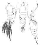 iIssued from : W. Giesbrecht in Systematik und Faunistik der Pelagischen Copepoden des Golfes von Neapel und der angrenzenden Meeres-Abschnitte. – Fauna Flora Golf. Neapel, 1892. Atlas von 54 Tafeln. [Taf.41, Figs.50, 54,5 6]. As Monops regalis. Male: 50, last thoracic segment (right) and urosome (dorsal); 54, habitus (dorsal); 56, habitus (right lateral side).
|
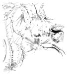 Issued from : W. Giesbrecht in Systematik und Faunistik der Pelagischen Copepoden des Golfes von Neapel und der angrenzenden Meeres-Abschnitte. – Fauna Flora Golf. Neapel, 1892. Atlas von 54 Tafeln. [Taf.26, Figs.1, 2, 3, 4, 5, 6, 7, 8, 9, 13]. As Monops regalis. Female: 2, A1 (ventral view); 4, distal part of the seta on lobe 4 of Mx2; 5, A2 (anterior view); 6 Md (mandibular palp, posterior view); 7, Md (masticatory edge, posterior view); 8, idem (anterior view); 9, Mx1 (distal part, anterior view); 14, P5 (anterior view). Male: 1, left A1, anterior view (median segments 13-15); 3, right A1; 13, P5 (anterior view).
|
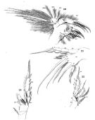 Issued from : W. Giesbrecht in Systematik und Faunistik der Pelagischen Copepoden des Golfes von Neapel und der angrenzenden Meeres-Abschnitte. – Fauna Flora Golf. Neapel, 1892. Atlas von 54 Tafeln. [Taf.26, Figs.20, 21, 22, 24]. As Monops regalis. Female: 20, Mx1 (posterior view); 21, Mxp (posterior view). Male: 22, P1 (anterior view); 24, P4 (posterior view).
|
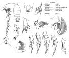 Issued from : T.S. Park in Fishery Bull. Fish Wild. Serv. U.S., 1968, 66 (3). [p.568, Pl.13, Figs.1-14]. Female: 1, habitus (dorsal); 2, last thoracic segment and urosome (dorsal); 3, A1; 4, A2; 5, Md (mandibular palp); 6, Md (masticatory edge); 7, Mx1; 8, Mx2; 9, Mxp; 10, P1; 11, P2; 12, P3; 13, P4; 14, P5. Nota: In the shape of the urosome the present specimen is not in full agreement with the description given by Giesbrecht (1892, tafel 41). According to Fleminger, however, it is not outside the variability shown by the species
|
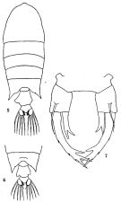 issued from: Q.-c Chen & S.-z. Zhang in Studia Marina Sinica, 1965, 7. [Pl.47, 5-7]. Female (from E China Sea): 5, habitus (dorsal); 6, urosome (another specimen), dorsal; 7, P5 (posterior).
|
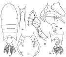 issued from : Q.-c Chen & C.-j. Shen in Studia Marina Sinica, 1974, 9. [p.130, Figs.23-28]. Female (from South China Sea): 23, habitus (dorsal); 24, rostrum (frontal view); 25, P5. Male: 26, last thoracic segment and urosome (dorsal); 27, right A1; 28, P5.
|
 issued from : E.G. Silas & P.P. Pillai in J. mar. biol. Ass. India, 1973 (1976), 15 (2). [p.839, Fig.29]. Female (from Indian Ocean): a, urosome (dorsal); b, P5. Nota: Genital segment enlarged and asymmetrically produced on left side into a distinct lobe; right lateral margin with no such modification, ventrally a median spine present near genital opening; a pair of short spinules present at base of genital segment and with a few short setae along its lateral margin. Outer lateral margin of exopod of P5 with 3 minute spuines and inner margin with 1 long spine; apically it is bifid subequally with outer branch being smaller. Male: c, urosome (dorsal); d, right A1 (geniculate part); e, P5 (terminal part of thumb, enlarged); f, P5. Nota: Right P5 chelate, with a broad hand and an elongated thumb; latter with a seta on its inner base; finger with bent tip and 2 marginal setae; it carries a conical process at its mid inner margin; 2 subequal setae prsent on basipod 2; left P5 with distal segments with 2 outer lateral spines and 2 subequal spines apically of which outer one is longest; penultimate drawn into a spine at outer distal margin; inner margin with a tuft of setae. Scale as in Calanopia minor.
|
 issued from : E.G. Silas & P.P. Pillai in J. mar. biol. Ass. India, 1973 (1976), 15 (2). [p.840]. The combination of characters in the modification of the female genital segment and P5 in both sexes agree to suggest two main types of infra-specific variations in P. regalis. Park (1968) has remarked that according to Fleminger (pers. comm.) this variation is not outside the usual variability shown by the species. However in view of the consistant differences shown by the two \"varieties\", the possibility of recognising Type-II as a distinct species from the former Type-I cannot be ruled out.
|
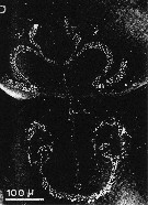 issued from : A. Ianora, A. Miralto & S. Vanucci in Mar Biol., 1992, 113. [p.403, Fig.2, D]. SEM micrographs. Female & Male: D, surface attachment structure (mass of fine setules arranged in three semicircles on a flattened area of the anterodorsal surface of the cephalosome).
|
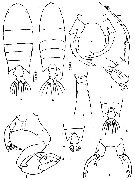 issued from : Z. Zheng, S. Li, S.J. Li & B. Chen in Marine planktonic copepods in Chinese waters. Shanghai Sc. Techn. Press, 1982 [p.91, Fig.51]. Female: a, habitus (dorsal); b, rostrum (frontal view); c, P5; d, urosme (dorsal, another specimen); e, urosome (dorsal, same individual); f, P5 (same individual). Male: g, habitus (dorsal); h, A1; i, P5. Scale bars in mm.
|
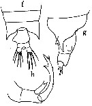 issued from : O. Tanaka in Publ. Seyo Mar. Biol. Lab., 1964, 12 (3). [p.268, Fig.240, f-h]. As Pontellopsis villosa. Female (from Izu Region, Japan): f-g, last thoracic segment and urosome (dorsal and lateral, respectively); h, P5)
|
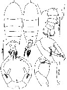 issued from : Mulyadi in Treubia, 2002, 32; [p.143, Fig.52]. Female (from Flores Sea): a, habitus (dorsal); b, P5. Male: c, habitus (dorsal); d, metasomal somite 5 and urosome (dorsal); e, P5.
|
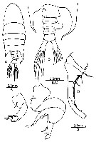 issued from : B.H.R. Othman & T. Toda in Coastal Mar. Sc., 2006, 30 (1). [p.317, Fig.22]. Male (from Sister's Island, Singapore): A, habitus (dorsal); B, posterior part of prosome and urosome (dorsal); C, P5; D, A1. Nota: Prosome to urosome length ratio 3.05 : 1. - Cephalon and 1st pedigerous somite separated, 4th and 5th pedigerous solites fused. Posterior thoracic corners asymmetrical, produced into acuminate processes, left extends betond genital segment, right directed posteriorly and curved inwards apically, reaching anal segment. - Urosome 5-segmented. - Genital segment broader than long. - Urosomal segment 3 with a prominent lobe on right margin, crowned with spinules at apex. - A1 geniculate, segment 13-17 enlarged, serrated plates present on anterior margins of segment 17 and 18, base of fused segments 19-21 with serially arranged spines. - P5 asymmetrical, right leg basis with 1 seta on inner margin; exopodal segment 1 (chela) short and broad with an elongated thumb, inner base of chela with 1 long seta; exopodal segment 2 (finger) with bent tip and 2 inner setae. Left leg, exopodal segment 1 with 1 strong distolateral spine; exopodal segment 2 with 2 outer and 2 unequal apical spines of which inner one shortest; inner margin hirsute. Remarks: There have been reports of small variations in the P5 of both sexes (see Silas & Pillai, 1973).
|
 issued from : P.P. Pillai in J. mar. biol. Ass. India, 1975, 17 (2). [p.136, Fig.3, i-L]. Female (from Indian Ocean): i, urosome (dorsal); j, P5. Male: k, P5; L, terminal portion of finger (enlarged). Scale: Single bars: 0.3 mm, double bars: 0.05 mm..
|
 Issued from : J.M. Bradford-Grieve, E.L. Markhaseva, C.E.F. Rocha & B. Abiahy in South Atlantic Zooplankton, edit. D. Boltovskoy. 1999, Vol. 2, Copepoda; [p.1073, Fig. 7.402: Pontellopsis regalis ]. Ur = urosome; r = right leg; l = left leg.; r P5= right fifth swimming leg. Female characters (from key, p.960): - Urosome distinctly of 2 somites. - Genital segment asymmetrical swollen on each side, each swelling bearing 3 spines; anal somite not projecting posteriorly. Male characters (from key, p.961): - Posterior prosome corners asymmetrical. - Left posterior prosome corner extending to posterior border of urosomal segment 2; right corner extending to posterior border of anal somite; urosomal segment 3 asymmetrically developed on right. - Urosomal segment 3 with spinulose swelling on right.
| | | | | Compl. Ref.: | | | Sewell, 1912 (p.376); 1914 a (p.239); 1948 (p.322, 357, 461); Demir, 1959 (p.176); Ganapati & Shanthakumari, 1962 (p.9, 16); Giron-Reguer, 1963 (p.54); V.N. Greze, 1963 a (tabl.2); Shmeleva, 1963 (p.141); Sherman, 1963 (p.216: fig.2, p.220: fig.9, p.223: fig.13, p.224: fig.14); Grice, 1963 a (p.496); De Decker & Mombeck, 1964 (p.13); Heinrich, 1964 (p.86: fig.4, p.89: fig.6); Anraku & Azeta, 1965 (p.13, Table 2, fish predator); Shmeleva, 1965 b (p.1350, lengths-volume-weight relation); Pavlova, 1966 (p.44); Mazza, 1966 (p.72); 1967 (p.326, 367, fig.65); Fleminger, 1967 a (tabl.1); Evans, 1968 (p.14); Champalbert, 1969 a (p.601); Sherman & Schaner, 1968 (p.583, fig.2); Apostolopoulou, 1972 (p.328, 369); Heinrich, 1974 (p.43, fig.1); Weikert, 1975 (p.139, carte); Carter, 1977 (1978) (p.36); Grice & Gibson, 1978 (p.23, tab.8, Rem.:?); Vives, 1982 (p.295); Kovalev & Shmeleva, 1982 (p.85); Dessier, 1983 (p.89, Tableau 1, Rem., %); Scotto di Carlo & al., 1984 (1045); Guangshan & Honglin, 1984 (p.118, tab.); Regner, 1985 (p.11, Rem.: p.38); Brinton & al., 1986 (p.228, Table 1); Madhupratap & Haridas, 1986 (p.105, tab.1); Lozano Soldevilla & al., 1988 (p.60); Hernandez-Trujillo, 1989 (tab.1); 1989 a (tab.1); Cervantes-Duarte & Hernandez-Trujillo, 1989 (tab.3); Heinrich, 1990 (p.19); Suarez & al., 1990 (tab.2); Baessa De Aguiar, 1991 (1993) (p.107); Suarez, 1992 (App.1); Hernandez-Trujillo, 1994 (tab.1); Shih & Young, 1995 (p.72); Hure & Krsinic, 1998 (p.103); Suarez-Morales & Gasca, 1998 a (p.111); Wong & al, 1998 (tab.2); Lavaniegos & Gonzalez-Navarro, 1999 (p.239, Appx.1); Fernandez-Alamo & al., 2000 (p.1139, Appendix); Suarez-Morales & al., 2000 (p.751, tab.1); El-Serehy & al., 2001 (p.116, Table 1: abundance vs transect in Suez Canal); Vukanic, 2003 (p.139, tab.1); Alvarez-Silva & al., 2005 (p.39); Hwang & al., 2006 (p.943, tabl. I); Dur & al., 2007 (p.197, Table IV); Neumann-Leitao & al., 2008 (p.799: Tab.II, fig.6, as P. regalis); Ayon & al., 2008 (p.238, Table 4: Peruvian samples); Mazzocchi & Di Capua, 2010 (p.427); Medellin-Mora & Navas S., 2010 (p.265, Tab. 2), Maiphae & Sa-ardrit, 2011 (p.641, Table 2, 3, Rem.); Tutasi & al., 2011 (p.791, Table 2, abundance distribution vs La Niña event); Lavaniegos & al., 2012 (p. 11, Appendix); in CalCOFI regional list (MDO, Nov. 2013; M. Ohman, comm. pers.); Tseng & al., 2013 (p.507, seasonal abundance); Lidvanov & al., 2013 (p.290, Table 2, % composition); Jerez-Guerrero & al., 2017 (p.1046, Table 1: temporal occurrence); El Arraj & al., 2017 (p.272, table 2); Palomares-Garcia & al., 2018 (p.178, Table 1: occurrence); Belmonte, 2018 (p.273, Table I: Italian zones) | | | | NZ: | 16 | | |
|
Distribution map of Pontellopsis regalis by geographical zones
|
| | | | | | | | | | | | 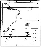 Issued from : A.K. Heinrich inTrudy Inst. Okeanol., 1974, 98 [p.44, Fig.1]. Issued from : A.K. Heinrich inTrudy Inst. Okeanol., 1974, 98 [p.44, Fig.1].
Distibution of three species of Pontellidae from SW Atlantic (Cruise of the R/V 'Akademik Kurchatov', 1971 and 1972).
Points = stations; cross = Labidocera acutifrons ; circle= Pontellopsis regalis ; triangle= Pontellopsis villosa . |
 issued from : A.A. Shmeleva in Bull. Inst. Oceanogr., Monaco, 1965, 65 (n°1351). [Table 6: 36]. Pontellopsis regalis (from South Adriatic). issued from : A.A. Shmeleva in Bull. Inst. Oceanogr., Monaco, 1965, 65 (n°1351). [Table 6: 36]. Pontellopsis regalis (from South Adriatic).
Dimensions, volume and Weight wet. Means for 50-60 specimens. Volume and weight calculated by geometrical method. Assumed that the specific gravity of the Copepod body is equal to 1, then the volume will correspond to the weight. |
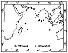 issued from : P. Pillai in J. mar. biol. Ass. India, 1975, 17 (2). [p.142, Fig.8]. issued from : P. Pillai in J. mar. biol. Ass. India, 1975, 17 (2). [p.142, Fig.8].
Occurrence of Pontellopsis regalis and P. macronyx collected from 32 stations in the Indian Ocean (See in Hand Book to the International Zooplankton Collections, volume I, 1969). |
| | | | Loc: | | | South Africa (E), off Angola, Tristan da Cunha Is., off Ascension Is., Brazil, off Trinidad Is., Cape Verde Is., off Morocco-Mauritania, Canary Is., off Azores (W & S), Barbados Is., Caribbean Colombia, G. of Mexico, Cuba, Florida, Bermuda, Woods Hole, Georges Bank, Medit. (Banyuls, G. of Lion, Ligurian Sea, Napoli, Strait of Messina, Adriatic Sea, Malta, Ionian Sea, Aegean Sea, Port Said), Suez Canal, Arabian Sea, ? Arabian Gulf, Is. Maldive Is., Sri Lanka, Natal, Indian, India (Lawson's Bay), G. of Bengal, Burma, Andaman Is., Nicobar Is., Nankauri Harbour, Straits of Malacca (Singapore), G. of Thailand, Indonesia (N Java : Lombok , Banda Sea, Eastern Indonesian waters, Philippines, Viet-Nam (Cauda Bay), Taiwan, NE Taiwan, China Seas (East China Sea, South China Sea), Nagasaki, Japan, Pacif. (W equatorial), Australia (Great Barrier, New South Wales), Pacif. (N central), Hawaii, off E Tuamotu, Galapagos, off NE Easter Is., off California, W Baja California, La Paz, Gulf of California, Bahia de los Angeles, Zihuatanejo Bay, G. of Tehuantepec, W Mexico, Central America, Bahia Cupica (Colombia), Galapagos-Ecuador, off Peru, Pacif. (SE tropical), Chile
Type locality: Sulu Sea | | | | N: | 108 ? | | | | Lg.: | | | (16) F: 4,08-3,75; (34) F: 3,47; (35) F: 3,6; (45) F: 4,5-4; M: 3,5-3,25; (46) F: 4,4-4; M: 3,5-3,4; (72) F: 3,2; (120) F: 3,65-3,37; M: 3,22-3,08; (187) F: 3,84-3,7; M: 3,35; (256) F: 4,22-3,39; M: 3,58-3,25; (290) F: 2,65-2,75; (394) F: 2,98-2,85; M: 2,76-2,72; (449) F: 4,4-4; M: 3,5-3,4; (485) F: 3,847-3,464; M: 3,379-3,123; (806) M: 3,3; 3,28; (1023) F: 3,26-3,56; 2,74-2,85; M: 2,67-3,37; (1086) M: 1,64; (1087) F: 3,40; M: 3,20; (1115) F: 2,74-3,16; M: 2,64-3,01; {F: 2,65-4,50; M: 1,64-3,58} | | | | Rem.: | epiplagic; oceanic-neritic
An important confusion reigns between the species described by Dana (1849) and Pontellopsis grandis Lubbock (1853), due to the established synonymy by Giesbrecht (1892). For Bradford-Grieve (1999 b, p.207) the species are distinct. The localisations for the two species are thus difficult to specify, all the more that their presence seems largely distributed in the three oceans.
Mulyadi (2002, p.144) notes by the authors the variations in the urosomal somite 1 of female and the form of P5 in both sexes. Giesbrecht (1892) reported three variations of urosomal somite 1 of female, but only one type of female P5 in that its terminal spines being subequal and in the male P5 the finger of terminal segment of the right leg has been shown to be without any modification. Tanaka (1968) described P. regalis having a swelling on the right side of the urosomal somite 1, and Park (1968) described the material from the North Pacific Ocean female having the urosomal somite 1 with each side projected posteriorly into a conical process and the P5 with similar terminal spines. Silas & Pillai (1973) described two types of ''variant'' observed in the female and male from the materials from the Laccadive Sea and Andaman Islands. The urosomal somite 1 of female has a distinct bulge on its left side and this is characteristically linked with the subequal terminal spines on P5. The male also shows the characteristic modification in the finger of terminal segment of right P5 and the presence of a slightly elevated flap-like structure behind its tip. | | | Last update : 24/10/2022 | |
|
|
 Any use of this site for a publication will be mentioned with the following reference : Any use of this site for a publication will be mentioned with the following reference :
Razouls C., Desreumaux N., Kouwenberg J. and de Bovée F., 2005-2026. - Biodiversity of Marine Planktonic Copepods (morphology, geographical distribution and biological data). Sorbonne University, CNRS. Available at http://copepodes.obs-banyuls.fr/en [Accessed January 05, 2026] © copyright 2005-2026 Sorbonne University, CNRS
|
|
 |
 |


