|
|
 |
|
Calanoida ( Order ) |
|
|
|
Pseudocyclopoidea ( Superfamily ) |
|
|
|
Pseudocyclopidae ( Family ) |
|
|
|
Pseudocyclops ( Genus ) |
|
|
| |
Pseudocyclops lakshmi Haridas, Madhupratap & Ohtsuka, 1994 (F,M) | |
| | | | | | | Ref.: | | | Haridas & al., 1994 (p.151, figs.F,M) | 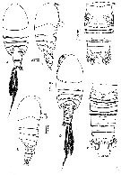 issued from : P. Haridas, M. Madhupratap & S. Ohtsuka in Proc. Biol. Soc. Wash., 1994, 107 (1). [p.152, Fig.1]. Female (from Agatti and Kadmat, Maldives): A-B, habitus (dorsal and lateral, respectively); C, urosome (dorsal). Nota: Cephalosome and 1st pedigerous somite separate, 4th and 5th completely separate. Rostrum pointed, triangular, with a pair of minute sensilla. Urosome 4-segmented, distal margins of first 2 somites lamellar. Anal somite small, telescoped into 3rd urosome somite. Caudal ramus with serrate posterior margin dorsomedially and with 1 dorsal, 4 terminal and 1 outer subterminal setae. Male: D-E, habitus (dorsal and lateral, respectively; F, urosome (dorsal). Nota: Dimorphism observed only in males. The differences between the two \"morphs\" are found in body length and Mx2
|
 issued from : P. Haridas, M. Madhupratap & S. Ohtsuka in Proc. Biol. Soc. Wash., 1994, 107 (1). [p.154, Fig.3]. Female: A1. Nota: A1 21-segmented, not quite reaching to posterior end of cephalosome; 1st segment with 3 large aesthetascs and 11 setae; 4th and 5th and 18th and 19th segments partly fused; terminal segment with 1 aesthetasc. Male: right A1. Nota: Right A1 18-segmented, geniculate between 14th and 15th segments; 1st segment with 3 large aesthetascs and 3 rows of minute spinules; 7th and 8th segments fused or separate; 14th segment with sinuous process along whole length of anterior margin; 16th segment produced distally into triangular process reaching midlength of 17th segment; terminal segment with 1 aesthetasc; Left A1 21-segmented, 4th and 5th, 18th and 19th segments incompletely fused (as in female).
|
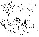 issued from : P. Haridas, M. Madhupratap & S. Ohtsuka in Proc. Biol. Soc. Wash., 1994, 107 (1). [p.155, Fig.4]. Female: A, A2; B, Md; C, Mx1; D, Mxp. Nota: A2 endopod 3-segmented, exopod 7-segmented (3rd to 5th segments incompletely fused. Mx1: praecoxal arthrite with 5 setae on posterior surface and 1 weak and 8 stout spine-like setae along inner margin; coxal and 2 basal endites having 3, 4 and 5 setae respectively; coxal epipodite with 9 setae of unequal lengths; basal exite with short, single seta; endopod 2-segmented, 1st segment bearing 5 middle and 5 terminal setae along inner margin, 2nd segment with 6 terminal setae; exopod 1-segmented with 11 setae. Mx2: Praecoxal and coxal endites (Fig.5B, female Mx2 similar to \"morph A\" of male) having 6, 3, 3 setae, respectivemy; basis completely sused with 1st endopod segment to form allobasis, furnished with 7 setae; endopod 3-segmented, 1st to 3rd segments having 2, 2 and 3 setae respectively
|
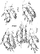 issued from : P. Haridas, M. Madhupratap & S. Ohtsuka in Proc. Biol. Soc. Wash., 1994, 107 (1). [p.158, Fig.6]. Female: A-E, P1 to P5. Nota: P1P4 of both \"morphs\" male similar to those of the female
|
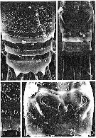 issued from : P. Haridas, M. Madhupratap & S. Ohtsuka in Proc. Biol. Soc. Wash., 1994, 107 (1). [p.153, Fig.2]. Female (SEM photomicrographs): A, urosome (dorsal); B, idem (ventral); C, distal margin of genital double-somite (dorsal); D, genital double-somite (ventral). All scale bars = 0.010 mm. Nota: Gonopores and copulatory pores paired, closed off by operculum-like P6.
|
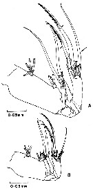 issued from : P. Haridas, M. Madhupratap & S. Ohtsuka in Proc. Biol. Soc. Wash., 1994, 107 (1). [p.156, Fig.5]. Male : A, Mx2 (\"morph B\"); B, Mx2 (\"morph A\"). Nota: Mx2 of \"morph\" B about twice as long as that of \"morph\" A; 2 serrate setae on basis (indicated by small arrows) much stouter in \"morph \" B than in \"morph\" A; seta on 2nd endopod segment (indicated by large arrow) longer and stouter in \"morph\" B than in \"morph\" A. A2, Md, Mx1 and Mxp of both morphs similar to those of the female. Rem: Although there remains a possibility that the two \"morphs\" of male may be two distinct species, the absence of females with large-sized Mx2 and the co-occurrence of both \"morphs\" of males led us to the conclusion that these \"morphs\" belong to the same species. It is possible that the feeding behaviors of these two \"morphs\" of male may be different because Mx2 plays an important role in feeding (see in Do & al., 1984).
|
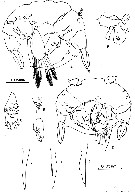 issued from : P. Haridas, M. Madhupratap & S. Ohtsuka in Proc. Biol. Soc. Wash., 1994, 107 (1). [p.159, Fig.7]. Male: P5: A, posterior view (left exopod segment 2 omitted); B, left exopod segment 2 ; C, left endopod; D, anterior view; E-F, right endopod variability; G-l, pointed process on basis of left leg showing variability in shape. Nota: P5 of both \"morphs\" similar to each other except being slightly smaller in \"morph\" A. Right P5 coxa and intercoxal sclerite fused; lefl P5 coxa incompletely fused with basis on both surfaces.
|
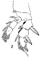 Issued from : V.N. Andronov in Russian Acad. Sci. P.P. Shirshov Inst. Oceanol. Atlantic Branch, Kaliningrad, 2014. [p.45, Fig.13, 2]. Pseudocyclops lakshmi after Haridas & al., 1994. Female P1.
| | | | | NZ: | 1 | | |
|
Distribution map of Pseudocyclops lakshmi by geographical zones
|
| | | | | | | Loc: | | | Maldive Is. (Kadmat & Agatti atolls) | | | | N: | 1 | | | | Lg.: | | | (775) F: 0,95-0,86; M: 0,95-0,85; {F: 0,86-0,95; M: 0,85-0,95} | | | | Rem.: | Surface hauls at night.
For Haridas & al., 1994 (p.160) the new species is one of the most primitive species of the genus Pseudocyclops, having 5 separate pedigerous somites, 21-segmented female A1 and 7-segmented A2 exopod; it has an outer basal spine on P3, 3-segmented endopod in P5 of female and 5 plumose setae on the left endopod of male P5. The present species, however, has several advanced morphological characters present on the urosome, P1 and female P5: The urosome is covered with minute prominences, the 2nd exopod segment of P1 has an outer bulbous process, and there are 4 setae on the 3rd endopod segment of the female P5 (6 setae in P. kulai, P. lepidotus, P. mathewsoni, P. rubrocinctus, P. steinitzi). The urosome with numerous minute prominences appears to be unique, although P. lepidotus has foliaceous scales on the urosome. P. lakshmi seems to be most closely related to P. australis from South Australia and Japan and P. gohari from the Red Sea in having 3-segmented endopod of the female P5 with 4 setae on the terminal segment and a bulbous process on the 2nd exopod segment of P1. In particular, the structures of P5 of both sexes of P. lakshmi resemble those of P. gohari. | | | Last update : 20/04/2016 | |
|
|
 Any use of this site for a publication will be mentioned with the following reference : Any use of this site for a publication will be mentioned with the following reference :
Razouls C., Desreumaux N., Kouwenberg J. and de Bovée F., 2005-2025. - Biodiversity of Marine Planktonic Copepods (morphology, geographical distribution and biological data). Sorbonne University, CNRS. Available at http://copepodes.obs-banyuls.fr/en [Accessed December 27, 2025] © copyright 2005-2025 Sorbonne University, CNRS
|
|
 |
 |


