|
|
 |
|
Calanoida ( Order ) |
|
|
|
Clausocalanoidea ( Superfamily ) |
|
|
|
Pseudocyclopiidae ( Family ) |
|
|
|
Stygocyclopia ( Genus ) |
|
|
| |
Stygocyclopia philippensis Jaume, Fosshagen & Iliffe, 1999 (F,M) | |
| | | | | | | Ref.: | | | Jaume & al., 1999 (p.405, figs.F,M) | 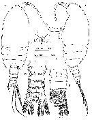 issued from : D. Jaume, A. Fosshagen & T.M. Iliffe in Sarsia, 1999, 84. [p.406, Fig.10]. Female (from 9°37'N, 123°47'E): A-B, habitus (dorsal and lateral, respectively); C, rostrum (ventral); ; D, urosome (dorsal); E, same (ventral). Nota ; Eyes absent. - Body robust, colourless, laterally compressed. - Prosome about 3 times longer than urosome. - Rostrum short and tapering, directed ventrally, with pair of long and slender distal filaments. 1st pedigerous somite incorporated into cephalosome, 4th and 5th pedigerous somites fused, symmetrical. - Dorsal fold of cuticular membrane, resembling posterior projection of cephalothorax, covering anterior dorsal half of 2nd pedigerous somite. 2nd and 3rd pedigerous somites with rounded lateral margins bearing row of sub-marginal setules along anterior half. - margins of 3rd somite with long and robust sensilla implanted sub-marginally in posterior half. - Urosome 4-segmented. Surface of somites covered with both lamellar and ordinary long spinules (spinules easily lost when manipulation). - Genital double-somite symmetrical, with faint, hyaline frill around dorsal rear margin ; smooth hyaline frill around ventral rear margin. Ovoid seminal receptacle on each side of somite, communicating to single gonopore opening ventrally ; operculum quadrangular. - Anal somite with smooth operculum. - Caudal rami symmetrical, short, about as long as wide, armed with 7 setae and with transverse row of long setules midway along medial margin ; Setae III to VI implanted distally, I, II and VII subdistally ; setae III to VI thick, with internal tissue finely corrugated ; seta I very much reduced. Rear margin of rami with broad, triangular projections ventrally.
|
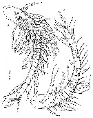 issued from : D. Jaume, A. Fosshagen & T.M. Iliffe in Sarsia, 1999, 84. [p.408, Fig.11]. Male: A, A1 (ventral). Female: B, A1 (ventral). Nota Female : A1 short, symmetrical, 23-segmented, directed ventrally,not reaching distal end of cephalothorax. 1st segment largest (representing ancestral segments I to IV) ; other compound segments, X-XI and XXVII-XXVIII. Nota Male : A1 symmetrical, 22-segmented ; segmentation pattern as in female, with additional compound segment involving ancestral segments XXII and XXIII. Segment 1 (corresponding to ancestral segmentsI to IV), 6 setae + 5 aesthetascs ; segment 2 (ancestral V), 2 setae + 2 aesthetascs ; segment3 (VI), 2 setae + 1 aesthetasc. 2 sensillae on dorsal surface of 1st segment and 1 sensilla on second segment.
|
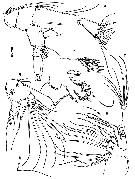 issued from : D. Jaume, A. Fosshagen & T.M. Iliffe in Sarsia, 1999, 84. [p.409, Fig.12]. Female: A, A2; B, Md (mandibular palp); C, Mx1 (ornamentation of spines on praecoxal arthrite omitted); D-E, Mx2. Nota : A2 biramous. Coxa with distal seta and patch of coarse pinules along inner margin ; basis with 2 distal setae on inner margin. Endopod 2-segmented, longer than exopod, with proximal segment completely separate from basis, elongate, bearing 2 unequal setae at 2/3 of distance along inner margin and short row of tiny spinules proximally ; distal segment short, bilobed ; inner lobe (representing ancestral segment II) with 9 setae ; outer lobe (ancestral segments II and IV) with 7 distal setae and 2 combs of spinules. Exopod 7-segmented ; segment 2 representing ancestral segmentsII-IV ; segment 7 representing segments IX-X, with intersegmental articulation partially expressed ; setal formuma 1, 3, 1, 1, 1, 1, (1+3). - Md coxal gnathobase heavily constructed, short, with cutting blade bearing sharp denticles. Palp with basis expanded, bearing 3 setae and 2 patches of spinules ; endopod shorter than exopod, 2-segmented, 1st segment bearing 1 seta, 2nd segment with 11 distal setae, 3 of them reduced, and comb of spinules. ; exopod apparently 4-segmented (close analysis revealing only partial expression of articulation between proximal segment, rather spirally configured, and 2nd segmen tresulting in actual 3-segmented condition ; setal formula (1+1), 1, 3 ; distal segment derived from ancestral segments IV and V. - Mx1 with segmentation not clearly defined ; praecoxal arthrite with 10 stout spines plus 4 setae ; coxal epipodite with 6 long and 1 reduced setae ; coxal endite with 2 setae ontip ; proximal basal endite discrete, with 4 distal setae ; distal endite incorporated into segment, bearing 5 marginal setae and row of sub-marginal spinules ; endopod fully incorporated into basis forming lobe with 12 long, distal setae ; exopod discrete, with row of 7 long and 1 reduced marginal setae.
|
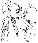 issued from : D. Jaume, A. Fosshagen & T.M. Iliffe in Sarsia, 1999, 84. [p.410, Fig.13]. Female: A, Mxp; B, P1 (anterior); C, P5 (posterior). Nota : Mxp powerfully developed, 8-segmented, reflexed distally. Syncoxa with 9 setae distributed in 4 groups along medial margin 1, 2, 3, 3 ; distal portion of medial margin produced into powerful lobe, lobe integument with 2 microtubules parches and row of short setules. ; basis about as long as syncoxa, slender, with 3 long setae along medial margin and sub-marginal row of coarse spinules ; endopod 6-segmented ; small 1st segment defined medially but not apparent laterally ; setal formula 2, 4, 4, 3, 3+1, 4 ; transverse row of coarse spinules on distal margins of 3rd and 4th segments : row of slender spinules along distal margin of 5th.
|
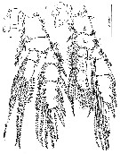 issued from : D. Jaume, A. Fosshagen & T.M. Iliffe in Sarsia, 1999, 84. [p.411, Fig.14]. Female: A, P2 (anterior); B, P3 (anterior).
|
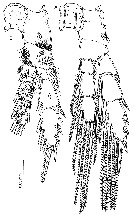 issued from : D. Jaume, A. Fosshagen & T.M. Iliffe in Sarsia, 1999, 84. [p.412, Fig.15]. Female: A, P4 (anterior); B; same (posterior).
|
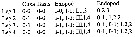 issued from : D. Jaume, A. Fosshagen & T.M. Iliffe in Sarsia, 1999, 84. [p.407]. Female: Spine (Roman numerals) and seta (Arabic numerals) formula for swimming legs P1 to P4.
|
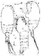 issued from : D. Jaume, A. Fosshagen & T.M. Iliffe in Sarsia, 1999, 84. [p.413, Fig.16]. Male: A-B, habitus (dorsal and lateral, respectively); C, urosome (dorsal); D, P5 (posterior). : Nota : Body similar to female excepet for A1, urosome segmentation and P5. - Urosome 5-segmented, - Genital somite asymmetrical, slifgtly produced and extended posteriorly on left side ; single gonopore opening ventrolaterally on left side close to posterior margin of somite. - P5 asymmetrical, uniramous, slender ; left longer than right, 5-segmented, with proximal segment completely fused to intercoxal sclerite ; segment 3 with tiny distal spine on outer margin ; segment 5 with 2 long and slender distal spines, inner longest ; both spines more than twice length of segment. Right leg exactly as in female. Both legs and intercoxal sclerite richly ornamented with setules and spinules.
| | | | | NZ: | 1 | | |
|
Distribution map of Stygocyclopia philippensis by geographical zones
|
| | | | Loc: | | | Philippines (Panglao Is.: Calingoob Cave) | | | | N: | 1 | | | | Lg.: | | | (837) F: 0,57-0,61; M: 0,52-0,55; {F: 0,57-0,61; M: 0,52-0,55} | | | | Rem.: | For Jaume & al. (1999, p.414) this species shows apparent differences, such as the 23-segmented condition of the female A1 compared to only 22-segmented in S. balearica, the presence of 9 setae on the inner lobe of the distal endopod segment of A2 (only 7 in S. balearica), the presence of 7 setae on the coxal epipodite, and of 12 on the endopod of Mx1 (6 and 11, respectively, in S. balearica), the presence of 4 setae on the proximal endopod segment and of 6 on the proximal syncoxal endite of Mx2 (only3 and 5, respectively, in S. balearica), and the armature of the 2nd endopod segment of Mxp (4 setae rather than only 3 in S. balearica) are largely the result of inaccuracy in the original description of the latter taxon by Jaume & Boxshall, 1995. The segmentation of the mandibular exopod in S. balearica is reinterpreted here as 3-segmented as in S. philippinensis rather than 4-segmented. The similarities are sufficient to justify its accomodation of S. pholippinensis in this genus. | | | Last update : 02/11/2015 | |
|
|
 Any use of this site for a publication will be mentioned with the following reference : Any use of this site for a publication will be mentioned with the following reference :
Razouls C., Desreumaux N., Kouwenberg J. and de Bovée F., 2005-2026. - Biodiversity of Marine Planktonic Copepods (morphology, geographical distribution and biological data). Sorbonne University, CNRS. Available at http://copepodes.obs-banyuls.fr/en [Accessed June 10, 2026] © copyright 2005-2026 Sorbonne University, CNRS
|
|
|
|


;)
;)
;)
;)
;)
;)
;)
