|
|
 |
|
Calanoida ( Order ) |
|
|
|
Clausocalanoidea ( Superfamily ) |
|
|
|
Pseudocyclopiidae ( Family ) |
|
|
|
Thompsonopia ( Genus ) |
|
|
| |
Thompsonopia mediterranea Jaume, Fosshagen & Iliffe, 1999 (F,M) | |
| | | | | | | Ref.: | | | Jaume & al., 1999 (p.394, figs.F,M); Vives & Shmeleva, 2007 (p.713, figs.F,M, Rem.) | 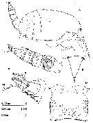 issued from : D. Jaume, A. Fosshagen & T.M. Iliffe in Sarsia, 1999, 84. [p.393, Fig.1]. Female (Mallorca, Balearic Islands): A, habitus (lateral); B, rostrum (frontal view); C, urosome (lateral); D, genital double-somite (ventral); E, detail of right caudal ramus (lateral). Nota: Ryes absent. - Body compact, with prosome laterally compressed. - Rostrum movable, tapering, with pair of apical filaments. - 1st pedigerous somite incorporated into cephalosome, 4th and 5th pedigerous somites completely fused. - Sensillae distributed on prosome (as figured). - Urosome 4-segmented with surface of somites richly ornamented with both ordinary and lamellar spinules (latter easily lost when manipulating animals). - Genital double somite symmetrical. Internal genital system not fully resolved: both seminal receptacles developed, forming dorsal lobe at each side, with serpentiform receptacle ducts; pair of oviducts emptying into separate gonopores also discerned. Ducts and gonopores opening into common mid-sentral atrium closed off to quadrangular operculum. 2 sac-like sclerotized folds similar to those displayed by Crassarietellus huysi (see Ohtsuka & al., 1994, fig.2a) positioned ventro-laterally near anterior margin of double-somite. Nota Male: Body similar to female except for segmentation and armature of A1 and armature of P4, morphology of P5. Urosome 5-segmentred. Genital somite slightly asymmetrical, with single, left gonopore opening posterolaterally on ventral side.
|
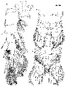 issued from : D. Jaume, A. Fosshagen & T.M. Iliffe in Sarsia, 1999, 84. [p.395, Fig.2]. Female: A, anal somite and caudal rami (dorsal); B, same (ventral); C, P1 (anterior). Nota: Anal somite with weakly developed, unarmed operculum. - Caudal rami symmetrical, short, with medial margin covered with setules; armature of 7 setae: Seta III to VI implanted distally, very thick, with internal tissue finely granulated; Seta I tiny, positioned laterally near insertion of seta II (arrowed in fig.1E; Setae II and VII reduced; seta II implanted dorsolaterally; seta VII displaced ventromedially.
|
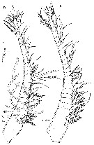 issued from : D. Jaume, A. Fosshagen & T.M. Iliffe in Sarsia, 1999, 84. [p.396, Fig.3]. Female: A, right A1 (ventral). Male: B, right A1 (ventral). - Nota Female: A1 short, symmetrical, 20-segmented, directed ventrally, not reaching distal end of cephalothorax; 1st segment largest (representing ancestral segments I to IV). Other compound segments involving ancestral segments X-XIV and XXVII-XXVIII. 2 sensillae on dorsal surface of 1st segment; 1 sensilla on 2nd segment. Nota Male: Right A1 20-segmented; segment 1 (ancestral segment I), 1 seta + aesthetasc; segment 2 (segments II-IV), 5 + 3 aesthetascs; segment 3 (V)), 2 + aexthetasc; segment 4 (VI), 2 setae; segment 5 (VII), 2 setae + 2 aesthtascs; segment 6 (VIII), 1 seta; segment 7 (IX), 2 setae; segment 8 (X-XIV, 6 setae + 2 aesthetascs; segment 9 (XV), 1 seta; segment 10 (XVI), 1 seta + aesthetasc; segments 11 to 14 (XVII to XX), 1 seta each; segment 15 (XXI), 1 seta + aesthetasc; segment 16 (XXII-XXIII, with intersegmental articulation partially expressed), 2 setae; segments 17 to 19 (XXIV to XXVI), 2 setae each; segment 20 (XXVII-XXVIII), 5 setae + aesthetasc. 1 short sensilla on dorsal surface of each of first three segments. LeftA1 as for the right except for the complete failure to express, instead of partial, of the articulation between segments XXII and XXIII.<
|
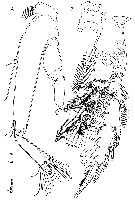 issued from : D. Jaume, A. Fosshagen & T.M. Iliffe in Sarsia, 1999, 84. [p.397, Fig.4]. Female: A, A2; B, P4 (posterior). Nota: A2 biramous. Coxa unarmed, with patch of long spinules among inner margin; basis completely fused to 1st endopd segment (forming elongate allobasis), 2 groups of 2 setae positioned along inner margin; free endopod segment elongate, bilobed, inner lobe (corresponding to ancestral endopod segment II) with 9 setae, outer lobe (corresponding to ancestral segments III-IV) with 7 distal setae; several combs of long spinules (as figured 4A). Exopod 6-segmented (segment 2 representing ancestral segments II-IV, plus partially incorporated segment V, segment 6 representing partially incorporated ancestral segment IX, elongate, bearring reduced seta) and X bearing 3 setae; exopod segmentation and setal formula 0, (0+1), 1, 1, 1, (1+3). Segmental homology deducted after comparison with segmental pattern and armature of Stygocyclopia philippinensis Jaume & al., 1999.
|
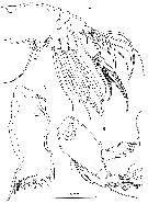 issued from : D. Jaume, A. Fosshagen & T.M. Iliffe in Sarsia, 1999, 84. [p.399, Fig.5]. Female: A, Md (mandibular coxal gnathobase); B, Md (mandibular palp); C, Mx2. Nota: Md with short, heavily built coxal gnathobase; cutting blade with major group of 4 triangular teeth , plus complex array of serrate teeth and spines dorsally. Md palp with basis expanded, bearing 2 long plus 1 reduced setae on inner margin plus patch of spinules proximally. Endopod aligned with main axis of basis, shorter than exopod, 2-segmented; 1st segment with single seta plus patch of tiny spinules; 2nd with 11 distal setae (2 of them reduced), and comb of long spinules. Exopod apparently 4-segmented (but close analysis revealing partial fusion between proximal segment, somewhat spirally configured, and 2nd and 3rd segments, resulting in actual 2-segmented conditions); setal formula (1+1+1), 3; distal segment derived from ancestral segments IV and V . (see Huys & Boxshall, 1991, p.24). - Mx2 comprising syncoxa, allobasis, and unsegmented endopod. Proximal syncoxal endite with 5 long marginal setae plus short, hyaline spiniform process; remaining syncoxal endites each with 3 setae, one of them shorter, plus bub-marginal row of spinules; soft process of uncertain homology between proximal and 2nd endite. Basal endite carrying 2 syout spines plus 2 more slender elements distally; tiny seta remnant of incorporated endopodal segment close to insertion of free endopod. Free endopod short, bearing 7 unequal setae.
|
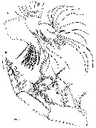 issued from : D. Jaume, A. Fosshagen & T.M. Iliffe in Sarsia, 1999, 84. [p.400, Fig.6]. Female: A, Mx1; B, Mxp (lateral). Nota: Mx1 with praecoxal arthrite carrying 9 stout marginal spines plus 4 more slender spines sub-marginally. Coxal epipodite with 7 long and 2 reduced setae; coxal endite discrete, with 2 setae distally; row of spinules along inner margin. Peoximal basal endite elongate, with 5 distal setae. Distal endite incorporated into basis, with 5 setae and row of marginal spinules. Endopod fully incorporated into basis, forming lobe armed with 11 long distal setae. Exopod discrete, also incorporated to basis, with row of 8 long marginal setae. - Mxp powerfully developed, 8-segmented, reflexed distally. Syncoxa with 9 setae in 4 groups along medial margin 1, 2, 3, 3; distal portion of medial margin produced into powerfull lobe with microtuberculate integument and patch of short setules proximally. basis slender, with 3 long setae along medial margin and sub-marginal row of spinules. Endopod 6-segmented, 1st segment somewhat reduced, setal formula 2, 4, 4, 3, 3+1, 4; transverse row of coarse spinules on distal margin on distal margin of segments 3 andd 4; 2 rows of spinules on 5th.
|
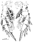 issued from : D. Jaume, A. Fosshagen & T.M. Iliffe in Sarsia, 1999, 84. [p.401, Fig.7]. Female: A, P2 (anterior); B, P3 (anterior). Nota: Legs richly ornamented with denticles, spinules and setules, especially on posterior surfaces of segments except for coxa of P4, which lacks any ornamentation posteriorly.
|
 issued from : D. Jaume, A. Fosshagen & T.M. Iliffe in Sarsia, 1999, 84. [p.398]. Female and male: Spine and seta formula of swimming legs P1 to P4. Nota Male: P4 as in female except for absence of inner coxal spine. Setal condition of armature elements on two proximal endopod segments unconfirmed.
|
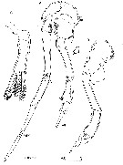 issued from : D. Jaume, A. Fosshagen & T.M. Iliffe in Sarsia, 1999, 84. [p.402, Fig.8]. Male: A, P5 (anterior); B, detail of left leg. Female: C, right P5 (posterior). Nota Female: P5 symmetrical, uniramous, very reduced. Legs 3-segmented, with 2 proximal segments unarmed, 1st fused to intercoxal sclerite . Distal segment about 1.7 times as long as wide, bearing 2 coarse, unequal bipinnate spines distally, innermost largest and incorporated basally to segment; additional coarse bipinnate spine sub-distally on outer margin. Nota Male: P5 uniramous, assymmetrical, elongate. Coxae of both legs fused, althrough retaining trnasverse suture line; patch of stout spinules posterolaterally of left coxa. Right leg longest, with short and slender basis bearing patch of tiny spinules posteromedially; segment 3 and 4 partially fused, about same length, with patch of 4 stout, plus 1 more slender spinule; 5th segment longest, spiniform, with lateral margin serrate distally; tiny spinule on posterior surface of segment.Left leg with basis expanded posteriorly into rounded lobe and bearing patch of tiny spinules posteromedially; 3rd segment longest reflexed proximally (fig.8B); 4th segment expanded mediodistally, with 3 clusters of spinules; 5th segment tapering, with ridges and short digitiform process; spinulose posteromedial lobe implanted proximally on segment.
| | | | | NZ: | 1 | | |
|
Distribution map of Thompsonopia mediterranea by geographical zones
|
| | | | | | | Loc: | | | W Medit.: Baleares (Mallorca: Cova de Na Mitjana) | | | | N: | 1 | | | | Lg.: | | | (837) F: 0,88; M: 0,76; {F: 0,88; M: 0,76} | | | | Rem.: | For Jaume & al. (1999, p.403) this species differs from T. stephoides from the North Sea and the coasts of Mauritania in the absence of the spiniform processes on the inner margin of the two proximal segments of the female P5. The right male P5 of the latter taxon has segments 3 and 4 completely fused, and segment 5 extremely elongate and bent; the right leg segment 4 is more elongate and constricted halfway compared to that of T. mediterranea. | | | Last update : 02/11/2015 | |
|
|
 Any use of this site for a publication will be mentioned with the following reference : Any use of this site for a publication will be mentioned with the following reference :
Razouls C., Desreumaux N., Kouwenberg J. and de Bovée F., 2005-2026. - Biodiversity of Marine Planktonic Copepods (morphology, geographical distribution and biological data). Sorbonne University, CNRS. Available at http://copepodes.obs-banyuls.fr/en [Accessed January 08, 2026] © copyright 2005-2026 Sorbonne University, CNRS
|
|
 |
 |


