|
|
 |
|
Calanoida ( Order ) |
|
|
|
Epacteriscoidea ( Superfamily ) |
|
|
|
Ridgewayiidae ( Family ) |
|
|
|
Ridgewayia ( Genus ) |
|
|
| |
Ridgewayia typica Thompson & Scott, 1903 (F,M) | |
| | | | | | | Syn.: | Suezia canalis Gurney, 1927 d (p.457, Descr.M, figs.M);
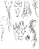
Ridgewayia canalis : M.S. Wilson, 1958 (p.145, Rem.); Humes & Smith, 1974 (p.130, Rem.) | | | | Ref.: | | | Thompson & Scott, 1903 (p.234, 245, Descr.F, figs.F); Wilson, 1958 (p.142, 143, 144, Rem.M); Ummerkutty, 1963 (p.16, Redescr.F, Descr.M, figs.F,M, Rem. p.23); Por, 1979 (p.14, figs.F,M, Rem.) | 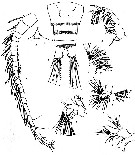 issued from : F.D. Por in Crusraceana, 1979, 37 (1). [p.15, Figs.1-8]. Female (from Di Zahav pool, Gulf of Elat): 1, urosome (ventral); 2, rostrum; 4, A2; 5, Md; 6, Mx1; 7, Mx2; 8, Mxp. Nota: Urosome 4-segmented, with last segment completely hidden under the peniltimate segment. Genital segment with a lateral, curved spine on the right side. Caudal rami2 times as long as broad and bear some setae on the distal part of their internal edges; there are 4 terminal setae and 1 spine in the external corner; 1 surface seta. A1 25-segmented, without groups of hairs on segments 7 to 13. Exopod of A2 8-segmented. Md with exopod 4-segmented and unclearly 3-segmented endopod. Basal segment of Mxp with 1 terminal seta. Male: 3, left A1; Nota: left A1 21-segmented, slightly genicukated between segments 18 and 19, segment 11 reduced; there is no serrulate lamina on the first of the four terminal segments, as found by Ummerkutty (1963); right A1 25-segmented (as in the female).
|
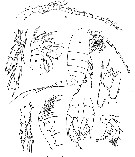
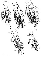 issued from : F.D. Por in Crusraceana, 1979, 37 (1). [p.17, Figs.9-13]. Female: 9, P1; 10, P2; 11, P3; 12 ,P4; 13, P5.
|
 issued from : F.D. Por in Crusraceana, 1979, 37 (1). [p.17, Figs.14-17]. With doubt. Male: 14, P5; 15, left P5 (other specimen); 16, idem (other specimen); 17, P5 (from Suez Canal specimen).
|
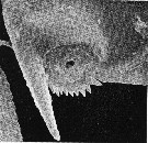
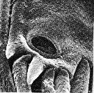 issued from : F.D. Por in Crusraceana, 1979, 37 (1). [Pl.2, Fig.62]. Female: last segments of exopod and endopod on P3 with \"tympani\"-like orifices on the anterior surface (Scanning electrom micrographs).
|
issued from : F.D. Por in Crusraceana, 1979, 37 (1). [Pl.3, Fig.63]. Female: last segment of endopod on P3 with \"tympanum\"-like orifice on the anterior surface (Scanning electrom micrographs).
|
 issued from : F.D. Por in Crusraceana, 1979, 37 (1). [Pl.4, Fig.65]. Male: orifice, surrounded by spines on the terminal surface of exopod of P5 (Scanning electrom micrographs).
|
 issued from : A.N.P. Ummerkutty in Bull. Dep. mar. Biol. Oceanogr., 1963, 1. [Pl.I, Figs.1-9]. Female: 1, habitus (dorsal); 2, urosome (dorsal); 3, genital segment (ventral)5, P1. Nota: Head and 1st pediger segment with indistinct separation. last thoracic segment, in dorsal view, terminates in angular corners and in lateral view, the inner edge of this segment is broken by a hook-like notch; the area between this notch and posterior tip is very thin and transparent with 2 minute hairs. Genital segment looks rather hexagonal in dorsal view and is asymmetrical with the right postero-lateral corner produced into a spine-like structure; A1 25-segmented, reacing to the posterior margin of the penultimate prosomal segment. Urosome 4-segmented. P1 shows several modifications: outer spines of exopod are very slender and sharp; outer apical spine is short, but inner apical spine is subequal to the last two exopod segments; distal to the spine, a flat digitiform process fringed all along the entire margins, is present in 1st and 2nd segments; in 3rd segment there is no indication of this specialised structure; a long sensory filament is present between the digitiform process and thespine of 2nd exopod segment; there is a curved, sickle-shaped spine on the inner distal margin of basipod 2 segment; 1st basipod segment does not bear any seta. Male: 4, urosome and part of prosome (dorsal); 6, right A1; 7, right A1 (9th segment, enlarged); 8, P5; 9, right leg of P5 exopod (terminal part, enlarged). Nota: left A1 25-segmented; right A1 geniculate 22-segmented. Exopod of right leg of P5 2-segmented, while in left leg 3-segmented; right endopod longer than the left. Urosome 5-segmented.
|
 issued from : A.N.P. Ummerkutty in Bull. Dep. mar. Biol. Oceanogr., 1963, 1. [Table I]. The structural difference between the three groups of species of the genus . The American species: R. marki, R. gracilis and R. shoemakeri.
|
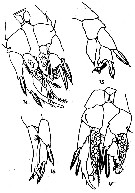
 Issued from : R. Gurney in Trans. zool. Soc. Lond., 1927, 22. [p.457, Fig. 109]. As Suezia canalis. Male (from Kabret and Ismaïlia): A, habitus (lateral); B, right A1; C, A2; D, Mx2; E, Mxp; F, P1; G, P2 (from behind); H, P5; I, P5 (left side). Nota: - Cephalothorax more than twice as long as the abdomen. - Pedigerous somite 1 and head separate, pedigerpous somites 3 and 4 distinct. - Pedigerous somite 5 with lateral wings, a small backwardly-directed tooth. - Rostrum downturned, ending in a slender point., undivided. - Abdomen of 4 somites, the 5th somite scarcely distinct.. - A1 21 or 22-segmented, segments rather indistinct; right A1 not prehensile. - A2 with exopod a litthle longer than inner and consisting of 7 segments. - Md palp large, biramate, exopod of 4 and endopod of 2 segments. - Mx2 very small, not longer than the 1st segment of Mxp, with 5 well-developed inner lobes (= endites) bearing setae, and a small terminal part of 2 segments. - Mxp with 2 long, slender, basal segments and a distal part consisting of 5 elongated segments.. - Legs with both branches 3-segmented; outer spines without hyaline membranes. - P5 biramous on both sides; exopod 2-segmented, much modified, and endopod 1-segmented.
| | | | | NZ: | 2 | | |
|
Distribution map of Ridgewayia typica by geographical zones
|
| | | | | | | Loc: | | | Suez Canal , Gulf of Aqaba (Di Zahav pool), Indian Ocan (Sri Lanka) | | | | N: | 3 | | | | Lg.: | | | (77) F: 0,85; (493) M: 0,72-0,74; (518) F: 0,82-0,8; M: 0,78-0,76; (905) F: 0,80; M: 0,73; {F: 0,80-0,85; M: 0,72-0,78} | | | | Rem.: | hyperbenthic (in nightly plankton).
For Figueroa (2011, p.158), Por (1979) likely incorrctly assumed that these males belong to R. typica (specimens of which were collected in the nearby Di Sahav Pool in the Gulf of Elat, Red Sea) and suggests that the observed differences in P5 structure are due to polymorphism within the species. Por neglects to describe the males from Bitter Lakes in detail beyond their P5 structure. | | | Last update : 07/05/2018 | |
|
|
 Any use of this site for a publication will be mentioned with the following reference : Any use of this site for a publication will be mentioned with the following reference :
Razouls C., Desreumaux N., Kouwenberg J. and de Bovée F., 2005-2026. - Biodiversity of Marine Planktonic Copepods (morphology, geographical distribution and biological data). Sorbonne University, CNRS. Available at http://copepodes.obs-banyuls.fr/en [Accessed June 15, 2026] © copyright 2005-2026 Sorbonne University, CNRS
|
|
|
|


;)
;)
;)
;)
;)
;)
;)
;)
;)

