|
|
 |
|
Calanoida ( Order ) |
|
|
|
Clausocalanoidea ( Superfamily ) |
|
|
|
Scolecitrichidae ( Family ) |
|
|
|
Amallothrix ( Genus ) |
|
|
| |
Amallothrix valida (Farran, 1908) (F,M) | |
| | | | | | | Syn.: | Scolecithrix valida Farran, 1908 b (p.55, Descr.F, figs.F); 1929 (p.244, Rem.); Hardy & Gunther, 1935 (1936) (p.164, Rem.); Brodsky, 1950 (1967) (p.261, figs.F,M); Bradford, 1973 (p.143);
Scolecithricella valida : A. Scott, 1909 (p.92, figs.F); Sewell, 1948 (p.526); Tanaka, 1962 (p.70, figs.F,M, Rem.); Grice & Hulsemann, 1965 (p.224); Park, 1970 (p.511, Rem.); Minoda, 1971 (p.29); 1972 (p.326); Park, 1980 (p.55); Hattori, 1991 (tab.1, Appendix); Chihara & Murano, 1997 (p.901, Pl.174,176: F,M); Park & Ferrari, 2009 (p.143, Table 4, Appendix 1, biogeography); Galbraith, 2009 (pers. comm.);
Scolecithricella (Amallothrix) valida : Vervoort, 1957 (p.107, Rem.); Tanaka, 1962 (p.70, figs.F,M); Björnberg, 1973 (p.333);
no Amallothrix valida : Sars, 1925 (p.185, figs.F); ? Jespersen, 1934 (p.90); Sewell, 1929 (p.217, figs.F); Rose, 1933 a (p.155); Sewell, 1948 (part., p.349, 502, 514, 546, 549); Mazza, 1966 (p.71);
Scaphocalanus validus : With, 1915 (p.198, figs.F,M); Wilson, 1932 a (p.78) | | | | Ref.: | | | Jespersen, 1934 (p.90); C.B. Wilson, 1942 a (p.171, fig.F); Brodsky, 1950 (1967) (p.261, figs.F,M, Rem.); Grice & Hulsemann, 1967 (p.16); Morioka, 1972 a (p.314); Deevey & Brooks, 1977 (tab.2); Séret, 1979 (p.122, fig.M); Bradford & al., 1983 (p.78, 86, figs.F,M, Rem.); Bradford-Grieve & al., 1999 (p.881, 931, figs.F,M); Vyshkvartzeva, 1999 (2000) (p.235, Rem.); Vives & Shmeleva, 2007 (p.733, figs.F,M, Rem.) | 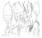 issued from : O. Tanaka in Publ. Seto Mar. Biol. Lab., 1962, X (1). [p.71, Fig.143]. As Scolecithricella valida. Female (from Sagami Bay): a, habitus (dorsal); b, forehead (left lateral side); c, last thoracic segment and urosome (left lateral side); d, rostrum (dorsal); e, P1; f, P2; g, proximal pair of terminal spine of exopod of P2; h, P5; i, P5 (other specimen). Male: j, habitus (dorsal); k, Mx2; l, P5; m, distal segment of exopod of right P5. Nota Female: - Cephalothorax about 3.11-3.16 times the abdomen length. - Posterolateral corners of the last thoracic segment rounded, but slightly produced on the posterior corner. - Rostrum bifurcate, with robust basal portion with terminal filaments. - Abdomen 4-segmented; segments and caudal rami in the proportional lengths 36 : 24 : 23 : 7: 10 = 100. - Genital segment not swollen ventrally. - Caudal rami 1.2 times as long as wide. - A1 23-segmented on left side ; 22-segmented on right side; extend to the middle of the 2nd abdominal segment. - A2 exopod 1.1 as long as endopod. - Mx1 with 9 setae on the outer lobe; 9 setae on exopod; 8 setae on endopod; 5 setae on the 2nd basal; 2 setae on the 2nd inner lobe; 4 setae on the 3rd inner lobe. - Mx2 with 5 bud-like and 3 worm-like sensory filaments on the distal segments. - Mxp with the 1st basal segment about as long as the 2nd; 1st basal with 1 amalliform filament on anterior margin about the middle. - P1 with a slender outer edge spine on the 1st and 2nd exopodal segments; the spine on the 1st segment is as long as the outer margin of the 2nd segment. - P2 as described by Farran; number of teeth on terminal spine of exopod about 41; and the spine forms fenestella (fig.g). The outer edge spine on the 1st exopodal segment reaches the middle of the outer margin of the 2nd segment, and the spine is curved (fig.f) - P3 with terminal spine of exopod similar in structure to that of P2. - P5 as described and figured by Farran. The inner marginal spine arises from about the middle of the distal segment. The spine is rather coarsely serrated on anterior margin and the teeth arranged in a slightly twisted line. Outer marginal spine small, located just to the opposite to the inner marginal spine. A specimen (4.07 mm) had the P5 furnished with usual spines, 1 small spine at the base of the apical spine (fig.i). Nota Male: - Cephalothorax about 2.22 times the abdomen length. - 4th and 5th thoracic segments with trace of articulation in lateral view. - Rostrum as in the female. - Abdominal segments and caudal rami in the proportional lengths 12 : 38 : 20 : 23 : 2 : 5 = 100. - 2nd to 4th abdominal segments fringed with fine teeth on distal margin. - Caudal rami divergent, as long as wide. - A1 20-segmented on right side, extends about to the end of cephalothorax. - A2 exopod 1.3 times as long as endopod; exopodal 1st segment furnished with short hairs on anterior proximal corner. - Md with a robust 2nd basal segment. - Mx1 as in female. - Mx2 with 3 long vermiform and 5 amalliform sensory filaments on distal segment; the 5th lobe with a long and strong spine (fig.k). - Mxp with basal segments more robust than those of the female; - P1 with outer edge spine on the 1st exopodal segment slender, reaching the base of the outer edge spine of the 2nd segment. - P2 with the outer edge spine on the 1st exopodal segment reaches the middle of the outer margin of the 2nd segment, and slightly curved; terminal spine of exopod with 48 teeth, the spine makes fenestella. - P3 with the terminal spine of exopod provided with fenestella. - P5 resemble to that of Scolecithricella gracilis Sars, 1905 (= Amallothrix gracilis Sars, 1925) but much shorter, reaching back to the middle of the 3rd abdominal segment. Endopod of the right leg short and furnished with a small spine at apex; exopod and endopod of left leg are subequal in lengths; terminal segment of exopod furnished with a small spine at apex and comb-like hairs on the inner margin. Distal segment of exopod of right leg lancet-form (fig.m).
|
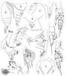 issued from : T. Park in Antarct. Res. Ser. Washington, 1980, 31 (2). [p.56, Fig.15]. As Scolecithricella valida. Female: a, habitus (lateral left side); b, last thoracic segment and urosome (lateral right side); c, forehead (lateral); d, genital segment (lateral left side); e, rostrum (anterior); f, A2; g, Md; h, Mx1; i, distal part of Mx2; j, Mxp; k, P1 (anterior); l, P2 (posterior); m, P3 (posterior); n, P5 (posterior).
|
 issued from : T. Park in Antarct. Res. Ser. Washington, 1980, 31 (2). [p.57, Fig.16]. As Scolecithricella valida. Male: a, forehead (lateral); b, last thoracic segments with P5 and urosome (lateral left side); c, P1 (anterior); d, P2 (posterior); e, P3 (posterior); f, P5 (posterior); g, P5 (right side).
|
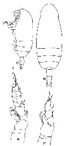
 Issued from : K.A. Brodskii in Calanoida of the Far Eastern Seas and Polar Basin of the USSR. Opred. Fauna SSSR, 1950, 35 (Israel Program for Scientific Translations, Jerusalem, 1967) [p.261, Fig.169]. Female (from NW Pacif.): habitus (dorsal and lateral right side); R, rostrum; Mp1, Mx2; S1, P1; S2, P2; S5, P5.
|
 Issued from : K.A. Brodskii in Calanoida of the Far Eastern Seas and Polar Basin of the USSR. Opred. Fauna SSSR, 1950, 35 (Israel Program for Scientific Translations, Jerusalem, 1967) [p.262, Fig.170]. Male (NW Pacif.): habitus (dorsal and lateral right side); Mp1, Mx2; S5, P5; S5Ri, right leg of P5.
|
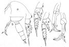
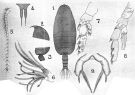 issued from : A. Scott in Siboga-Expedition, 1909, XIX a. [Plate XXXII, Figs.1-9]. As Scolecithricella valida. Female (from Halmahera Sea and Banda Sea): 1, habitus (dorsal); 2, forehead (lateral); 3, last thoracic and genital segments (left side); 4, rostrum; 5, A1; 6, Mx2 (distal portion); 7, P2; 8, P4; 9, P5.
|
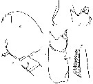
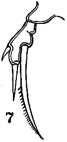 issued from : C.B. Wilson in Scientific Results of Cruise VII of the Carnegie during 1928-1929. Biology-I. Carnegie Inst. Wash. Publ. 536, 1942. [p.219, Fig.7]. With doubt. Female (from SE Pacific]: 7, P5.
|
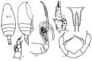
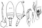
 issued from : G.P. Farran in Fish. Ire. Sci. Invest., 1906, II [1908]. [Pl. V, Figs.14-17]. As Scolecithrix valida. Female (from 53°7'N, 15°6'W): 14-15, habitus (dorsal and lateral, respectively; not at the same scale); 16, P2; 17, P1.
|
 issued from : G.P. Farran in Fish. Ire. Sci. Invest., 1906, II [1908]. [Pl. VI, Fig.7] As Scolecithrix valida. Female (from 53°7'N, 15°6'W): 7, P5.
|
 issued from : C. With in The Danish Ingolf-Expedition, Copepoda I, 1915, III, 4. [p.198, Text-fig. 62]. As Scaphocalanus validus. Female (from 61°30'N, 17°08'W): a, forehead (lateral); b, rostrum (from the right); c, last thoracic segment with P5 and genital segment (left lateral); d, basis of left P2 (anterior view); e, left P3 (coxa and basis); f, terminal seta of left P2. Nota: A1 do not reach the end of the caudal rami; segments 24-25 well separated.
|
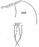 Issued from : C. Séret in Thesis 3ème Cycle, UPMC, Paris 6. [Pl. XXI]. Male (from off Kerguelen Is.): 209, forehead (lateral); 210, rostrum (frontal view).
|
 issued from : O. Tanaka in Publ. Seto Mar. Biol. Lab., 1962, X (1). [p.70]. As Scolecithricella valida. Female: Proportional lengths of segments of A1 on left side. Nota: A1 23-segmented on the left side; 22-segmented on the right side. Extend to the middle of the 2nd abdominal segment.
|
 issued from : O. Tanaka in Publ. Seto Mar. Biol. Lab., 1962, X (1). [p.72]. As Scolecithricella valida. Male: Proportional lengths of segments of A1 on the right side: A1 20-segmented on the right side; extends about the end of cephalothorax. Distal segment missing. Segments 8-12 completely fused, 20 and 21 fused on the right side, the segments 14 and 15 form obtuse angle.
| | | | | Compl. Ref.: | | | ? Wilson, 1942 a (p.171, figs.F); ? Unterüberbacher, 1964 (p.25); Mazza, 1966 (p.71); Grice & Hulsemann, 1967 (p.16); Björnberg, 1973 (p.384); Harding, 1974 (p.141, Table 2, gut contents); Deevey & Brooks, 1977 (p.256, Table 2, station "S"); Vives, 1982 (p.292); Kovalev & Shmeleva, 1982 (p.84); Pakhomov & McQuaid, 1996 (p.271, abundance, distribution, seabirds); Voronina & Kolosova, 1999 (p.71); Lapernat, 2000 (tabl.3, 4); Holmes, 2001 (p.58); Yamaguchi & al., 2002 (p.1007, tab.1); Kuriyama & Nishida, 2006 (p.299: Tab.II; p.309: Tab.III, fig.7, 10, vertical distribution, Rem.: p.313); Ikeda & al., 2006 (p.1791,Table 2); Gaard & al., 2008 (p.59, Table 1, N Mid-Atlantic Ridge); Homma & Yamaguchi, 2010 (p.965, Table 2); Homma & al., 2011 (p.29, Table 2, 3, abundance, feeding pattern: detritivores). | | | | NZ: | 13 + 1 doubtful | | |
|
Distribution map of Amallothrix valida by geographical zones
|
| | | | | | | | | | | | 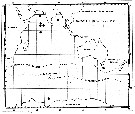 Issued from : C. Séret in Thesis 3ème Cycle, UPMC, Paris 6. 1979, Annexe. [p.41]. Issued from : C. Séret in Thesis 3ème Cycle, UPMC, Paris 6. 1979, Annexe. [p.41].
Geographical occurrences of Amallothrix valida in the Indian Ocean and Antarctic zone. [after publications from: Brady, 1883, 1918; Thompson, 1900; Wolfenden, 1908, 1911; With , 1915; Rosendorn, 1917; Farran, 1929; Sewell, 1929, 1947; Brady & Gunther, 1935; Steuer, 1929, 1392, 1933; Ommaney, 1936; Vervoort, 1957; Tanaka, 1960; Brodsky, 1964; Seno, 1966; Andrews, 1966; Grice & Hulsemann, 1967; Seno, 1966; Frost & Fleminger, 1968; Voronina, 1970; Zverva, 1972].
Nota: C. S"ret notes yje occirrence of this species at station 56°S, 70°E. |
| | | | Loc: | | | Antarct. (Weddell Sea, SW Atlant., Indian, S Kerguelen Is., SW & SE Pacif.), South Georgia, sub-Antarct. (SW Atlant., Indian, SW & SE Pacif. ), ? Namibia, off Mauritania, Canary Is., Sargasso Sea, off Bermuda (Station "S"), off E Cape Cod, Woods Hole, S Bay of Baffin, S Iceland, Faroe Is., off W Ireland, W Mediterranean Sea (Algiers), Indian, Indonesia-Malaysia, Japan, off Sanriku, off SE Hokkaido, Station Knot, Okhotsk Sea, Bering Sea, S Aleutian Basin, S Aleutian Is., off British Columbia, Chile | | | | N: | 39 | | | | Lg.: | | | (5) F: 3,24; (7) F: 3,95; (22) F: 4-3,9; M: 4,5; (24) F: 3,9-3,8; (25) F: 3,87-2,8; (35) F: 4,5; (117) F: 4,36; M: 4,64; (208) F: 4,07; M: 5,35; (246) F: 2,1-4,6; M: 4,64; (866) F: 2,1-4,4; M: 4-4,64; (1000) F: 4,0 ± 0,4; M: 4,0 ± 0,5; {F: 2,10-4,60; M: 4,00-5,35}
(246) Lg prosome: 3,17 mm. | | | | Rem.: | meso-bathypelagic. Sargasso Sea: 1000-2000 m (Deevey & Brooks, 1977, station "S").
Distributional range (m) from Grice & Hulsemann (1965): 2000-3000 (in Kuriyama & Nishida, 2006).
For Vervoort (1957, p.107) this species is differentiated from Scolecithricella (Amallothrix) polaris (= Pseudoamallothrix emarginata) by the structure of the P1-P2 and the spinulation of the internal spine of the terminal segment of P5, which is very coarse in valida; the spinules, as has already been observed by Farran, are not placed in a straight line but are arranged along a slightly twisted line. Both Vervoort's specimens from 'Banzare' collection (66°-77°S, 73°-147°E) lack a spinule at the exrernal margin ot the the free segment of P5 as has been figured by Farran.
For Tanaka (1962, p.73) the species shows a considerable variation in size: Farran's specimen from the North Atlantic, 3.8-3.9 mm; those from the Antarctic, 4.06 mm; A. Scott's from the Malay Archipelago, 3.24 mm; With's from the North Atlantic, 3.95 mm; Sewell's from the Indian Ocean, 2.66 mm. The specimen from Japan (Sagami Bay), though much larger in size4.36 mm for female and 4.64 mm for the male, agrees well with Farran. Sometimes exist any differences in the proportional lengths of the abdominal segments and caudal rami: For Sars' sspecimen : 41 : 21 : 17 : 4 : 7. Sars' specimen appears somewhat differs from those of Farran or Sewell in minute points of structure compared with Scolecithricella paravalida (= Amallothrix paravalida).
Locality records uncertain because of possible confusion between this species and both Amallothrix paravalida and Scolecithrix valens by some authors.
According to Park (1980, p.58) Wilson's (1942) record is accompanied by two figures of the female P5, which are not only different from each other but also different from the P5 as figured by Farran (1908). | | | Last update : 19/01/2021 | |
|
|
 Any use of this site for a publication will be mentioned with the following reference : Any use of this site for a publication will be mentioned with the following reference :
Razouls C., Desreumaux N., Kouwenberg J. and de Bovée F., 2005-2026. - Biodiversity of Marine Planktonic Copepods (morphology, geographical distribution and biological data). Sorbonne University, CNRS. Available at http://copepodes.obs-banyuls.fr/en [Accessed March 27, 2026] © copyright 2005-2026 Sorbonne University, CNRS
|
|
|
|


;)
;)
;)
;)
;)
;)
;)
;)
;)
;)
;)

;)
