|
|
 |
|
Calanoida ( Order ) |
|
|
|
Clausocalanoidea ( Superfamily ) |
|
|
|
Scolecitrichidae ( Family ) |
|
|
|
Scaphocalanus ( Genus ) |
|
|
| |
Scaphocalanus farrani Park, 1982 (F,M) | |
| | | | | | | Syn.: | ? Amallophora impar Wolfenden, 1911 (p.263, figs.F);
Scaphocalanus impar : Tanaka, 1961 a (p.172, figs.F);
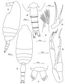
Scaphocalanus brevicornis : Farran, 1929 (p.209, 248, figs.F, Rem.); Hardy & Gunther, 1935 (p.165); Vervoort, 1951 (p.113, Rem., figs.F); 1957 (p.107, figs.F,M, Rem.); Tanaka, 1960 (p.42, figs.F, Rem.); ? Tanaka, 1961 a (p.169, figs.F,M); Bradford, 1970 a (p.356, fig.M); 1971 b (p.23, figs.81, 82: F,M); Bradford & al., 1983 (p.95); ? Voronina & Kolosova, 1999 (p.71) | | | | Ref.: | | | Park, 1982 (p.95, Descr.F,M, figs.F,M, Rem.); Razouls, 1994 (p.118, figs.F,M, Rem.); Mazzocchi & al., 1995 (p.196, figs.F,M, Rem.); Bradford-Grieve & al., 1999 (p.881, 932, figs.F,M, Rem.); Markhaseva & Renz, 2011 (p.67, Fig.M) | 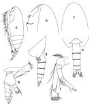 issued from : T. Park in Biology of the Antarctic Seas XI, Antarct. Res. Ser, 1982, 34. [p.96, Fig.11]. Female: a, habitus (lateral left side); b, forehead (lateral); c, idem (dorsal); e, last thoracic segment and urosome (dorsal); f, A2; g, Md. Nota: Rostrum of 2 slender filaments. Urosome about 28/100 length of prosome.
|
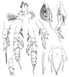 issued from : T. Park in Biology of the Antarctic Seas XI, Antarct. Res. Ser, 1982, 34. [p.98, Fig.12]. Female: a, Mx1; b, distal part of Mx2; c, Mxp; d, P1 (anterior); e, exopod of P1 (posterior); f, P2 (posterior); g, P3 (posterior); h, P4 (two distal exopodal segments missing), posterior; i, P5 (posterior); j, distal part of inner spine of P5.
|
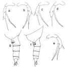 issued from : T. Park in Biology of the Antarctic Seas XI, Antarct. Res. Ser, 1982, 34. [p.99, Fig.13]. Female: a-e, P5 showing variation; f, last thoracic segment of thorax and urosme of a small specimen (lateral left side); idem from a large specimen.
|
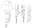 issued from T. Park in Biology of the Antarctic Seas XI, Antarct. Res. Ser, 1982, 34. [p.100, Fig.14]. Male: a, forehead (lateral); b, last thoracic segment and urosome including P5 (lateral right side); c, P1 (anterior); d, P2; e, P5 (anterior view). Nota: Urosome about half the length of prosome.
|
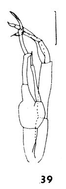 issued from : J.M. Bradford in N.Z. Jl Mar. Freshw. Res., 1970, 4 (4). [p.356, Fig. 39]. As Scaphocalanus brevicornis. Male (off Kaikoura, New Zealand): 39, P5. Scale bar represents 0.1 mm. Nota: The male agree with Vervoort's (1957) description.
|
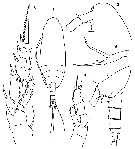
 Issued from : M.G. Mazzocchi, G. Zagami, A. Ianora, L. Guglielmo & J. Hure in Atlas of Marine Zooplankton Straits of Magellan. Copepods. L. Guglielmo & A. Ianora (Eds.), 1995. [p.197, Fig.3.35.1]. Female: A, habitus (lateral left side); B, urosome (dorsal); C, 1st inner lobe of Mx1; D, free segment of P5. Nota: Rostrum with two slender filaments. Proportional lengths of urosomites and furca 33:20:18:12:17 = 100. 1st inner lobe of Mx1 with 3 posterior spines. Male: E, habitus (dorsal); F, furca; G, P5. Nota: Proportional lengths of urosomites and furca 6:38:20:23:3:10 = 100
|
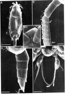 Issued from : M.G. Mazzocchi, G. Zagami, A. Ianora, L. Guglielmo & J. Hure in Atlas of Marine Zooplankton Straits of Magellan. Copepods. L. Guglielmo & A. Ianora (Eds.), 1995. [p.198, Fig.3.35.2]. Female (SEM preparation): A, habitus (dorsal); B, forehead with rostrum (ventral); C, A1 showing detail of transparent strip (arrows); D, urosome (lateral left side); E, P5. Bars: A 0.500 mm; B, E 0.050 mm; C, D 0.100 mm.
|
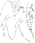
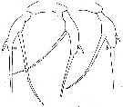
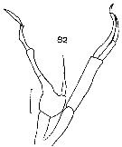
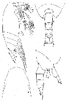 issued from : W. Vervoort in B.A.N.Z. Antarctic Reseach Expedition, Report-Ser. B, Vol. III, 1957 [Fig.97]. As Scaphocalanus brevicornis. Female (from 64°55'S, 54°35'E): a, habitus (lateral); b, d, posterior part cephalothorax and urosome (dorsal and lateral, respectively); c, forehead (dorsal). Nota: Head and 1st thoracic somite, 4th and 5th, completely fused. basal portion of rostrum thickened, apex bifurcated, each carries a fine filiform appendage Proportional lengths of cephalothorax and abdomen as 42:13. Postero-lateral thoracic border produced into triangular flaps, that cover the beginning of the genital somite, apex of the flap rounded. Abdomen 4-segmented; proprtional length of somites and caudal rami 27:23:23:13:14 = 100. Genital somite perfectly symmetrical in dorsal aspect; in lateral view no genital tubercle appears; the genital area is not noticeably elevated; a genital flap has not been observed. Distal part of the 1st to 3rd abdominal somites fringed by a row of plate shaped teeth. Anal somite with almost straight anal flap. Caudal rami about twice as long as broad, rami parallel, there are 4 marginal setae on each ramus, plus 1 appendicular seta (seen in lateral aspect). A1 about as long as the cephalothorax, with flattened segments.
|
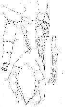
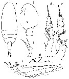
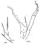
 issued from : W. Vervoort in B.A.N.Z. Antarctic Reseach Expedition, Report-Ser. B, Vol. III, 1957 [Fig.98]. As Scaphocalanus brevicornis. Female: a, head (lateral); b, left P2 (posterior); c, P5; d, P5 (another specimen). Nota: The variability of P5 make the identification of the species extremely difficult. In such cases the only solution is dissection of the specimen. The P2 do not appear to show much variability.
|
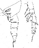
 issued from : W. Vervoort in B.A.N.Z. Antarctic Reseach Expedition, Report-Ser. B, Vol. III, 1957 [Fig.99]. As Scaphocalanus brevicornis. Female (from 66°30'S, 61°08'E): abnormal P5. Male (from 65°27'S, 54°35'E): a, posterior part cephalothorax and urosome (lateral); b, left P2 (posterior); c, P5.
|
 issued from : R.N. Wolfenden in Die Marinen Copepoden der Deutschen Südpolar-Expedition 1901-1903, 1911. [264, Fig.38]. As Amallothrix impar. With doubt. Female (Antarctic Continent): a, habitus (lateral); b, urosome (dorsal); c, P2; d, P5.
|
 issued from : W. Vervoort in Verh. K. ned. Akad. Wet., Afd. Natuurk., 1951, (Sect. 2) 47 (2). [p.114, Fig.60]. As Scaphocalanus brevicornis. Female (from 66°58'S, 16°03'.5W): a, posterior part cephalothorax and urosome (lateral); b, right P2 (posterior).
|
 issued from : W. Vervoort in Verh. K. ned. Akad. Wet., Afd. Natuurk., 1951, (Sect. 2) 47 (2). [p.115, Fig.61]. As Scaphocalanus brevicornis. Femelle: a, P5; b, left P5 (another specimen).
|
 issued from : O. Tanaka in Publ. Seto Mar. Biol. Lab., 1961, IX (1). [p.170, Fig.117]. As Scaphocalanus brevicornis. Synonymy doubtful. Female (from Japan: Izu Region): a, habitus (dorsal); b, forehead (lateral); c, last thoracic segment and genital segment (lateral, left side); d, P2 e, P3; f, P5. Nota: Cephalothorax and urosome in proportional lengths 76:24. Head and 1st thoracic segment fused, Th4 and Th5 fused. Last thoracic segment with lateral corner obtusely rounded in lateral view. Rostrum with an inflated base to which fairly long filaments attached. Urosome 4-segmented, urosomal segments and caudal rami in proportional lengths (measured dorsally) 34:21:19:10 16 = 100 Caudal rami 2 times as long as broad, the first 3 segments are fringed with fine teeth on the distal margin. A1 22-segmented (segments 8-9-10 fused, 24-25 fused; the 1st segment furnished with a row of minute spine on the posterior distal margin, segments 8, 12, 14 and 18 each with 2 setae, segment 10 with a distal seta); extends to the posterior margin of the last thoracic segment; segments with proportional lengths 71:100:33:29:29:25:29:67:21:21:33:37:42:50:50:50:46:42:46:46:58:18 = 1000. Endopod of A2 1.3 times as long as the exopod. Terminal segment on Mx2 with 3 long vermiform and 5 amalliform sensory filaments, of which 2 are larger, the setae on the 5th lobe are slender. Mxp with a row of fine spinules on the anterior proximal margin. Terminal spine of exopod of P2 with 23-27 teeth. Terminal spine of exopod of P3 with 18-21 teeth. Terminal spine of exopod of P4 with 33 teeth P4. P5 2-segmented, distal segment with 3 spines, the inner marginal spine about 1.5 times as long as the apical spine, and rather coarsely denticulated on the distal outer margin (number of teeth about 17); terminal spine about as long as the segment itself; the outer marginal spine arises opposite to the inner marginal spine, and divides the outer margin of the segment in the proportions 3:1. Male: g, 1st segment of exopod of P2; h, P5. Nota: Cephalothorax and urosome in proportional lengths 64:36. Urosome 5-segmented; urosomal segments and caudal rami in proportional lengths 6:37:20:24:5:8 = 100. A1 20-segmented, extends to the end of the 3rd thoracic segment, segments 20 and 21 fused on the right side. P5 reaches the distal end of the 2nd urosomal segment; exopod of the left leg 3-segmented, endopod 2-segmented; endopod of the right leg styletform, reaches about distal end of the 2nd basal segment of the left leg.
|
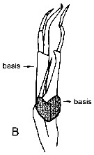 issued from : E.L. Markhaseva & J. Renz in Zootaxa, 2011, 2889. [p.67, B]. Changed and schematized after Park (1982). Male: B, P5 (Nota: right basis in grey).
|
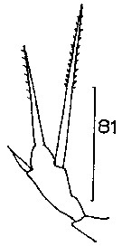 Issued from : J.M. Bradford in N.Z. Oceanogr. Inst., 1971, 206, Part 8, No 59. [p.22, Fig.81]. As Scaphocalanus brevicornis. Female (from 73°56'S, 176°30'W): 81, P5. Scale bar: 100 µm.
|
 Issued from : J.M. Bradford in N.Z. Oceanogr. Inst., 1971, 206, Part 8, No 59. [p.22, Fig.82]. As Scaphocalanus brevicornis. Male (from 73°56'S, 176°30'W): 82, P5. Scale bar: 100 µm.
|
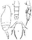
 Issued from : O. Tanaka in Spec. Publs. Seto mar. biol. Lab., 10, 1960 [Pl. XVIII ]. As Scaphocalanus brevicornis. Female (from 67°03.5', 40°49'E): 1, habitus (dorsal); 2, forehead (lateral); 3, last thoracic segment and urosome (lateral); 4, P1; 5, P2; 6, P56. Same scale in figs. 4, 5 and 6. Nota: Cephalothorax and abdomen in the proportional lengths 77 to 23. Basal part of the rostrum thickened, apex bifurcate with slender filaments. Posterior thoracic margin broadly rounded and slightly emarginate. Abdomen 4-segmented; segments and caudal rami in the proportional lengths 34 : 20 : 19 : 11 : 16 = 100. Genital area not produced below; genital, 2nd and 3rd segments fringed with fine teeth on the distal border. Caudal rami 2 times as long as broad; an appendicular seta arises from the posterior margin of the ramus. A1 23-segmented, extends about to the posterior end of the thoracic segment. P1 with a small outer edge spine on the 2nd exopodal segment. P2 with a fairly long outer edge spine on the 1st exopodal segment, which exceeds the middle of the outer margin of the next segment; terminal spine with 26 teeth along the outer margin. P5 2-segmented; distal segment with 3 spines, of which the inner proximal is long and serrated along the inner edge; middle spine about 1/3 the length of the distal spine.
|
 Issued from : J.M. Bradford-Grieve, E.L. Markhaseva, C.E.F. Rocha & B. Abiahy inSouth Atlantic Zooplankton, Edit. D. Boltovskoy, 1999, Vol. 2. [p.1026, Fig. 7.200]. Female: P5. Male: P5 (l = left leg; r = right leg).
|
 Issued from : C. Razouls in Ann. Inst. océanogr., Paris, 1994, 70 (1). [p.118]. Caractéristiques morphologiques de Scaphocalanus farrani femelle et mâle adultes. Terminologie et abbréviations: voir à Calanus propinquus.
| | | | | Compl. Ref.: | | | Hopkins, 1985 (p.197, Table 1, gut contents); Hopkins & Torres, 1988 (tab.1); Ward & al., 1995 (p.195, Table 2); Errhif & al., 1997 (p.422); Razouls & al., 2000 (p.343, tab. 5, Appendix); Schnack-Schiel & al., 2008 (p.1046: Tab.2); Park & Ferrari, 2009 (p.143, Table 3, 7: common deep water species, figs.1, 2, Appendix 1, biogeography); Michels & al., 2012 (p.369, Table 1, occurrence frequency); Sano & al., 2013 (p.11, Table 9, food habits) | | | | NZ: | 4 | | |
|
Distribution map of Scaphocalanus farrani by geographical zones
|
| | | | | | | | |  Issued from : E.T. Park & F.D. Ferrari in A selection from Smithsonian at the Poles Contributions to International Polar year. I. Krupnik, M.A. Lang and S.E. Miller, eds., Publs. by Smithsonian Institution Scholarly Press, Washington DC., 2009. [p.167, Fig.2]. Issued from : E.T. Park & F.D. Ferrari in A selection from Smithsonian at the Poles Contributions to International Polar year. I. Krupnik, M.A. Lang and S.E. Miller, eds., Publs. by Smithsonian Institution Scholarly Press, Washington DC., 2009. [p.167, Fig.2].
Distribution of selected pelagic calanoids Scaphocalanus farrani of the Southern Ocean and the closest relative in the subarctic region of the Arctic Ocean. |
| | | | Loc: | | | Antarct. (Weddell Sea, SW & SE Atlant., Indian, SW & SE Pacif., Croker Passage), sub-Antarct. (SW Atlant., South Georgia, Indian, SW & SE Pacif., S Indian (subtropical convergence), S Tasmania, Straits of Magellan | | | | N: | 21 | | | | Lg.: | | | (25) F: 2,84-2,3; M: 2,88-2,79; (31) F: 2,7-2,52; (35) F: 2,62-2,4; (66) F: 2,6; (76) F: 2,92-2,4; M: 3,28-2,68; (102) F: 2,5; M: 2,75; (313) M: 2,38-2,60; {F: 2,30-2,92; M: 2,38-3,28} | | | | Rem.: | meso & bathypelagic.
Sampling depth (Antarct., sub-Antarct.) :200-1000 m.
For Tanaka (1961a, p.171) the species resembles closely S. major, differing from it in having a long outer edge spine on the 1st segment of the exopod of P2. Vervoort (1957) illustrated an abnormal P5 female with rudimentary endopod.
Park (1982, p.100) considers that the antiboreal forms of Scaphocalanus brevicornis correspond to this new species (See remaks S. brevicornis) | | | Last update : 22/03/2017 | |
|
|
 Any use of this site for a publication will be mentioned with the following reference : Any use of this site for a publication will be mentioned with the following reference :
Razouls C., Desreumaux N., Kouwenberg J. and de Bovée F., 2005-2025. - Biodiversity of Marine Planktonic Copepods (morphology, geographical distribution and biological data). Sorbonne University, CNRS. Available at http://copepodes.obs-banyuls.fr/en [Accessed June 04, 2025] © copyright 2005-2025 Sorbonne University, CNRS
|
|
|
|


;)
;)
;)
;)
;)
;)
;)
;)
;)
;)
;)
;)
;)
;)
;)
;)
;)
;)
;)
;)

