|
|
 |
|
Calanoida ( Order ) |
|
|
|
Clausocalanoidea ( Superfamily ) |
|
|
|
Scolecitrichidae ( Family ) |
|
|
|
Scaphocalanus ( Genus ) |
|
|
| |
Scaphocalanus magnus (T. Scott, 1894) (F,M) | |
| | | | | | | Syn.: | Scolecithrix ( Amallophora ) magna T. Scott, 1894 b (p.55, Descr.F, figs.F); Norman, 1903 b (p.139: Rem.);
Amallophora magna : Sars, 1902 (1903) (p.51, figs.F,M); Pearson, 1906 (p.17, Rem.); Damas & Koefoed, 1907 (p400, tab.II, III); Wolfenden, 1911 (p.262, Rem.); Gundersen, 1953 (p.1, 26, seasonal abundance);
? Amallophora gracilis : Wolfenden, 1911 (p.265, figs.F);
Scaphocalanus acrocephalus Sars, 1900 (p.36, figs.F,M); Mrazek, 1902 (p.522); Sirenko & al., 1996 (p.349); Farran, 1908 b (p.51); van Breemen, 1908 a (p.76); Kosobokova & Hirche, 2000 (p.2029, tab.2); Kosobokova & al., 2007 (p.919, Tab.2, fig.2); Park & Ferrari, 2009 (p.143, fig.2, biogeography); Bucklin & al., 2010 (p.40, Table 1, Biol mol.); Kosobokova & al., 2011 (p.29, Table 2, figs.4, 8, Rem.: Arctic Basins);
? Scolecithrix cristata Giesbrecht, 1895 c (p.252, figs.F); Giesbrecht & Schmeil, 1898 (p.48, Rem.F, fig.F); Thompson, 1903 a (p.21, figs.M);
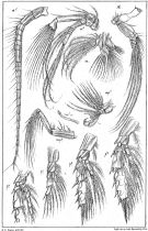
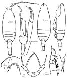
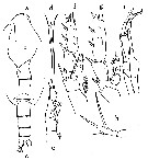
Scolecithrix magna : Esterly, 1906 a (p.66, figs.F); Farran, 1908 b (p.51) | | | | Ref.: | | | A. Scott, 1909 (p.97); With, 1915 (p.189, figs.F,M, Rem.: p.192); Lysholm & Nordgaard, 1921 (p.20); Sars, 1925 (p.169); Farran, 1926 (p.257); Sewell, 1929 (p.207); Wilson, 1932 a (p.77, figs.F,M); Rose, 1933 a (p.147, figs.F,M); Jespersen, 1934 (p.87, fig.M); 1940 (p.37, fig.4); Wilson, 1942 a (p.207); Lysholm & al., 1945 (p.27); Sewell, 1947 (p.144, figs.F, Rem.: 2 forms); Davis, 1949 (p.42, figs.F, Rem.F,M); C.B. Wilson, 1950 (p.328, ? part.); Brodsky, 1950 (1967) (p.248, figs.F,M, Rem.); Vervoort, 1957 (p.111, Rem.); Tanaka, 1961 a (p.158, figs.F,M, Rem.); Paiva, 1963 (p.51); Vervoort, 1965 (p.62, Rem.); Owre & Foyo, 1967 (p.66, figs.F,M); Ramirez, 1969 (p.67, fig.M, Rem.); Tanaka, 1969 (p.276); Vaupel-Klein, 1970 (p.4, 19); Minoda, 1971 (p.28); Vidal, 1971 a (p.14, 24, 118, figs.F,M); Silas, 1972 (p.647); Bradford, 1973 (p.143); Séret, 1979 (p.115, figs.M, Rem.M); Björnberg & al., 1981 (p.637, figs.F); Park, 1982 (p.76, 77, 79, figs.F, p.82: Rem.); Gardner & Szabo, 1982 (p.288, figs.F,M); Bradford & al., 1983 (p.93, 100, figs.F,M, Rem.); Roe, 1984 (p.357); Heinrich, 1990 (p.17, 22, figs.F); Bradford-Grieve & al., 1999 (p.881, 932, figs.F); Vives & Shmeleva, 2007 (p.762, figs.F,M, Rem.) | 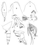 issued from T. Park in Biology of the Antarctic Seas XI, Antarct. Res. Ser, 1982, 34. [p.80, Fig.1]. Female: a, habitus (lateral left side); b, forehead (lateral); c, idem (dorsal); d, posterior part of thorax and urosome (lateral left side); e, A2; f, Md; g, Mx1; h, distal part of Mx2.
|
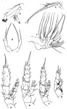
 issued from T. Park in Biology of the Antarctic Seas XI, Antarct. Res. Ser, 1982, 34. [p.81, Fig.2]. Female: a, Mxp; b, P1 (anterior view); c, P2 (posterior); d, P3 (posterior); e, P4 (posterior); f, P5 (posterior)
|
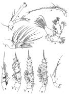
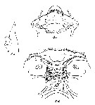
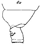
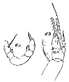
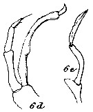
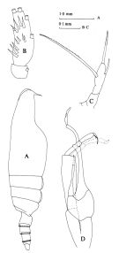 Issued from : J.M. Bradford, L. Haakonssen & J.B. Jillett in Mem. N.Z. oceanogr. Inst., 1983, 90. [p.100, Fig.59]. Female: A, habitus (lateral right side); B, endopod of P2; C, P5. Male: D, P5.
|
 issued from : G.O. Sars in An Account of the Crustacea of Norway. Vol. IV. Copepoda Calanoida. Published by the Bergen Museum, 1903. [Pl. XXXIV]. As Amallophora magna. Female & Male. Nota: C = forehead (lateral left side); mp2 = Mxp.
|
 issued from : G.O. Sars in An Account of the Crustacea of Norway. Vol. IV. Copepoda Calanoida. Published by the Bergen Museum, 1903. [Pl. XXXV]. As Amallophora magna. Female. Nota: M = Md; m = Mx1; mp1 = Mx2; mp2 = Mxp.
|
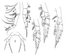
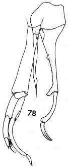 issued from : F.C. Ramirez in Contr. Inst. Biol. mar., Buenos Aires, 1969, 98. [p.62, Lam. XI, figs.78 ]. Male (from off Mar del Plata): 78, P5.
|
 issued from : R.B.S. Sewell in The John Murray Expedition, 1933-34, Scientific Reports, VIII (1), 1947. [p.145, Fig.35]. As Scaphocalanus magnus forma major. Female (from S Arabian Sea): A, A1; B, A2; C, Mx1; D, Mx2; E, P1; F, P2; G, P3; H, P4; I, P5. Nota: The proportional lengths of the various segments of the body (cephalon to caudal rami) as 507:91:65:78:81:71:53:19:35 = 1000. Head and 1st pediger segment fused, 4th and 5th fused. There seems to be a definite reduction in the proportional length of the fused cephalon-thoracic segment 1 as we pass from the smaller to the larger form, and this is to some extent compensated by an increase in the proportional length of thoracic segment 2 (small form); on the other hand, at the posterior end of the body there is a small reduction in the lengths of the anal segment and the caudal rami in large form.
|
 issued from : R.B.S. Sewell in The John Murray Expedition, 1933-34, Scientific Reports, VIII (1), 1947. [p.147, Fig.36]. As Scaphocalanus magnus forma minor. Female (from Arabian Sea): A, A1; B, A2; C, Mx2; D, P1; E, P2; F, P3; G, P4; H, P5. Nota: The proportional lengths of the cephalothorax and abdomen as 76 to 24. The proportional lengths of the various segments of the body (cephalon to caudal rami) as 526:79:66:75:84:61:51:21:37 = 1000. In Mx2 the postrior margin of the basal segment is produced backwards in a sharply rounded prominence. In Swimming legs the groups of minute spinules on the anterior aspect of the rami of P2 to P4 and on the 2 nd basal segment of P3 andP4 are almost completely absent.
|
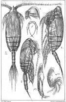
 Issued from : K.A. Brodskii in Calanoida of the Far Eastern Seas and Polar Basin of the USSR. Opred. Fauna SSSR, 1950, 35 (Israel Program for Scientific Translations, Jerusalem, 1967) [p.249, Fig.156]. Female (from NW Pacif.): habitus (dorsal and lateral left side); forehead (lateral); R, rostrum (Arctic specimen): S1, P1 (Arctic specimen); S5, P5 (Arctic specimen). Male (from NW Pacif.): habitus (dorsal); forehead (lateral); S5, P5.
|
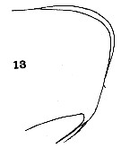 issued from : C.O. Esterly in Univ. Calif. Publs Zool., 1906, 3 (5). [Pl.9, Fig.13]. As Scolecithrix magna. Female (from San Diego, California): 13, forehead (lateral).
|
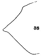 issued from : C.O. Esterly in Univ. Calif. Publs Zool., 1906, 3 (5). [Pl.11, Fig.38]. As Scolecithrix magna. Female: 38, outline of last thoracic segment (lateral).
|
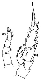 issued from : C.O. Esterly in Univ. Calif. Publs Zool., 1906, 3 (5). [Pl.12, Figs.52, 64]. As Scolecithrix magna. Female: 52, exopod of P1; 64, P2.
|
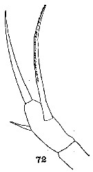 issued from : C.O. Esterly in Univ. Calif. Publs Zool., 1906, 3 (5). [Pl.13, Fig.72]. As Scolecithrix magna. Female: 72, P5.
|
 issued from : W. Giesbrecht in Bull. Mus. comp. Zool. Harv., 25 (12). [Taf. II, Figs.6-8]. As Scolecithrix cristata. Female (from 35°N, 125°W): 6, P2; 7-8, forehead (dorsal and lateral, respectively).
|
 issued from : W. Giesbrecht in Bull. Mus. comp. Zool. Harv., 25 (12). [Taf. III, Figs.1-5]. As Sclecithrix cristata. Female): 1, Mx1; 2, urosome (lateral, right side); 3, exopod of P1; 4, P5.
|
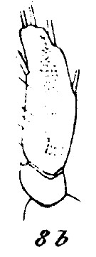
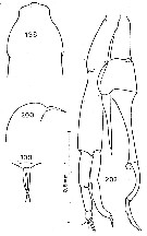
 issued from : C.C. Davis in Univ. Wash. Publs Biol., 1949, 14. [Pl.7, Figs.87-88]. Female (from NE Pacific): 87, P5; 88, last thoracic segment and urosome (lateral). Nota : The fused head and 1st thoracic segment occupy fully half the length of the entire body. Head with a well-defined crest. 4th and 5th thoracic segments fused and produced into blunt points. Genital segment only slightly longer than the 2nd urosomal segment, which in turn is slightly longer than the 3rd ; anal segment quite short. Caudal rami about as long as broad. A1 22-segmented. P5 imperfectly 3-segmented, the last two segments being partially fused ; the terminal segment bears 1 very long spine on the inner border, 1 rather short spine on the outer border directly opposite the inner spine, and a 3rd spine, which is intermediate in length, apically ; the outer spine is naked, while the other tw oare finely plumose. P5 rather small, and when attached to the animal scarcely reach beyond the middle ofthe urosome.
|
 issued from : O. Tanaka in Publ. Seto Mar. Biol. Lab., 1961, IX (1). [p.158, Fig.113]. Female (from Japan: Suruga Bay): a, forehead (lateral); b, last thoracic segment and genital segment (lateral, left side); c, last thoracic segment and urosome (dorsal); d, rostrum (frontal view); e, P1; f, P2; g, P3; h, P5. Nota: Cephalothorax and abdomen in proportional lengths 77 : 23. - Head with a median crest. - Last thoracic segment produced triangularly and narrowly rounded at the apex. - Rostral filaments long and slender. - Urosome 4-segmented; urosomal segments and caudal rami in proportional lengths 36 : 24 : 20 : 6 : 15 = 100. - Genital segment not produced below on the ventral surface; 1st to 3rd urosomal segments are fringed with fine teeth on the distal margin. - Caudal rami 1.7 times as long as wide. - A1 extends to the distal margin of the genital segment; 1st segment with a row of short hairs on the posterior distal margin. - Endopod of A2 about 1.4 times as long as the exopod (72:53). - Mx2 with 8 filaments on the distal segments of which 3 are long and worm-like, and the remainings bud-like. - In P5 the outer marginal spine reaches the distal margin of the segment; terminal spine about as long as the 2nd segment; inner marginal spine 2.5 times as long as the 2nd segment, the spine is furnished with fine spinules on the distal 2/3 of the spine. Male (from Japan: Sagami Bay): i, P5. Nota: Cephalothorax and abdomen in proportional lengths 67 : 33. - Head without median crest. Last thoracic segment rounded in lateal view. - Urosome 5-segmented; urosomal segments and caudal rami in proportional lengths 13 : 33 : 21 : 24 : 2:7 = 100. - Caudal rami about as long as wide. - A1 extends to the distal end of the last thoracic segment, segments 8 to 12 and segments 24-25 fused; segments 20 and 21 fused on the right side; 1st segment with a row of fine spinules on the posterior distal margin. - Exopod of A2 about as long as the endopod.. - Md palp conspicuous, exopod 2 times as long as the endopod. - Mx1 with 7+2 setae on the outer lobe, 7 setae on the exopod. Mx2 feeble. - P1 to P4 as in female. - P5 reaches back to the distal margin of the 2nd urosomal segment; endopod of right leg extends a little beyond the distal margin of the 2nd basal segment of the left leg; exopod of left leg is about 4/5 the length of the endopod of the same leg; endopod of the left leg 2-segmented, but there is a notch on the proximal 1/3 of the inner margin of the endopod.
|
 issued from : C. With in The Danish Ingolf-Expedition, Copepoda I, 1915, III (4). [p.190, Text-Fig.58, a-i]. Female (from Arctic-subarctic): a-i, left lateral corner of the distal thoracic segment in 8 different adult females.
|
 issued from : C. With in The Danish Ingolf-Expedition, Copepoda I, 1915, III, 4. [Pl. VII, Figs. 8a, c, d]. Female (from 61°30'N, 17°08'W): 8a, head left (lateral) with mouth; 8c, labrum (anterior view); 8d, same (oral view). Nota : The elevation in front of the labrum (corresponding to the antennal segment, is slightly raised ; the labrum proper, which is produced in front as a rounded protuberance ; is by an anteriorly convex line divided into an anterior and a posterior smaller part ; the labrum proper is beset with a number of shorter and longer bristles, the somewhat complicated arrangement of which is most easily underwtood by studying in fig.8c. The oral surface of the labrum (fig.8d) shows a rather characteristic structure ; the longitudinal series on each side consists anteriorly of an oblique group of fairly long and strong spines ; behind this two or three groups of comparatively long and slender setae are found, followed by a more medially placed, almost square, area of short spines ; behind the last mentioned group, which is only separated from the corresponding one of the other side by a narrow smooth area, an oblique one covered with delicate hairs is found. The lamina labialis seems to be represented by a very short transverse plate ; in front of it and the serrula 6-dentata a medial short row of fairly long setae and a longer curved lateral one of shorter setae are found. Behind the lamina labialis and between the serrulae is found on each side a short group of strong spines, almost fused with the corresponding one of the other side, as well as a more posterior and lateral group.
|
 issued from : C. With in The Danish Ingolf-Expedition, Copepoda I, 1915, III, 4. [Pl. VII, Fig. 8b]. Female: 8b, endopodite of P2. Nota : The first inner segment of P2 has a small outer tooth, which was, however, wanting in another specimen. Nota : The first inner segment of P2 has a small outer tooth, which was, however, wanting in another specimen.
|
 issued from : C. With in The Danish Ingolf-Expedition, Copepoda I, 1915, III, 4. [Pl. VIII, Fig. 6a]. Female: 6a, last thoracic segment andgenital segment (laft lateral).
|
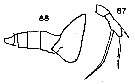
 issued from : C. With in The Danish Ingolf-Expedition, Copepoda I, 1915, III, 4. [Pl. VIII, Figs. 6b, c]. Female: 6b, left P4 in an abnormal specimen; 6c, P5 in an abnormal specimen (posterior view).
|
 issued from : C. With in The Danish Ingolf-Expedition, Copepoda I, 1915, III, 4. [Pl. VIII, Figs. 6d, e]. Male: 6d, left P5; 6e, right P5. Nota : Cephalosome and 1st pedigerous somite separate ; 4th and 5th separate. The cpmparative length of the abdominal somites and the caudal rami 15 : 83 : 47 : 52 : 10 : 20. A1 extend somewhat beyond the end of thorax ; the proximal portion forms an obtuse angle with the more attenuated distal portion. Segments 8-9 completely fused with segments 10-12, segment 13 well separated. With (1915, p.191) notes that the young males stage V are much more common than the young females (34 vs. 5), probably because the development of the last copepodite is more fast than one of females.
|
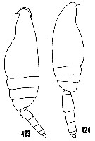 issued from : H.B. Owre & M. Foyo in Fauna Caribaea, 1, Crustacea, 1: Copepoda. Copepods of the Florida Current. [p.67, Figs.423-424]. After Sars (1902). Female: 423, habitus (lateral). Male: 424, habitus (lateral).
|
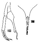 issued from : H.B. Owre & M. Foyo in Fauna Caribaea, 1, Crustacea, 1: Copepoda. Copepods of the Florida Current. [p.24, Figs.103, 108]. Female: 108, rostrum. Male: 103, P5.
|
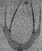 issued from : H.B. Owre & M. Foyo in Fauna Caribaea, 1, Crustacea, 1: Copepoda. Copepods of the Florida Current. [p.25, Fig.115]. Female: 115, P5.
|
 Issued from : C. Séret in Thesis 3ème Cycle, UPMC, Paris 6. [Pl. XXXIII, Figs.198-201]. . Male (from off Kerguelen Is.): 198, forehead (dorsal); 199, rostrum (ventral view); 200, same (lateral); 201, P5.
|
 issued from : O. Tzanaka in Publ. Seto Mar. Biol. Lab., 1961, IX (1). [p.159]. Male: Proportional lengths of the segments of A1.
| | | | | Compl. Ref.: | | | Oliveira, 1945 (p.191); Sewell, 1948 (p.329, 496, 502, 507, 508, 512, 514, 516, 521, 525, 533, 539, 546, 549, 557, 566); Østvedt, 1955 (p.15: Table 3, p.69); Minoda, 1958 (p.253, Table 1, abundance); Fagetti, 1962 (p.24); Grice, 1962 a (p.101); 1963 a (p.495); M.W. Johnson, 1963 (p.89, Table 1, 2); De Decker & Mombeck, 1964 (p.14); Mazza, 1966 (p.70); Grice & Hulsemann, 1965 (p.224); Harding, 1966 (p.17, 66); Furuhashi, 1966 a (p.295, vertical distribution in Oyashio region, Table 6); Grice & Hulsemann, 1967 (p.16); Fleminger, 1967 a (tabl.1); Dunbar & Harding, 1968 (p.319, 320); Morris, 1970 (p.2301); Park, 1970 (p.476); Shih & al., 1971 (p.207); Roe, 1972 (p.277, tabl.1, tabl.2); 1972 b (p.532); Björnberg, 1973 (p.331, 389); Harding, 1974 (p.141, tab.2, gut contents); Vives & al., 1975 (p.43, tab.II, XII); Deevey & Brooks, 1977 (p.256, tab.2, Station "S"); Pipe & Coombs, 1980 (p.223, figs. 1, 2, table 1, vertical distribution); Buchanan & Sekerak, 1982 (p.41, vertical distribution, Rem.: p.48); Vives, 1982 (p.292); Kovalev & Shmeleva, 1982 (p.84); Brenning, 1984 (p.5, Rem.); Guangshan & Honglin, 1984 (p.118, tab.); Stephen, 1984 (p.161, 169, Distribution vs thermocline & geographic); Brenning, 1985 a (p.28, Table 2); Groendahl & Hernroth, 1986 (tab.1); Rudyakov, 1986 (tab.2); Lozano Soldevilla & al., 1988 (p.59); Kosobokova, 1989 (p.27); Shih Marhue, 1991 (tab.3); Mumm, 1993 (tab.1, fig.2); Richter, 1994 (tab.4.1a); Shih & Young, 1995 (p.73); Hirche & Kwasniewski, 1997 (p.299, Table 5); Padmavati & al., 1998 (p.349); Kosobokova & al., 1998 (tab.2); Suarez-Morales & Gasca, 1998 a (p.111); Mumm & al., 1998 (p.189, Fig.6); Voronina & Kolosova, 1999 (p.71); Lapernat, 2000 (tabl. 3, 4); Razouls & al., 2000 (p.343, tab. 5, Appendix); Holmes, 2001 (p.59); Yamaguchi & al., 2002 (p.1007, tab.1); Auel & Hagen, 2002 (p.1013, tab.2); Kazmi, 2004 (p.229); Kuriyama & Nishida, 2006 (p.300: Tab.II; p.309: Tab.III, fig.7, 10, vertical distribution); Ikeda & al., 2006 (p.1791,Table 2); Hop & al., 2006 (p.182, Table 4); Blachowiak-Samolyk & al., 2007 (p.2716, Table 2); Blachowiak-Samolyk & al., 2008 (p.2210, Table 3, biomass); Gaard & al., 2008 (p.59, Table 1, N Mid-Atlantic Ridge); Darnis & al., 2008 (p.994, Table 1); Galbraith, 2009 (pers. comm.); Dvoretsky & Dvoretsky, 2010 (p.991, Table 2); Kosobokova & Hopcroft, 2010 (p.96, Table 1, fig.7); Homma & Yamaguchi, 2010 (p.965, Table 2); Medellin-Mora & Navas S., 2010 (p.265, Tab. 2); Homma & al., 2011 (p.29, Table 2, 3, abundance, feeding pattern: detritivores); Tutasi & al., 2011 (p.791, Table 2, abundance); Matsuno & al., 2012 (Table 2); Zizah & al., 2012 (p.79, Tableau I, Rem.: p.86); Takenaka & al., 2012 (p.1669, fig.2, 3, Table 1, bioluminescence assay); in CalCOFI regional list (MDO, Nov. 2013; M. Ohman, comm. pers.); Sano & al., 2013 (p.11, Table 9, food habits); Lidvanov & al., 2013 (p.290, Table 2, % composition); Bonecker & a., 2014 (p.445, Table II: frequency, horizontal & vertical distributions); Bode & al., 2015 (p.268, Table 1, chemical components, trophic level, geographic zone); Smoot & Hopcroft, 2016 (p.1, fig.7, vertical distribution); El Arraj & al., 2017 (p.272, table 2, seasonal composition); Chaouadi & Hafferssas, 2018 (p.913, Table II: occurrence); Dias & al., 2019 (p.1, Table II, IV, occurrrence, vertical distribution). | | | | NZ: | 22 | | |
|
Distribution map of Scaphocalanus magnus by geographical zones
|
| | | | | | | | | | | | | | | 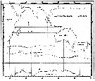 Issued from : C. Séret in Thesis 3ème Cycle, UPMC, Paris 6. 1979, Annexe. [p.38]. Issued from : C. Séret in Thesis 3ème Cycle, UPMC, Paris 6. 1979, Annexe. [p.38].
Geographical occurrences of Scaphocalanus magnus in the Indian Ocean and Antarctic zone. [after publications from: Brady, 1883, 1918; Thompson, 1900; Wolfenden, 1908, 1911; With , 1915; Rosendorn, 1917; Farran, 1929; Sewell, 1929, 1947; Brady & Gunther, 1935; Steuer, 1929, 1392, 1933; Ommaney, 1936; Vervoort, 1957; Tanaka, 1960; Brodsky, 1964; Seno, 1966; Andrews, 1966; Grice & Hulsemann, 1967; Seno, 1966; Frost & Fleminger, 1968; Voronina, 1970; Zverva, 1972].
Nota: C. Séret notes the occurrence at station 56°S, 70°E. |
| | | | Loc: | | | ? Cosmopolite : Antarct. (Weddell Sea, Indian, Drake Passage), sub-Antarct. (Indian, SW & SE Pacif.), South Africa, Atlant. (tropical),Brazil (The Campos Basin), Caribbean Colombia, off Bermuda: Station S (32°10N, 64°30W), off E Cape Cod, off Moroccan coast, W Medit. (Habibas Is., Algiers, Balearic Sea), Indian (Arabian Sea, Bay of Bengal), Pacif., Iceland, W Baffin Bay, Greenland Sea, Fram Strait, Spitsbergen, Wyville Thomson Ridge, Kongsfjorden, Norwegian Sea, Barents Sea, Arctic (Polar Basin), Nansen Basin, Canadian abyssal plain, Laptev Sea, Arct. (Fletcher's Ice Is., Chukchi Sea, SE Beaufort Sea, Canada Basin), Bering Sea (Aleutian Basin), S Aleutian Is., Japan (S. Hokkaido), China Seas (South China Sea), Galapagos-Ecuador | | | | N: | 122 | | | | Lg.: | | | (5) F: 5-4,5; M: 4,5; (7) F: 5,23; M: 4,74; (9) F: 5,4-4,3; M: 4,6; (11) F: 4,97-3,55; (16) F: 4,35-4,02; (17) F: 4,4; (22) F: 5,2-4,5; M: 5-4,5; (29) F: 4,9; (38) F: 4,56; (43) F: 4,7-4,55; (45) F: 5,25-3,7; M: 4,75-4,5; (47) F: 4,7-4,55; (59) F: 5,6-3,7; M: 4,8-4,5; (65) F: ± 5; M: ± 4,5; (73) F: 4,42; (76) F: 4,75-4,25; (105) F: 4,43; (108) F: 4,85-4,39; M: 4,65; (199) F: 4,94-3,8; M: 4,56-4,26; (208) F: 5,6-4,95; M: 5,1; (246) M: 5,28; (1000) F: 4,65-4,55; (1001) F: 4,6 ±0,5; (1111) F: 4,26; {F: 3,55-5,60; M: 4,02-5,28} | | | | Rem.: | epi-meso-hadopelagic.
Sampling depth (Antarct., sub-Antarct.) : 700-1000 m. Sargasso Sea: 500-2000 m (Deevey & Brooks, 1977, station "S"). 500-580 m (Pipe & Coombs, 1980, Arctic: 60°N).
Distributional range (m) from Roe (1972) Day: 570-950, Night: 410-960; from Grice & Hulsemann (1965): 450-1000 (in Kuriyama & Nishida, 2006).
The species shows a certain variability (cf. in With, 1915; Brodsky, 1967, p.250).
See the remark on Scaphocalanus cristatus.
Kosobokova & Hirche (2000) do not recognize the synonymy between S. variabilis and S. magnus. For Kosobokova, Hopcroft and Hirche (2011, p.35) Scaphocalanus acrocephalus is maintened, as in Park & Ferrari (2009, p.167, fig.2 where this form is the closest relative from S. parantarcticus in the Southern Ocean.
For Bradford & al. (1983, p.100) S. magnus shows some variation.
There are differences in the shape of the posterolateral border of the metasome and female P5 (Vervoort, 1957, p.111); in the 'Banzare' specimen the border is slightly thickened into a rounded knob; the P5 on each side consist of a single free segment attached to a basal portion; the free segment may show a more or less distinct articulation in the basal part; it carries 3 spiniform setae; there is a long internal spine, serrate along the inner edge, a shorter terminal spine and a minute external spine on the terminal segment; there is some variation in the lengths of these spiniform setae; some specimens may also show a rudimentary endopod (With, 1915, p.191). The variability in length induced Sewell (1947, p.144) to distinguish two forms: major and minor, also characterized by small structural differences. The male is easily recognized by the peculiar shape of the head and P5. | | | Last update : 25/10/2022 | |
|
|
 Any use of this site for a publication will be mentioned with the following reference : Any use of this site for a publication will be mentioned with the following reference :
Razouls C., Desreumaux N., Kouwenberg J. and de Bovée F., 2005-2025. - Biodiversity of Marine Planktonic Copepods (morphology, geographical distribution and biological data). Sorbonne University, CNRS. Available at http://copepodes.obs-banyuls.fr/en [Accessed August 23, 2025] © copyright 2005-2025 Sorbonne University, CNRS
|
|
|
|


;)
;)
;)
;)
;)
;)
;)
;)
;)
;)
;)
;)
;)
;)
;)
;)
;)
;)
;)
;)
;)
;)
;)
;)
;)
;)
;)

;)
