|
|
 |
|
Calanoida ( Order ) |
|
|
|
Clausocalanoidea ( Superfamily ) |
|
|
|
Scolecitrichidae ( Family ) |
|
|
|
Scolecithricella ( Genus ) |
|
|
| |
Scolecithricella nicobarica (Sewell, 1929) (F,M) | |
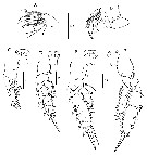
| | | | | | | Syn.: | Scolecithrix nicobarica Sewell, 1929 (p.209, figs.F,M); Tanaka, 1962 (p.36, figs.F,M); Chen & Zhang, 1965 (p.60, figs.F,M); Silas, 1972 (p.647); Bradford, 1973 (p.141); Carter, 1977 (1978) (p.36); Zheng & al., 1982 (p.41, figs.F); Bradford & al., 1983 (p.113); Furuhashi, 1966 a (p.295, vertical distribution in Kuroshio region, Table 10) ; Guangshan & Honglin, 1984 (p.118, tab.); Yoo, 1991 (tab.1); Gopalakrishnan & Balachandran, 1992 (p.167, figs.1, 5, Table 1, 2); Kim & al., 1993 (p.270); Shih & Young, 1995 (p.73); Go & al., 1997 (tab.1); Noda & al., 1998 (p.55, Table 3, occurrence); Lo & al., 2001 (1139, tab.I); Kazmi, 2004 (p.229); Wang & Zuo, 2004 (p.1, Table 2, dominance, origin); Zuo & al., 2006 (p.163: tab.1, 3, fig.9G, spatial abundance, fig.8: stations group); Hwang & al., 2006 (p.943, tabl. I); Dur & al., 2007 (p.197, Table IV); Fernandes, 2008 (p.465, Tabl.2); C.-Y. Lee & al., 2009 (p.151, Tab.2); Sun & al., 2010 (p.1006, Table 2) | | | | Ref.: | | | Farran, 1936 a (p.97, figs.F); Sewell, 1948 (p.323); Grice & Hulsemann, 1967 (p.17); Fleminger, 1967 a (tabl.1); Vyshkvartzeva, 1999 (2000) (p.234); Soih & al., 2013 (p.102, figs.F,M) | 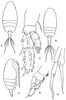 issued from: Q.-c Chen & S.-z. Zhang in Studia Marina Sinica, 1965, 7. [Pl.18, 8-13]. As Scolecithrix nicobarica. Female (from E China Sea): 8, habitus (dorsal); 9, idem (lateral right side); 10, right P1 (anterior); 11, left P3 (posterior). Male: 12, habitus (dorsal); 13, P5 (posterior).
|
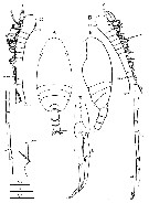
 issued from : R.B.S. Sewell in Mem. Indian Mus., 1929, X. [p.210, Fig.78]. As Scolecithrix nicobarica. Female (from Nankauri Harbour): a, habitus (dorsal); b, A1; c, P1; d, P2; e, P3; f, P4. Male: g, P5.
|
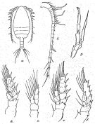
 issued from : Z. Zheng, S. Li, S.J. Li & B. Chen in Marine planktonic copepods in Chinese waters. Shanghai Sc. Techn. Press, 1982 [p.42, Fig.24]. As Scolecithrix nicobarica. Female: a-b, habitus (dorsal and lateral, respectively); c, P1; d, P3. Scale bars in mm.
|
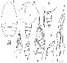
 Issued from : H.Y. Soh, S.Y. Moon & J.H. Wi in Invertebrate Fauna of Korea (eds) Incheon: NIBR, 2013, 21 (28). [p.103, Fig.59]. Female (from Korean waters): A-B, habitus (dorsal and lateral, respectively); C, rostrum; D, A1; E, A2; F, Md; G, Mx1. Scale bars: A, B = 300 µm; C-E = 10 µm; F, G = 20 µm.
|
 Issued from : H.Y. Soh, S.Y. Moon & J.H. Wi in Invertebrate Fauna of Korea (eds) Incheon: NIBR, 2013, 21 (28). [p.104, Fig.60]. Female: A, Mx2; B, Mxp; C, P1; D, P2; E, P3; F, P4. Scale bars: A, B = 20 µm; C-F = 10 µm.
|
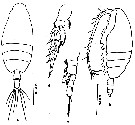
 Issued from : H.Y. Soh, S.Y. Moon & J.H. Wi in Invertebrate Fauna of Korea (eds) Incheon: NIBR, 2013, 21 (28). [p.105, Fig.61]. Male: A-B, habitus (dorsal and lateral, respectively); C, D, A1; E, P5. Scale bars: A, B = 300 µm; C, D = 20 µm; E = 10 µm.
|
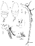
 issued from : O. Tanaka in Publ. Seto Mar. Biol. Lab., 1962, X (1). [p.36, Fig.127]. As Scolecithrix nicobarica. Female (Sagami Bay): a, habitus (dorsal); b, head (lateral left side); c, last thoracic segment and urosome (lateral left side); d, P1; e, P2; f, endopod of P3; g, basal segments of P4. Male: h, last thoracic segment and urosome (lateral left side); i, P5. Nota Female: - Cephalothorax about 4.13 times the abdomen length. - Head and 1st pediger segment fused, 4th and 5th pedigers fused, line of fusion not observed. - Posterolateral corners of the last thoracic segment slightly indented on margin. - Rostrum with 2 strong spines terminating into fine point. - Abdomen 4-segmented; segments and caudal rami in the proportional lengths 43 : 18 : 18 : 9 : 12 = 100. - Genital segment not swollen ventrally. - Anal segment short. - Caudal rami as long as wide. - A2 20-segmented, extends to the posterior margin of the 2nd abdominal segment. Segments 1-2, 8-9-10, 24-25 are fused., segments 12 and 13 partially fused. - A2 exopod slightly longer than endopod (7 : 5). - Mx1 with 7 long and 2 short setae on outer lobe; 7 setae on exopod; 6 setae on endopod; 3 setae on the 2nd basal; 3 setae on the 3rd inner lobe; 2 setae on the 2nd inner lobe; 9 setae on the 1st inner lobe (= arthrite). - Mx2 with 3 long vermiform and 5 bud-like sensory filaments on endopod. - Mxp with the 1st basal segment slightly shorter than the 2nd. - P1 exopod 3-segmented and endopod 1-segmented; exopodal segment 1 without marginal spine; coxa with a row of spinules. - P2 with exopod 3-segmented and endopod 2-segmented; the 2nd and 3rd exopodal segments and the 2nd endopodal segment are furnished with rows of spinules on the posterior surface; coxa xery finely denticulated on proximal half of the outer margin; basis with a small spine on inner margin about the middle. - P3 and P4 with exopod and endopod each 3-segmented. - P4 with coxa finely serrated on outer distal margin; there is a small protuberance near the outer distal margin; the segment has a transverse row of spinules at the base of the small inner marginal seta on posterior surface; basis with a rounded process near the inner distal margin where the endopod articulates. - P5 absent. Nota Male (from Suruga Bay): - Cephalothorax about 2.95 the abdomen length. - Urosome 5-segmented; abdominal segments and caudal rami in the proportional lengths 17 : 33 : 17 : 21 ; 5 : 7 = 100. - Caudal rami as long as wide. - A1 18-segmented on the left side, reaches back to the distal end of the 2nd abdominal segment; segments 8-13, 20-21 and 24-25 fused, segment 12 and 13 fused on the posterior margin. - A2 exopod slightly longer than endopod. - Md robust. - Mx1 ith 7+2 setae on outer lobe. - Mx2 with long vermiform filaments on endopod. - Mxp robust. - P1 to P4 as those of the female. - P5 reaches back to the caudal rami; left leg with endopod very small; exopod 3-segmented, the distal segment carries a thin lamellous plate on the apex. Endopod of right leg triangular in shape, carries a slender spine at apex; distal segment of exopod slender.
| | | | | Compl. Ref.: | | | Brinton & al., 1986 (p.228, Table 1); Chen Y.-Q., 1986 (p.205, Table 1: abundance %, Table 2: vertical distribution); Madhupratap & Haridas, 1986 (p.105, tab.1); Suarez-Morales & Gasca, 1998 a (p.111); Kuriyama & Nishida, 2006 (p.300: Tab.II; p.309: Tab.III, fig.7, 10, vertical distribution); in CalCOFI regional list (MDO, Nov. 2013; M. Ohman, comm. pers.). | | | | NZ: | 6 | | |
|
Distribution map of Scolecithricella nicobarica by geographical zones
|
| | | | | | | | | 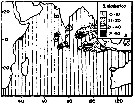 issued from : T.C. Gopalakrishnan & T. Balachandran in Oceanogr. Indian Ocean, 1992. [p.171, fig.5, A]. As Scolecithrix nicobarica. issued from : T.C. Gopalakrishnan & T. Balachandran in Oceanogr. Indian Ocean, 1992. [p.171, fig.5, A]. As Scolecithrix nicobarica.
Distribution of Scolecithricella nicobarica in the Indian Ocean.
Nota: Main occurences between 25° N - 20° S. |
| | | | Loc: | | | South Africa (E), Natal, Indian (W & N), Pakistan, Bay of Bengal, Nicobar Is.: Nankauri Harbour, China Seas (Yellow Sea, East China Sea, South China Sea), Taiwan, Okinawa, S Korea, Japan, Sagami Bay, Kuchinoerabu Is., Pacif. (W equatorial), Australia (Great Barrier), California, Gulf of California | | | | N: | 32 | | | | Lg.: | | | (29) F: 1,08; M: 1,08; (34) F: 1,37-1,35; (117) F: 1,446; M: 1,5; (290) F: 1,45-1,5; M: 1,4-1,5; (1000) F: 1,5 ± 0.0; M: 1,5 ± 0.0; (1023) F: 1,45; (1174) F: 1,21-1,23; M: 1,15-1,17; {F: 1,08-1,50; M: 1,08-1,50} | | | | Rem.: | epi-mesopelagic. 298-526 m at Station N-1 (SW Bösö, E middle Japan) from Furuhashi (1966 a).
After Tanaka (1962, p.38) the specimen from the Izu region (Japan) differs from Sewell in the structure of the A2. | | | Last update : 18/01/2021 | |
|
|
 Any use of this site for a publication will be mentioned with the following reference : Any use of this site for a publication will be mentioned with the following reference :
Razouls C., Desreumaux N., Kouwenberg J. and de Bovée F., 2005-2026. - Biodiversity of Marine Planktonic Copepods (morphology, geographical distribution and biological data). Sorbonne University, CNRS. Available at http://copepodes.obs-banyuls.fr/en [Accessed April 15, 2026] © copyright 2005-2026 Sorbonne University, CNRS
|
|
|
|


;)
;)
;)
;)
;)
;)
;)

;)
