|
|
 |
|
Calanoida ( Order ) |
|
|
|
Clausocalanoidea ( Superfamily ) |
|
|
|
Scolecitrichidae ( Family ) |
|
|
|
Scolecithricella ( Genus ) |
|
|
| |
Scolecithricella propinqua (Sars, 1920) (F,M) | |
| | | | | | | Syn.: | Amallothrix propinqua : Sars, 1925 (p.178, figs.F); ? Wilson, 1942 a (p.171, fig.M: abnormal); Sewell, 1948 (p.349, 502, 567, 568); C.B. Wilson, 1950 (p.162, fig.F); Grice & Hulsemann, 1965 (p.223); El-Maghraby, 1965 (p.54, Appendix); Björnberg, 1973 (p.384); Deevey & Brooks, 1977 (p.256, tab.2, Station "S"); Carter, 1977 (1978) (p.35); Vives, 1982 (p.292); Kovalev & Shmeleva, 1982 (p.84); Kuriyama & Nishida, 2006 (p.293, 299: Tab.II; p.309: Tab.III, fig 10, vertical distribution); Belmonte, 2018 (p.273, Table I: Italian zones);
Scolecithricella (Amallothrix) propinqua : Vervoort, 1965 (p.65, Rem.); Björnberg, 1973 (p.332); Vives & Shmeleva, 2007 (p.729, figs.F,M, Rem.) | | | | Ref.: | | | Sars, 1920 c (p.9, Rem.F); Tanaka, 1962 (p.63, figs.F,M); Bradford, 1973 (p.143); Bradford & al., 1983 (p.78, Rem); Vyshkvartzeva, 1999 (2000) (p.235) | 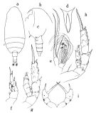 issued from : O. Tanaka in Publ. Seto Mar. Biol. Lab., 1962, X (1). [p.64, Fig.140]. Female (from Izu region): a, habitus (dorsal); b, forehead (left lateral side); c, last thoracic segment and urosome (left lateral side); d, rostrum (dorsal view); e, Mx2; f, P1; g, P2; h, P3; i, P5. Nota Female: - Cephalothorax about 4 times the abdomen length. 4th and 5th pedigers fused. - Posterolateral corners of the last thoracic segment rounded but there is a sudden change in outline along the dorso-lateral corner. - Rostrum with rami robust, terminate in fine filaments. - Abdominal segments and caudal rami proportional lengths 39 : 21 : 19 : 7 : 14 = 100. - Genital segment as long as wide, slightly swollen ventrally; there is a transverse row of minute spinules on the proximal part near junction with the cephalothorax. - Caudal rami slightly longer than wide. - A1 23-segmented; extends to the distal end of the caudal rami.. - A2 exopod 1.2 times as long as endopod. - Mx1 with 7 long and 2 short setae on the outer lobe; 9 setae on exopod; 5+3 setae on endopod; 5 setae on the 2nd basal; 4 setae on the 3rd inner lobe; 2 setae on the 2nd inner lobe; 13 setae on the 1st inner lobe (= arthrite). - Mx2 has 5 bud-like sensory filaments of about equal in size, and 3 long worm-like ones on the distal segments. - Mxp with a tuft of hairs at base of the bud-like sensory filament on the 1st basal segment, which arises from the middle of the external margin; 4th lobe with, beside 3 usual setae, a small rounded process near the distal border. - P1 with the 1st exopodal segment 1 outer edge spine reaching the distal margin of 2nd segment. - P2 with the 1st exopodal segment a long and curved outer edge spine, exceeding the middle of the outer margin of the 2nd segment. Terminal spine of exopod with 32 teeth. - P3 with 22 teeth on terminal spine of exopod. - P5 3-segmented. Distal segment with a strong inner marginal spine, coarsely denticulated along the anterior margin; terminal spine short, about 1/3 the length of the inner marginal spine; there is on the outer margin 1 small spine at base ot the terminal spine, and 1 small outer marginal spine opposite to the inner marginal spine. Posterior surface of segments furnished with a group of spines.
|
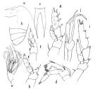 issued from : O. Tanaka in Publ. Seto Mar. Biol. Lab., 1962, X (1). [p.65, Fig.141]. Male (from Izu region): a, forehead (left lateral side); b, thoracic segments (left lateral side); c, rostrum (dorsal view); d, A2; e, Mx2; f, P1 (posterior view); g, P2; h, P3; i, P4; j, P5. Nota Male: - Cephalothorax about 2 times the abdomen lenth. - 4th and 5th pedigers incompletely fused, line of fusion clearly seen in lateral aspect. - Rostrum as in female, but the basal rami much stronger. - Urosome 5-segmented; segments and caudal rami in proportional lengths 9 : 32 : 27 : 25 : 2 : 5 = 100. - 2nd and 3rd abdominal segments fringed with fine teeth on the distal border. - Caudal rami divergent, a little wider than long. - A1 20-segmented on left sie, 19-segmented on right side; extends to the distal end of the 2nd abdominal segment. - A2 exopod 1.2 times as long as endopod; 1st exopodal segment furnished with short hairs along the anterior margin. - Md and Mx1 as in female. - Mx2 bears 5 bud-like sensory filaments and 4 worrm-like on the distal segments, one of the latter is much larger than the others. - P1 with 1 outer edge spine on , extending to the middle of the 2nd segment. - P2 with 1 outer edge spine on the 1st exopodal segment, curved considerably at the distal end. Terminal spine of exopod with 32 teeth. - P3 with on the 2nd basal segment (= basis) rows of spinules along anterior distal border. Terminal spine of exopod with 20 teeth.
|
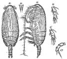 Issued from : G.O. Sars in Résult. Camp. Scient. Prince Albert I, 69, pls.1-127 (1924). [Pl.XLIX, figs.22-27]. As Scolecithrix propinqua. Female: 22, habitus (dorsal); 23, idem (lateral left side); 24, rostrum; 25, endopodal segments of P2; 26, endopodal segments of P3; 27, P5. Nota Female (as Amallothrix propinqua): - Posterolateral corners of the last thoracic segment slightly obtusely rounded. - Rostrum comparatively shorter than A. gracilis, but of the same structure. - Urosome length about 1/4 of prosome. - Genital segment as long as two following segments together, slightly swollen ventrally. - Caudal rami small and slightly divergent. - A1 reaches end of caudal rami. - A2, mouthparts and swimming legs similar to those A. gracilis. - P5 resembles those A. gracilis, distinctly biarticulate; proximal segment shorter than distal and hairies outside at end; distal segment furnished with of number rigid hairies outside, segment obliquely truncate at yje extremity and with 2 spines, 1 inner very strong and 1 smaller at apex.
|
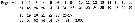 issued from : O. Tanaka in Publ. Seto Mar. Biol. Lab., 1962, X (1). [p.63]. Female: Proportional lengths of segments of A1. -A1 23-segmented. Segments 14 and 18 with each a proximal seta.
|
 issued from : O. Tanaka in Publ. Seto Mar. Biol. Lab., 1962, X (1). [p.66]. Male: proportional lengths of segments on the left side of A1. - A1 20-segmented on the left side; 19-segmented on the right side. Segments 14, 18, 21, 22 and 25 have each a long seta. Proximal segments with well-developed aesthetascs.
| | | | | Compl. Ref.: | | | Roe, 1972 (p.277, tabl.1, tabl.2); Belmonte, 2018 (p.273, Table I: Italian zones) | | | | NZ: | 8 | | |
|
Distribution map of Scolecithricella propinqua by geographical zones
|
| | | | | | | | | | Loc: | | | South Africa (E, Natal), G. of Guinea, off Mauritania, Canaries, off NW Canary Is., off Portugal, off Bermuda: Station S (32°10N, 64°30W), Sargasso Sea, Medit. (S Adriatic Sea, Lebanon Basin, Egypt coast), Philippines (in C. B. Wilson, 1950), Japan, N-S Chile (in Björnberg, 1973) | | | | N: | 14 | | | | Lg.: | | | (1) F: 2,3; (16) F: 3,6-2,8; M: 3,41-3,04; (117) F: 2,95; M: 2,98; (199) F: 3,27-3,12; (1000) F: 3,1 ± 0,1; {F: 2,30-3,60; M: 3,04-3,41} | | | | Rem.: | Bathypelagic. Sargasso Sea: 1000-1500 m (Deevey & Brooks, 1977, station "S").
Distributional range (m) from Roe (1972) Day: 940, Night: 910-960; from Grice & Hulsemann (1965): 1000-2000 (in Kuriyama & Nishida, 2006).
Probable in the Genus Amallothrix.
For Sars (1925, p.179) this species resembles much to Amallothrix gracilis except P5 which show in the latter a structure unisegmented and 1 spine on the outer margin.
For Vervoort (1965, p.66) the specimenns from the Gulf of Guinea agree well with Tanaka's account but for some variation in the degree of curvature of the head, at times approaching the condition observed in Scolecithrix (Amallothrix) arcuata Sars, 1920, and variability in the length of the terminal spine of P5 in the female. | | | Last update : 19/01/2021 | |
|
|
 Any use of this site for a publication will be mentioned with the following reference : Any use of this site for a publication will be mentioned with the following reference :
Razouls C., Desreumaux N., Kouwenberg J. and de Bovée F., 2005-2026. - Biodiversity of Marine Planktonic Copepods (morphology, geographical distribution and biological data). Sorbonne University, CNRS. Available at http://copepodes.obs-banyuls.fr/en [Accessed March 22, 2026] © copyright 2005-2026 Sorbonne University, CNRS
|
|
|
|


;)
;)
;)

