|
|
 |
|
Calanoida ( Order ) |
|
|
|
Spinocalanoidea ( Superfamily ) |
|
|
|
Spinocalanidae ( Family ) |
|
|
|
Mimocalanus ( Genus ) |
|
|
| |
Mimocalanus heronae Damkaer, 1975 (F,M) | |
| | | | | | | Syn.: | Mimocalanus cultrifer : Tanaka,1956 c (p.387,figs.F,M);
no M. distinctocephalus : Boucher & de Bovée, 1970 (p.527);
no M. heronae : Hure & al., 1980 (p.297); Scotto di Carlo & al., 1984 (p.1042); Scotto di Carlo & al., 1991 (p.270); Hure & Krsinic, 1998 (p.41, 100); Mazzocchi & Di Capua, 2010 (p.427); Belmonte, 2018 (p.273, Table I: Italian zones) | | | | Ref.: | | | Damkaer, 1975 (p.80, figs.F, Rem.); Brodsky & al., 1983 (p.317, figs.F, M, Rem.); Bode & al., 2017 (p.600, Table I, III, fig. 2, 3, 4, morphology vs genetic). | 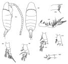 issued from : D.M. Damkaer in NOAA Technical Report NMFS CIRC-391, Seattle, 1975. [p.81, Fig.216-223; p.63, Fig.163]. Female: 163, terminal segments of A1; 216, habitus (right lateral side); 217, idem (dorsal view); 218, masticatory edge of Md; 219, Mxp; 220, P1; 221, P2; 222, P3; 223, P4.
|
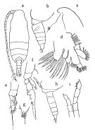 issued from : O. Tanaka in Publ. Seto Mar. Lab., 1956, V (3). [p.86, Fig.13]. Female: a, habitus (dorsal); b, last thoracic segment and urosome (left lateral side); c, head (left); d, Mx1; e, P1; f, P2; g, basipod segment 1 of P4. Male: h, last thoracic segment and urosome (left lateral side); i, P5.
|
 Issued from : M. Bode, S. Laakmann, P. Kaiser, W. Hagen, H. Auel & A. Cornils in J. Plankton Res., 2017, 39 (4). [p.603, Table I]. Species identified via morphological and molecular analyses, including the number of specimens used for each identification method (MI: morphological identification, COI: cytochrome c oxydase subunit I gene fragment, MS: MALDI-TOF analysis, 18 S: ribosomal 18 S gene fragment). Cryptic lineages were revealed only after molecular analysis. Sampling depth, latitude, total length (TL) and Prosome/Urosome ratio (Pr:Ur) of taxon were recorded. morphological characters are only listed here, if the identification was ambiguous due to missing body parts or if cryptic or pseudocryptic lineages were revealed during molecular analyses. For diagnostic character of species refer to the references: Park, 1970; Grice, 1971; Damkaer, 1975; Schulz, 1989, 1996. C = coxa; End = endopodite; P = swimming legs.
| | | | | Compl. Ref.: | | | Suarez-Morales & Gasca, 1998 a (p.111) | | | | NZ: | 5 | | |
|
Distribution map of Mimocalanus heronae by geographical zones
|
| | | | Loc: | | | Japan (Sagami), N off Marquesas Is., off Baja California, SE Atlant. (11-3°N, 25°S,) | | | | N: | 3 | | | | Lg.: | | | (13) F: 1,95-1,23; M: 1,28; (55) F: 1,5; M: 1,28; (1252) F: 1,62-1,80, 1,72, 1,38-1,40; {F: 1,23-1,95; M: 1,28}
(1252) F: Pr/Ur = 3,3-4,6. | | | | Rem.: | Depth: 600-50000 m (in Bode & al., 2017) | | | Last update : 25/03/2020 | |
|
|
 Any use of this site for a publication will be mentioned with the following reference : Any use of this site for a publication will be mentioned with the following reference :
Razouls C., Desreumaux N., Kouwenberg J. and de Bovée F., 2005-2025. - Biodiversity of Marine Planktonic Copepods (morphology, geographical distribution and biological data). Sorbonne University, CNRS. Available at http://copepodes.obs-banyuls.fr/en [Accessed June 04, 2025] © copyright 2005-2025 Sorbonne University, CNRS
|
|
|
|


;)
;)
