|
|
 |
|
Calanoida ( Order ) |
|
|
|
Clausocalanoidea ( Superfamily ) |
|
|
|
Stephidae ( Family ) |
|
|
|
Stephos ( Genus ) |
|
|
| |
Stephos margalefi Riera, Vives & Gili, 1991 (F,M) | |
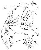
| | | | | | | Syn.: | Stephos balearensis Carola & Razouls,1996 (p.344, figs. F,M, Rem.); Bradford-Grieve, 1999 a (p.25, Rem.: Table 1); Vives & Shmeleva, 2007 (p.849, figs.F,M, Rem.) | | | | Ref.: | | | Riera & al., 1991 (p.318, figs.F,M); Bradford-Grieve, 1999 a (p.25, Table1: Rem.); Vives & Shmeleva, 2007 (p.853, figs.F,M, Rem.); Jaume & al., 2008 (p.514, figs.F, M, Rem.: emend.) | 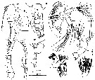 issued from : Carola & Razouls in Bull. Mar. Sci., 1996, 58 (2). [p.346, Fig.1, A-H]; As Stephos balearensis. Female (from Sa Gamba Cave, Minorca): A, habitus (dorsal); B, urosome with spermatophore (ventral); C, last thoracic segment and genital segment + 2nd urosomal segment (lateral); D-D\", A1; E, A2; F, Md; G, Mx1; H, Mx2; I, Mxp. Scales: A (a); B-D\" (b); E-I (c). Nota: A1 24-segmented, reaches posterior margin of caudal rami. A2 exopod longer than endopod (ratio = 1.5); coxa and basis completely separated, armed with 1 and 2 setae, respectively; endopod 2-segmented: segment 1 with 1 seta, segment 2 with 9 setae on subapical lobe and 7 on apical lobe; exopod 6-segmented, segment 1 with 1 seta, segment 2 with 3 setae, segments 3 to 5 (very difficulut to differenciate each segment) with 1 seta each, segment 6 with 1 marginal and 3 distal setae; Distal margin of gnathobase of Md with 7 cuspidate teeth and one exterior stronger; palp biramous: basis with 3 inner setae; endopod 2-segmented, proximal segment with 4 setae, distal segment with 9 setae; exopod 4-segmented (very difficulut to differenciate each segment), segment 1 to 3 each with 1 inner seta, segment 4 with 3 apical setae. Mx1 with precoxa produced into powerful endite bearing 5 stout spines and 5 setae; coxa with 3 setae on endite, outer lobe (epipodite) represented by 9 setae; basis fused to endopod, with 1 outer seta; proximal endite bearing 4 setae; distal endite, incorporated into segment, represented by 5 setae; fused endopod armed with one group of 5 setae on inner margin; free apical segment with 3 setae; exopod 1-segmented, with 8 setae along distal and lateral margins. Mx2 small, comprising precoxa, coxa and basis each armed with 2 endites, and a 3-segmented endopod; both precoxal endites with 3 slender setae; proximal coxal endite with 3 slender setae; distal coxal endite with 1 stout and 3 slender setae; proximal endite of basis powerful, bearing 3 slender setae, distal endite with 1 slender seta; endopod segments 1 and 2 without seta, segment 3 with 2 setae.
|
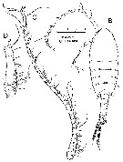
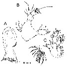
 issued from : Carola & Razouls in Bull. Mar. Sci., 1996, 58 (2). [p.346, Fig.1, J-Q]; As Stephos balearensis. Female: J, P1; K, P2; L, P3; M, P4; N, P5; O, idem (lateral view). Male: P, P5; Q, urosome (dorsal). Nota: P5 uniramous, 2-segmented; 1st segment unarmed with convex inner margin, distal segment with transverse row of spines across middle part, and row of distal spinules, fine setae and strong lateral denticle on distal margin.
|
 issued from : D. Jaume, G.A. Boxshall & F. Gràcia in Systematics and Biodiversity, 2008, 6 (4). [p.512, Fig.9, B-D]. Male (from Majorca): B, habitus (dorsal); C, right A1 (ventral); D, detail of proximal part of A1. Nota : Assymmetry of last prosomite pronounced, with left side extended posteriorly, subterminal spines on lest prosomite stout and conspicuous. Urosomie with patch of spinules anterodorsally on left side of genital somite (right in S. vivesi) and of blunt recurved outgrowth ventrodorsally on left side of 1st free abdominal somite.
|
 issued from : D. Jaume, G.A. Boxshall & F. Gràcia in Systematics and Biodiversity, 2008, 6 (4). [p.513, Fig.10, A-E]. Male: A, detail of last prosomal and genital somites (dorsal view showing functional genital duct on left side only); B, last two somites plus caudal rami (dorsal); C, P5 (postrrior); D, detail of distal segment of right P5 (anteromedial); E, same for left P5 (anterolateral). Nota: P5 symmetrical, uniramous, 3-segmented with proximal segment fused to intercoxal sclerite. Legs truncate distally, although appearing misleadingly pointed when view in lateral aspect. 1st and 2nd segments unarmed, 2nd with pore anteromedially ; 3rd segment slightly longer than preceding segment, with fan-like comb of spinules along truncate distal margin, seta, 2 prallel rows of spinules, and pore positioned on anterior surface about miway of segment ; seta implanted towards lateral margin, not extending beyond segment distally. Left P5 with proximal segment bearing pore on anterior surface.
|
 issued from : D. Jaume, G.A. Boxshall & F. Gràcia in Systematics and Biodiversity, 2008, 6 (4). [p.514, Fig.11]. Male: A, body (left lateral aspect); B, detail of proximal part of urosome and last prosomite (lateral view from left side); C, urosome (ventral). Scale bar : 0.25 mm (A); 0.05 (B, C). Nota : Urosome with presence of patch of spinules anterodorsally on left side of genital somite (right in S. vivesi) and of blunt outgrowth ventrolaterally on left side of free abdominal somite. Hyaline anal operculum faintly serrate. Caudal rami with 3 trnsverse rows of spinules dorsomedially ; seta V longer than rest ; seta II apparently retained, partially hidden within patch of spinules at ventrolateral corner of ramus.
|
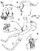
 issued from : D. Jaume, G.A. Boxshall & F. Gràcia in Systematics and Biodiversity, 2008, 6 (4). [p.515, Fig.12]. Male: A, left A2 (lateral); B, right Mx2 with disarticulated free endopod (lateral); C, right Mxp (medial).
|
 issued from : D. Jaume, G.A. Boxshall & F. Gràcia in Systematics and Biodiversity, 2008, 6 (4). [p.516, Fig.13, A-C]. Male: A, Md (gnathobase; note drops of unidentified substance extruded from pores on cutting edge); B, Md (mandibular palp); C, right Mx1 (anterior). Nota : Mandibular gnathobase with spinules of subdistal row on ventral margin (shorter than in S. vivesi) ; patch of triangular denticles about midway along dorsal margin. Cutting edge comprising 9 unequal teeth, either uni-bi- or tricuspidate ; 4 more dorsal teeth each with tubular extension ending in apparent secretory pore (still retaining drops of unidentified substance (stippled in figure A) ; ; 3 teeth situated more ventrally with hyaline tip , 2 pores and several rows of spinules present proximally to ventral teeth. The function of the pores is unknown (venom or aids to digestion ?). Mandibular palp with 1 additional terminal seta on endopod than in S. vivesi. Mx1 differing from S. vivesi in presence of 1 additional spine on praecoxal arthrite, in expression of basal exite seta, and in 1 additional seta subdistally on endopod. Integumentary ornamentation of segments differing also : 2 conspicuous oblique rows of stout spinules on anterior surface of distal basal endite ; 3 clusters of tiny denticles near insertion of 4 setae of praecoxal arthrite, on posterior surface.
|
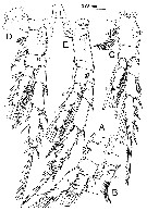 issued from : D. Jaume, G.A. Boxshall & F. Gràcia in Systematics and Biodiversity, 2008, 6 (4). [p.517, Fig.14]. Male: A, right P1 (anterior); B, detail of posterior ornamentation on inner basal seta of latter; C, left P2 (anterior); D, left P3 (anterior); E, left P4 (anterior). Nota : Swimming legs 1-4 with armature formula as in S. vivesi. Differing mainly in details of integumentary ornamentation.
|
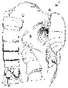
 issued from : D. Jaume, G.A. Boxshall & F. Gràcia in Systematics and Biodiversity, 2008, 6 (4). [p.518, Fig.15]. Female (from Majorca): A, habitus (dorsal); B, detail of genital double-somite (ventral); C, right A1 (ventral); D, detail of proximal part of latter (ventral). Scale bar: 0.25 mm (A); 0.05 mm (B-D). Nota : A1 differing from male in displaying fewer aesthetascs (only 1 on segment 1, and none on segments 2, 6, 16, 19 and 20) and in presence of 2 spinulose combs on segment 7. In addition, distal spinulose comb on segment 1 is not positioned as far distally as in male, and 2 proximal setae on this segment are not as reduced.Urosome 4-segmented. Genital double-somite asymmetrical, with ventrolateral bulge on right side and 3 rows of stout spinules. Only single genital opening expressed externally, on the left side, with single copulatory pore almost completely exposed anteriorly on margin of genital area, plus operculum placed ventrolaterally covering gonopore.
|
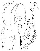
 issued from : D. Jaume, G.A. Boxshall & F. Gràcia in Systematics and Biodiversity, 2008, 6 (4). [p.516, Fig.13, D-E)]. Female: D, lateral view of last prosomal somite and left P5; E, P5 (anterior). Nota: 3rd segment of P5 truncate distally, although appearing misleadingly pointed when viewed in lateral aspect; proximal segment fused to intercoxal sclerite; 1st and 2nd segments unarmed, 2nd with pore anteromedially; 3rd segment slightly longer than preceding segment, with fan-like comb of spinules along truncate distal margin, seta, 2 parallel rows of spinules, and pore positioned on anterior surface about midway of segment; seta implanted towards lateral margin, not extending beyond segment distally.
| | | | | NZ: | 1 | | |
|
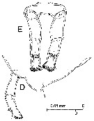
Distribution map of Stephos margalefi by geographical zones
|
| | | | | | | Loc: | | | W Medit.: Baleares (Majorca, Minorca: caves) | | | | N: | 3 | | | | Lg.: | | | (509) Minorca: F: 0,849-0,835; M: 0,761-0,665; (558) Majorca: F: 0,8-0,77; M: 0,74-0,73; (1017) F: 0.77-0.85; M: 0.74-0.80; {F: 0,77-0,85; M: 0,73-0,80}
The mean female size is 0.812 mm (n = 6; SD = 0.0375), and the mean male size is 0.739 mm (n = 6; SD = 0.0442). The size ratio (male : female) is 0.91. | | | | Rem.: | Types lost (Riera, pers. comm.).
A doubt subsists on the synonymy possible with S. balearensis taking into account the similar localities of occurrence.
The original form emended by Jaume & al., 2008) from specimens in the locality type.
According to Jaume & al. (2008, p.519) this species seems to be a strict thalasssostygobiont inhabiting caves with water that scarcely differs from the full marine salinity. It has never been caught outside caves. The disjunct distribution of this species, embracing three different Balearic islands, does not pose any biogeographical problem since all three comprised a single island as recently as 17 000 yr BP, at the time of the last glacial maximum when a global marine regression of up to 140 m took place.
In the Mediterranean Sea, the closest relative to S. margalefi is the Sicilian S. cryptospinus; it differs in the morphology of the distal segment of both left and right male P5 (right bifid, left with about 14 hyaline lamellae in C. cryptospinus; vs. right not bifurcate, left with only 4 hyaline lamellae in S. margalefi), among other features. S. canariensis from the nearby Canary Islands and which belongs to the same group of species, differs from S. margalefi in the bifid distal segment of the male right P5 , and in the lack of hyaline lamellae on the distal segment of the left P5.
After Bradford-Grieve (1999 a, p.25) this species belongs to type III (see Genus Stephos). | | | Last update : 23/01/2016 | |
|
|
 Any use of this site for a publication will be mentioned with the following reference : Any use of this site for a publication will be mentioned with the following reference :
Razouls C., Desreumaux N., Kouwenberg J. and de Bovée F., 2005-2025. - Biodiversity of Marine Planktonic Copepods (morphology, geographical distribution and biological data). Sorbonne University, CNRS. Available at http://copepodes.obs-banyuls.fr/en [Accessed December 02, 2025] © copyright 2005-2025 Sorbonne University, CNRS
|
|
|
|


;)
;)
;)
;)
;)
;)
;)
;)
;)
;)

