|
|
 |
|
Cyclopoida ( Order ) |
|
|
|
Oithonidae ( Family ) |
|
|
|
Oithona ( Genus ) |
|
|
| |
Oithona nishidai McKinnon, 2000 (F,M) | |
| | | | | | | Ref.: | | | McKinnon, 2000 (p.100, 108, figs.F,M) | 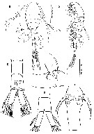 issued from : A.D. McKinnon in Plankton Biol. Ecol., 2000, 47 (2). [p.101, Fig.1]. Female (from 19°28'S, 147°03'E): a-b, habitus (dorsal and lateral, respectively); c, urosome somites 1 and 2 (dorsal); d, urosome somite 4 and anal somite (dorsal); e, ananl somite (ventral); f, P5 and genital complex (lateral). Nota : Rostrum produced antero-ventrally, strong, simple. A Thoracic segments 4 and 5 hirsute, short hairs covering the postero-dorsal margin and lateral faces in particular. Vento-posterior margin of urosomal somite 4 and anal somite with setules, comb extending on to the dorsal surface of anal somite. Caudal ramus length 3 times width. Proportional urosomites (with caudal ramu) 0.86 :1.83 :0.89 :0.82 :1.0 :1.0. P5 comprises dorsal seta of basis and exopod segment with crown seta.
|
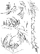 issued from : A.D. McKinnon in Plankton Biol. Ecol., 2000, 47 (2). [p.102, Fig.2]. Female: a, A1; b, A2; c, Md (mandibular palp); d, Mx1; e, Mx2; f, Mxp. Nota : A1 symmetrical, 14-segmented. A2 basipod with 1 seta on outer margina t mid-length, another seta more distally, inner surface with 2 setae medially ; 1st endopod segment with 1 proximal, 1 medial, 3 terminal setae, 2nd with 7 terminal setae. Md basis bearing 2 sharp recurved spines, each armed with spinules ; endopod with 4 setae, outermost hirsute ; exopod 3-segmented, with1, 1, 3 setae. Mx1 praecoxal endite with 8 spiniform setae, the most distal very long ; coxal endite with plumose seta ; 1st basal endite with 3 setae , 2 plumose ; 2nd basal endite with seta ; endopod with 3 setae ; exopod with 4 ; praecoxal epipodite with plumose seta.
|
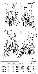 issued from : A.D. McKinnon in Plankton Biol. Ecol., 2000, 47 (2). [p.103, Fig.3]. Female: a-d, P1 to P4. below: Swimming legs P1 to P4 with armament formulae (Roman numerals = spines; Arabic numerals = setae). Nota : Two inner setae of P4 endopod segment 2 and proximal seta of endopod segment 3 modified with denticulate membrane flange on medial tip. Intercoxal plate of P4 with comb of spines on posterior surface.
|
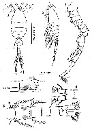 issued from : A.D. McKinnon in Plankton Biol. Ecol., 2000, 47 (2). [p.104, Fig.4]. Male: a-b, habitus (dorsal and lateral, respectively); c, urosome (dorsal); d, anal somite and caudal rami (ventral); e, genital somite and P5 and P6 (lateral, right side). Nota : Forehead broad and flat in dorsal view, blunt in lateral view. A1 17-segmented. Urosome 6-segmented ; urosome somite 5 distal margin coarsely denticulate, anal somite with comb of spines both dorsally and ventrally. Caudal ramus length 1.79 times width anal somite.
|
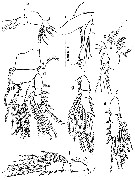 issued from : A.D. McKinnon in Plankton Biol. Ecol., 2000, 47 (2). [p.106, Fig.6]. Male: a, A2; b, Md (mandibular palp); c, Mx1; d, P1; e, P2 exopod; f, P3 exopod; f, P4 exopod. Nota : A2 similar to female, but endopode elongate, fused into one segment. Md endopod 1 distinctly articulated from basis, bearing 2 elongate denticulate spines. P1 to P4 as in female, but with inner marginal seta on exopod segment 1 reduced, and without modified setae on P4 endopod. P5 similar to female, P6 a single seta originating from ventro-lateral process on genital somite.
|
 issued from : A.D. McKinnon in Plankton Biol. Ecol., 2000, 47 (2). [p.108, Table 2]. Morphological characteristics useful in the discrimination of the females of co-occuring species of Oithona from coastal waters of Northern Australia. PR : UR = prosome : urosome length ratio; CR L : W = caudal ramus length : width ratio; Md B2 = mandible palp basipod armament; Md Ri = armature of mandible endopod; L1-L4 = armament of legs P1-P4; B = blunt; P = pointed; Ri = endopod; Re = exopod.
|
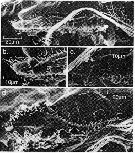 issued from : A.D. McKinnon in Plankton Biol. Ecol., 2000, 47 (2). [p.105, Fig. 5]. Male: Cephalosome flap organ. a, lateral; b, posterior margin; c, nested shield-like integumental organs containing sensilla; d, antero-lateral, another specimen showing anterior sensilla. Nota : Cephalosome-flap organ extending beyond thoracic segment 2 ; pore signature pattern belonging to type B of Nishida (in J. Plankton Res., 1986, 8 : 907), with 12 colums of 6-8 nested intergumental organs, each comprising a raised shield-like structure surronding long cilia. The dorsal margin of the organ has a row of protuberances, each terminating in a cilium. The integumental organs on the ventral margin comprises simpler pits, each containing a cilium.
| | | | | NZ: | 1 | | |
|
Distribution map of Oithona nishidai by geographical zones
|
| | | | Loc: | | | E Australia (N Queensland: mangrove) | | | | N: | 1 | | | | Lg.: | | | (847) F: 0,55-0,66; M: 0,52-0,60; { F: 0,55-0,66; M: 0,52-0,60} | | | | Rem.: | For McKinnon (2000, p.104) this species is very closely related to O. australis Nishida (1986) but differs in having 3 setae rather than 2 on the endopod of Mx1 of both sexes. O. nishidai is one of a group of species characterised by the possession of an elongate distal seta on the praecoxal arthrite of Mx1 as O. aruensis, O. attenuata, O. australis, O. davisae, O. wellershausi. The occurrence of this character in O. attenuata implies that it has been secondarily derived in this species, given its close relationship to O. nana. However, for the remaining species this character and the presence of 2 long spinulose setae on the 2nd endopodal segment of Mxp are probably indicative of membership of an Indo-West Pacific species group found principally in riverine systems (see Nishida, 1986 a; Ferrari & Orsi, 1984). O. nishidai is the dominant oithonid and often dominant copepod in mangrove estuaries from Townsville/ | | | Last update : 08/12/2016 | |
|
|
 Any use of this site for a publication will be mentioned with the following reference : Any use of this site for a publication will be mentioned with the following reference :
Razouls C., Desreumaux N., Kouwenberg J. and de Bovée F., 2005-2025. - Biodiversity of Marine Planktonic Copepods (morphology, geographical distribution and biological data). Sorbonne University, CNRS. Available at http://copepodes.obs-banyuls.fr/en [Accessed January 01, 2026] © copyright 2005-2025 Sorbonne University, CNRS
|
|
 |
 |


