|
|
 |
|
Cyclopoida ( Order ) |
|
|
|
Thaumatopsyllidae ( Family ) |
|
|
|
Caribeopsyllus ( Genus ) |
|
|
| |
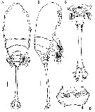
Caribeopsyllus chawayi Suarez-Morales, 1998 (F) | |
| | | | | | | Ref.: | | | in Suarez-Morales & Castellanos, 1998 (p.199, Descr.F, figs.F); Suarez-Morales & Tovar, 2004 (p.223, figs. juv.1-5, F, Rem.: p.243); Boxshall & Halsey, 2004 (p.691) | 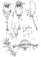 issued from : E. Suarez-Morales & I.A. Castellanos in J. Crustacean Biol., 1998, 18 (1). [p.200, Fig.1]. Female [? Juv. stage 5 ] (from Mahahual reef area, Mexico): A-C, habitus (dorsal, ventral and latteral, respectively); D, caudal rami (ventral); E, first urosomites (lateral); F, right A1 (anterior view); G, head (ventral). Nota: Body cyclopiform. Prosome almost 54 % of total body length. Head and 1st pedigerous somite fused, with lateral protuberance on midsection of prosome (probably vestige of intersomital division). Cephalosome rounded anteriorly, produced anteroventrally into broad, unarmed, low rostrum. Oral opening located about 27 % of way posteriorly along ventral surface of prosome, with scar of vestigial labrum. Ocelli present, pigment cups rounded, medially conjointed in dorsal view; cephalosome with several ventral cuticular processes around mouth area, forming transverse ridges extending to bases of A1; other cuticular structures including several groups of small pores on dorsal surface: 1 group of 5 pores aligned in single row about midlength of prosome, 2 pairs on 2nd pedigerous somite, and 1 pair on succeeding 3rd somite. A1 6-segmented, almost 16 % of total body length, and 29 % of prosome length; 1st antennular segment with several protuberances on anterior surface (probably result of fusion of several segments), 3rd segment with 2 rounded cuticular processes on anterior face; length ratio of antennular segments 28.6:9.4:22:18:13.9 = 100. Urosome consisting of 4 somites (5th pedigerous, genital double-somite, 1 free somite and elongated anal somite; ratio of length of urosomites 7.7:6.8:6.6:78.5 = 100. Anal somite representing about 35.5 % of total body length. Caudal rami slightly divergent, almost 2.3 times longer than wide, bearing 3 well-developed terminal setae with almost same length and breadth; outer margin with 2 protuberances, 1 on proximal 1/3 (probably at insertion of lateral seta), and other subterminal; 2 small spinules on ventral surface, near base of outermost furcal seta.
|
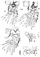 issued from : E. Suarez-Morales & I.A. Castellanos in J. Crustacean Biol., 1998, 18 (1). [p.201, Fig.2]. Female [? Juv. stage 5]: A, P1 (anterior view); B, P2 (anterior); C, P3 (anterior); D, P4 (anterior); E, P5 and genital double-somite (ventral). Nota: P5 uniramous, unsegmented, armed with 1 short subterminal spine; socket on tip of each ramus possibly indicating position of long seta (as in other 3 genera); P6 represented by anterolateral process with 2 curved,
subequal spines.
|
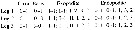 issued from : E. Suarez-Morales & I.A. Castellanos in J. Crustacean Biol., 1998, 18 (1). [p.202]. Female: Armament of swimming legs P1 to P3.
|
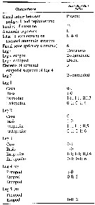 issued from : J.-s. Ho, M. Dojiri, G. Hendler & G.B. Deets in J. Crust. Biol., 2003, 23 (3). [p.589, Table 1]. Characters of female; Armature formulae for legs are presented as outer margin first, with Roman numerals for the number of spines, and Arabic numerals for setae. Data and armature formulae of the legs based on information from Suarez-Morales & Castellanos (1998, pers; comm.). Asterrisk (*) denotes cases where formulae were corrected for missing (probably broken) setae.
|
 Issued from : E. Suarez-Morales & E. Tovar in Sarsia, 2004, 89. [p.235, Fig.7]. Female copepodid V (from Mahahual reef lagoon, Yucatan): A-B, habitus (dorsal and lateral, respectively); C, urosome (ventral); D, genital field (ventral). Scale bars = 0.1 mm.
|
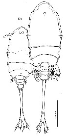 Issued from : E. Suarez-Morales & E. Tovar in Sarsia, 2004, 89. [p.238, Fig.9 part.]. Female copepodid V and adult (from Mahahual reef lagoon, Yucatan). Nota adult female: - Prosome length almost 51% of total body length (measured from anterior head to end of anal somite). - Oral opening located about 40% of way back along ventral surface of prosome. - Eye-shaped, with posterior scar of vestigial labrum. - Cephalosome with ventral cuticular processes and wrinkles around mouth area, forming transverse and vertical ridges which extend to the base of A1. - Cuticular structures include, mostly paired, groups of small pores on dorsal surface (as described for CV). - A1 6-segmented (armed with 4;1;0;2;2;8 setae plus single aesthetasc), almost 9% of total body length, and 17% of prosome length; 1st segment with several protuberances bearing each at least 1 seta on internal surface (probably the result of fusion of several proximal segments). - P4 biramous, basipod wide, intercoxal plate undefined; endopod partially fused with basis, represented by small, rounded process, with 1 long distal seta; exopod 1-segmented, armed with 5 setal elements (3 terminal, 2 subterminal), in some specimens this pattern is asymmetrical, with1 subterminal inner seta substitued by short spiniform seta on one side. - P5 uniramous, unsegmented, consisting of an elongate lobe rounded distally, armed with 1 short subterminal spine and single terminal seta. - P6 represented by anterolateral process with 2 curved, subequal spines (as in the copepodid V). - Urosome of 5 somites(4th and 5th pedigerous, genital double-somite, small post-genital somite, and elongated anal somite. - Genital double-somite with ventral genital field consisting of inverted V-shaped vestibulum, with 1 lateral canal on each side, almost transverse, with central copulatory pore (see in fig.7D). - Anal somite representing about 41% of total body length; width at its middle portion about 55% as wide as posterior margin, and about 35% of anterior margin; posterior margin of anal somite with a pair of small spines medially on ventral surface, near the base of each caudal rami. Caudal rami slightly divergent, over 3.4 times longer than wide, bearing 4 well-developed terminal setae with almost the same length and breadth; outer margin with lateral seta, dorsal seta slender, about 0.75 times as long as caudal ramus.
| | | | | Compl. Ref.: | | | Suarez-Morales & Gasca, 1998 a (p.112); Ho, Dojiri, Hendler & Deets, 2003 (p.589, tab.1) | | | | NZ: | 1 | | |
|
Distribution map of Caribeopsyllus chawayi by geographical zones
|
| | | | Loc: | | | E Mexico (Caribbean coast, Mahahual Reef) | | | | N: | 2 | | | | Lg.: | | | (163) F**: 1,78; (894) F*: 2,06-2,02; {F: 2,02-2,06}
** Lg = 1,57 mm: Caudal rami excluded. | | | | Rem.: | The description of the female in 1998 would be in fact a juvenile stage 5.
After Suarez-Morales & Tovar (2004, p.243) the morphology of the adult females recorded in Mahahual is identical to that of the holotype specimen (Suarez-Morales & Castellanos, 1998). However, three points should be considered: 1- the holotype has damaged parts and new morphological data are added . 2- the holotype specimen seems to be assignable to the CV stage. 3- the sharp differences in size between the adult and CV (the holotype measures 1.54 mm and the average size of the CV individuals was 1.66 mm, thus far from the 2.16 mm average length reached by fully mature adult females. The adult-like development of the CV genital field is, however, intriguing; explanations of this are: 1- the possibility of having polymorphic adults, in which the moult to the adult does not occur during the terminal moult; 2- the CV genital apparatus is not functional but becomes functional at CVI; 3- there are two closely related species mixed in Mahahual, which seems unlikely because there are no morphological differences other than size. | | | Last update : 29/04/2015 | |
|
|
 Any use of this site for a publication will be mentioned with the following reference : Any use of this site for a publication will be mentioned with the following reference :
Razouls C., Desreumaux N., Kouwenberg J. and de Bovée F., 2005-2026. - Biodiversity of Marine Planktonic Copepods (morphology, geographical distribution and biological data). Sorbonne University, CNRS. Available at http://copepodes.obs-banyuls.fr/en [Accessed April 08, 2026] © copyright 2005-2026 Sorbonne University, CNRS
|
|
|
|


;)
;)
;)
;)
;)
