|
|
 |
|
Harpacticoida ( Order ) |
|
|
|
Cervinioidea ( Superfamily ) |
|
|
|
Aegisthidae ( Family ) |
|
|
|
Aegisthus ( Genus ) |
|
|
| |
Aegisthus mucronatus Giesbrecht, 1891 (F,M) | |
| | | | | | | Syn.: | Aegisthus longirostris T. Scott, 1894 b (p.104, figs.F,M);
Aegisthus atlanticus Wolfenden, 1902 (p.364); Fowler, 1903 (p.124); Pearson, 1906 (p.36, Rem.) Rose, 1933 a (p.294); Klie, 1943 a (n°4, p.3, Rem.F); Sewell, 1948 (p.483); Gaudy, 1972 (1973 a) (p.948);
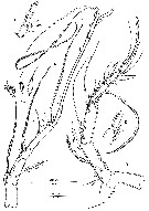
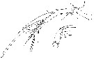
A. dubius Sars, 1916 (p.14, figs.M); Farran, 1926 (p.301, Rem.); Rose, 1933 a (p.294, figs.M); Sars, 1938 (p.51, figs.M); Gaudy, 1963 (p.31); Deevey & Brooks,1977 (p.265); Vives, 1972 (p.230, figs.F); Björnberg, 1973 (p.370); Vives & al., 1975 (p.56, tab.II, XII); Deevey & Brooks, 1977 (p.256, tab.2, Station "S"); Vives, 1982 (p.295); Kovalev & Shmeleva, 1982 (p.85) | | | | Ref.: | | | Giesbrecht, 1892 (p.573, 577, 770, Descr.F, figs.F); Canu, 1896 (p. 435, Rem.); Farran, 1908 b (p.91, Rem.); A. Scott, 1909 (p.234, Rem.); Wolfenden, 1911 (p.363); Farran, 1926 (p.300); Wilson, 1932 a (p.304, figs.F); Rose, 1933 a (p.293, figs.F); Farran, 1936 a (p.123); Johnson, 1937 (p.508, fig.M); Klie, 1943 a (n°4, p.3, figs.F); Sewell, 1947 (p.292); Lang, 1948 a (p.175, figs.F,M); Davis, 1949 (p.71, figs.F); Vervoort, 1957 (p.149); Marques, 1958 a (p.139, fig.M, Rem.); Owre & Foyo, 1967 (p.104, figs.F,M); Ramirez, 1969 (p.101, figs.F); Bradford, 1970 a (p.362, figs.F, M, Rem.); Chiba & Hirakawa, 1972 (p.75, figs. juv.5); Boxshall, 1979 (p.204, figs.F,M); Björnberg & al., 1981 (p.679, figs.F,M); Vives, 1982 (p.295); Gamô, 1983 (p.45, figs.F); Huys, 1988 (p.117, figs.F,M, Rem.); Bradford-Grieve & al., 1999 (p.887, 968, figs.F); Conroy-Dalton & Huys, 1999 (p.429, Rem.); Boxshall & Halsey, 2004 (p.229: figs.F,M); Vives & Shmeleva, 2010 (p.88, 90, figs.F,M, Rem.) | 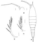 issued from : J.M. Bradford in N.Z. Jl Mar. Freshw. Res., 1970, 4 (4). [p.361, Figs 78-81]. Female (off Kaikoura, New Zealand): 78, P5; 81, habitus (dorsal). Male: 79, P5; 80, forehead and left A1. Scale bars represent 0.1 mm. Nota: The furcal setae in the New Zealand specimens were longer than in Giesbrecht's (1892) description (five times the length of the body as against \"more than 3.5 times as long at the body\").
|
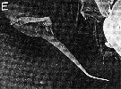
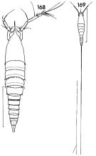 issued from : F.C. Ramirez in Contr. Inst. Biol. mar., Buenos Aires, 1969, 98. [p.98, Lam. XX, figs. 168, 169]. Female (from off Mar del Plata): 168, habitus (dorsal); 169, idem (including caudal setae). Scale bar in mm: 0.8 (168); 2.0 (169). Nota: caudal setae about 6 times length of cephalothorax, with bifid end.
|
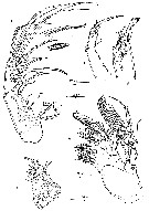
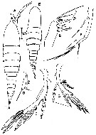 issued from : G.A. Boxshall in Bull. Br. Mus. nat. Hist. (Zool.), 1979, 35 (3). [p.205, Fig.1, A-G] Female (from 18°N, 25°W): A, habitus (dorsal); B, Mxp; C, P5; D, P6. Nota: Prosome without reticulate chitinous markings on dorsal surface. Male: E, habitus (dorsal); F, A2; G, P5. Nota: Prosome without reticulate chitinous markings on dorsal surface. Scales 0.1 mm unless otherwise indicated.
|
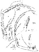
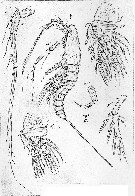 issued from : G.O. Sars in Res. Camp. scient. Prince Albert Ier Monaco, 1938. [Pl. II, Fig.B]. As Aegisthus dubius. Male. mp2 = Mxp.
|
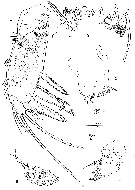
 issued from : R. Huys in Bijdr. Dierk., 1988, 58 (1). [p.119, Fig.1]. Female (18°N, 25°W): B, A1 (arrow indicating tube pore1; D, detail of distal part of segment 6. Nota : A1 6-segmented. 1st segment widening anteriorly and forming medially a strong hook-like process from which a slender seta arises . 2 nd segment with several spinules along outer margin ; furnished with 2 minute plumose, 1 strong bipinnate and 3 small smooth setae in proximal half, anterior part with 2 bare setae and a small tube pore. 3 rd segment with 10 minute setae (7 plumose, 3 smooth) and 2 extremely long setae ; distal part forming a distinct process with a long aesthete in addition to a minute bare seta and a longer bipinnate one. 4 th segment with 4 setae along the inner margin and 1 bi-articulated slender seta at the outer subdistal corner. 5 th segment smallest and bearing 2 setae (this segment is the equivalent of fused segments 4 and 5 in the 7-segmented A1 of A. aculeatus and A. spinulosus). Outer side of apical segment armed with 4 bi-articulated setae ; distal part with slender seta and long aexthetasc fused at base with bipinnate seta. Male: A, A1; C, detail of distal part of segment 8. a, b, c, characteristic spines of segment 4. Nota : A1 very long and slender, 8-segmented, haplocer, geniculation between segments 6 and 7. Segment 2 furnished with 10 minute setae along the inner margin and 1 long outer plumose seta ; proximal half with a long aesthetasc covered at base by a small operculum. Segment 4 long ; proximal half with 3 minute setae abd a slender one standing on a small process ; distal half with 1 bare and 2 characteristic pinnate spines in addition to a long aesthete which is fused with minute seta at its base. Segment 6 with 2 minute setae and 1 pinnate spine and forming haplocer apparatus with following segment (in A. aculeatus segments 5 and 6 are fused). Distal segment bearing a slender seta and a very long aesthetasc at the top ; inner margin with 4 bare setae ; outer margin with 5 bi-articulated setae.
|
 issued from : R. Huys in Bijdr. Dierk., 1988, 58 (1). [p.120, Fig.2]. Female: A, A2; a: detail of exopodite; b: detail of distal setae. Nota : A2 : coxa well defined, with several spinules along the outer margin. Basis and proximal endopodite segment forming elongated allobasis ; showing a distinct spinular pattern in proximal third and several longitudinal spinular rows distally ; furnished with inner bipinnate seta near aticulation with distal endopodite segment. Exopodite 2-segmented ; segment 1 provided with several fine spinules and a bipinnate seta at the inner distal corner ; segment 2 very small, wider than long, bare, with a long plumose seta. Endopodite with 4 groups of long spinules along the outer edge ; with 3 setae at about middle of inner margin ; with 6 pinnate setae (of which 2 fused at base) apically. Male: B, A2; c: detail of distal part of endopodite; d: detail of exopodite; e: detail of inner seta of endopodite. Nota : A2 : Coxa forming a swollen process armed with long spinules. Basis and 1st endopodite segment partially fused (forming an elongated allobasis), spinulated along the outer margin. Exopodite 2-segmented ; proximal segment long and bare, having a small seta at the inner subdistal corner ; distal segment indistinctly fused with the long apical seta. Distal endopodite segment furnished with 2 groups of long spinules along the outer side ; with at about middle of inner edge an aesthetasc-like seta showing a small basal process (rudimentary seta ?) ; terminal part having an articulating aesthetasc-like seta in addition to some minute spinules and fused with 1 bipinnate outer seta and 2 long aesthetasc-like setae.
|
 issued from : R. Huys in Bijdr. Dierk., 1988, 58 (1). [p.121, Fig.3]. Female: A, Mx1; C, Mx2 (arrows indicating tube pore and apical pore of claw). Nota : Mx1 well developed. Praecoxal arthrite with 10 stout bipinnate spines at the margin and 2 strong plumose setae on the anterior surface ; posterior surface with minute spinules. Coxa without epipodite ; with some diminutive spinules and a weakly defined endite bearing 1 bipinnate and 1 plumose seta. Basis rectangular, without endites or any trace of exo- or endopodite ; furnished with 4 bipinnate setae apically. Mx2 strongly developed, prehensie. Syncoxa long, integument pitted in a distinct pattern ; furnished with 4 ( ?) endites (it is difficult to decide whether the distalmost one is standing on the syncoxa or the basis), middle 2 rudimentary and with 2 setae , proximal and distal ones subcylindrical and with 4 and 3 setae, respectively. Basis forming a hollow, curved claw which is finely spinulated and opening at the tip ; furnished with 2 bipinnate setae, 2 serrate spines and a distinct tube pore at the basis of the claw. Endopodite 3-segmented ; proximal segment with 1geniculate seta, middle segment with 2 geniculate setae, distal segment with 2 geniculate and 2 bipinnate setae. Male: B, Mx1; D, Mx2; E, Mx2 (detail of maxillar basis and endopodite). Nota : Mx1 embedded in wrinkled tegument of postlabial area ; small, rudimentary, tegument wrinkled at the basal part. As in the female the praecoxal arthrite bears 10 marginal setae and 2 surface setae. Coxa and basis fused and represented by a common elongate process having 2 apical setae. Mx2 : Syncoxa smooth, lacking endites. Basis forming a spinulated curved claw which is slightly swollen in the middle ; furnished proximally with a small bilobed baso-endite with 2 setae, distally with some long spinules and 1 minute seta. Endopodite insistinctly 3-segmented, thin-walled ; segment 1 with 1 seta ; segment 2 with 2 setae and partially fused with segment 3 which is bearing 4 setae.
|
 issued from : R. Huys in Bijdr. Dierk., 1988, 58 (1). [p.123, Fig.4]. Female: A, Mxp; C, Md (mandibular gnatoobase); D, Md (detail of masticatory edge). Md : Palp absent, represented by well-developed coxa only. Gnathobasis strong, with several stout teeth and a bipinnate seta at the dorsal side ; ventral part with long hair-like setules. Labrum well developed, divided in two parts, each bearing numerous recurved denticles ; deeper area furnished with long spinules. Mxp stenopodial, strong ; indistinctly 3-segmented.Praecoxa not entirely free, fused along the inner side with the coxa (forming a syncoxa), armed with a few spinules. Coxal part with numerous spinules of different shape and length, most of them occurring at the anterior surface, armed with 3 enlarged spines (proximal ne bipinnate, distal two serrate) in addition to a slender bipinnate seta. Distal segment rectangular and possibly derived from the fused basis and endopodite ; outer margin with long spinules ; furnished with 1 subapical plumose seta and 2 long bipinnate setae apically. Male: B, Mxp; E, Md. Nota : Md small, rudimentary, withou palp. Tegument of coxa wrinkled ; gnathobasis with at least 8 spiny processes and a distinct seta which is fused at the basis. Labrum : Prelabral area wrinkled as well as lateral zones in which the Md are embedded. Oral aperture without any ornamentation. Labium without hairs ; tegument wrinkled ; paragnaths not differentiated. Mxp small, rudimentary, without any trace of segmentation (tegument wrinkled) ; distal part an elongate process, forming a minute lobe proximally, widening and armed with 2 setae distally. A long vestibulum is present between the Mxp and P1.
|
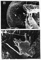
 issued from : R. Huys in Bijdr. Dierk., 1988, 58 (1). [p.125, Fig.6, B]. Female (SEM micrograph): B, genital double-somite with P6 (lateral view).
|
 issued from : R. Huys in Bijdr. Dierk., 1988, 58 (1). [p.125, Fig.6, D]. Female (SEM micrograph): D, spinule on caudal ramus.
|
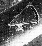
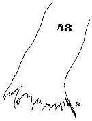
 issued from : R. Huys in Bijdr. Dierk., 1988, 58 (1). [p.127, Fig.7, B, C]. Female (SEM micrographs): B, rostrum (anterolateral view); C, proximal segments of A1 (arrow indicating inner process). Nota : Rostrum very long, round in transverse section, and anteriorly directed, slightly curved downwards ; sensillae absent.
|
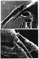
 issued from : R. Huys in Bijdr. Dierk., 1988, 58 (1). [p.131, Fig.9, B, E]. Male (SEM micrographs): B, rostrum (anterior view; arrows indicating integumental pores); E, Md. Nota : Rostrum not well defined, confluent with anterior margin cephalosome ; provided with 2 sets of 3 pores ventrally. Ventral wall of preantennular area of cephalosome exhibing numerous minute perforations.
|
 issued from : R. Huys in Bijdr. Dierk., 1988, 58 (1). [p.129, Fig.8, E]. Male (SEM micrograph): E, P6.
|
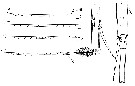
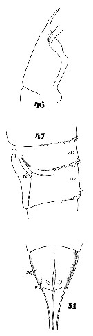 Issued from : W. Giesbrecht in Systematik und Faunistik der Pelagischen Copepoden des Golfes von Neapel und der angrenzenden Meeres-Abschnitte. Fauna Flora Golf. Neapel, 1892, 19 , Atlas von 54 Tafeln. [Taf.46, Figs.46, 47, 51]. Female: 46, head (lateral); 47, thoracic segment 5 and abdominal segments 1 and 2 (lateral); 51, anal segment and anterior portion of caudal rami (ventral).
|
 Issued from : W. Giesbrecht in Systematik und Faunistik der Pelagischen Copepoden des Golfes von Neapel und der angrenzenden Meeres-Abschnitte. Fauna Flora Golf. Neapel, 1892, 19 , Atlas von 54 Tafeln. [Taf.46, Fig.48]; Female: 48, Md.
|
 Issued from : W. Giesbrecht in Systematik und Faunistik der Pelagischen Copepoden des Golfes von Neapel und der angrenzenden Meeres-Abschnitte. Fauna Flora Golf. Neapel, 1892, 19 , Atlas von 54 Tafeln. [Taf.46, Fig.49]. Female: P5.
|
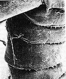
 issued from : R. Huys in Bijdr. Dierk., 1988, 58 (1). [p.124, Fig.5]. Female: Caudal ramus. Nota: The length of the caudal rami is averaging 5-7 times the body length. The caudal rami are fused along a considerable distance.Each ramus is provided with 1 bare outer seta at about the middle third but the insertion place is not symmetrical in both rami; this seta is probably homologous with the anterolateral seta (II) in other families. The distal end is armed with 2 setae; the largest is tri-articulate at the base, furnished with long spinules, directed outward and undoubtedly the equivalent of the dorsal seta (VII); the second one is shorter, implanted subterminally and probably representing the posterolateral seta (III); The surface of each ramus exhibits a bilateral pattern of minute, flattened triangular spines. In males the same setal configuration is observed. The caudal rami are pressed closely together along the proximal fourth but not fused.
|
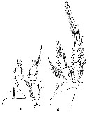
 Issued from : W. Giesbrecht in Systematik und Faunistik der Pelagischen Copepoden des Golfes von Neapel und der angrenzenden Meeres-Abschnitte. Fauna Flora Golf. Neapel, 1892, 19 , Atlas von 54 Tafeln. [Taf.49, Figs.2, 3]. Female: 2, A1 (ventral view); 3, Mxp.
|
 Issued from : W. Giesbrecht in Systematik und Faunistik der Pelagischen Copepoden des Golfes von Neapel und der angrenzenden Meeres-Abschnitte. Fauna Flora Golf. Neapel, 1892, 19 , Atlas von 54 Tafeln. [Taf.49, Figq.6, 10]. Female: 6, P4 (posterior view); 10, P1 (posrerior view).
|
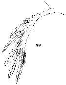
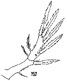 issued from : H.B. Owre & M. Foyo in Fauna Caribaea, 1, Crustacea, 1: Copepoda. Copepods of the Florida Current. 1967. [p.104, Fig.757]. Male: P5.
|
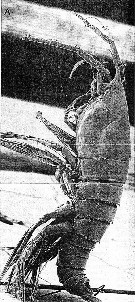 issued from : R. Huys in Bijdr. Dierk., 1988, 58 (1). [p.125, Fig.6, A]. Female (SEM micrographs) from 18°N, 25°W: A, habitus (lateral view).
| | | | | Compl. Ref.: | | | Cleve, 1904 a (p.185); Pearson, 1906 (p.36); Lysholm & al., 1945 (p.45); Sewell, 1948 (p.346); C.B. Wilson, 1950 (p.158, Rem.); King & Hida, 1955 (p.11); Grice, 1963 a (p.496); Gaudy, 1963 (p.31, Rem.); Unterüberbacher, 1964 (p.34); De Decker & Mombeck, 1964 (p.11); Furuhashi, 1966 a (p.295, vertical distribution in Oyashio/Kuroshio transitional area, Table 8, 10); De Decker, 1968 (p.45); Delalo, 1968 (p.139); Björnberg, 1973 (p.371, 384); Vives & al., 1975 (p.56, tab.II); Deevey & Brooks, 1977 (p.256, tab.2, Station "S"); Vives, 1982 (p.295); Kovalev & Shmeleva, 1982 (p.85); Guangshan & Honglin, 1984 (p.118, tab.); Lozano Soldevilla & al., 1988 (p.61); Shih & Young, 1995 (p.75); Böttger-Schnack, 1996 (p.1088); Errhif & al., 1997 (p.422); Padmavati & al., 1998 (p.349); Suarez-Morales & Gasca, 1998 a (p.113); Lapernat, 2000 (tabl.3, 4); Razouls & al., 2000 (p.343, Appendix); Lopez-Salgado & al., 2000 (tab.1); Sameoto & al., 2002 (p.11); Fernandes, 2008 (p.465, Tabl.2); Galbraith, 2009 (pers. comm.); Williamson & McGowan, 2010 (p.273, Table 3, Pacific central gyres: N and S); Hsiao S.H. & al., 2011 (p.475, Appendix I); Zizah & al., 2012 (p.79, Tableau I, Rem.: p.86); in CalCOFI regional list (MDO, Nov. 2013; M. Ohman, pers. comm.); Lidvanov & al., 2013 (p.290, Table 2, % composition); Bonecker & a., 2014 (p.445, Table II: frequency, horizontal & vertical distributions); El Arraj & al., 2017 (p.272, table 2, spatial distribution); | | | | NZ: | 19 | | |
|
Distribution map of Aegisthus mucronatus by geographical zones
|
| | | | | | | | | | | | | | | 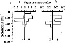 Issued from : P.-E. Lapernat in DEA Océanogr. Biol., Univ. P. & M. Curie, Paris VI. July 5, 2000. [Fig.9 d ]. Issued from : P.-E. Lapernat in DEA Océanogr. Biol., Univ. P. & M. Curie, Paris VI. July 5, 2000. [Fig.9 d ].
Verical distribution of Aegisthus mucronatus at an eutrophic site (off Mauritanian coast: 20°32 'N, 18°36' W) in females (F) and males (M) (ind. per m3) in the day (white circle) and night (black circle).
Nota: Sampling in the water column 0-1000 m, one during the day and another during the night with BIONESS multiple-net: 0-75; 75-150; 150-250; 250-350; 350-450; 450-550; 550-700; 700-850; 850-965 m. In May-June 1992. |
| | | | Loc: | | | sub-Antarct. (Indian), South Africa (E & W), off Tristan da Cunha (N & E), Namibia, Angola, off S Ascension Is., G. of Guinea, off E St. Paul Is., off Cape Verde Is. , off Mauritania-NW Cape Verde Is., off Mauritania, Mauritania-Morocco , Canary Is., Moroccan coast, off Madeira, Argentina, S & central Brazil, G. of Mexico, Caribbean, Florida, Sargasso Sea, off Bermuda (Station "S"), Georges Bank, off E Nova Scotia, Newfoundland, W Ireland, Porcupine Bank, off N Scotland, North Sea, Bay of Biscay, off SW Portugal, Azores, Baie Ibéro-marocaine, W Medit. (Alboran Sea, Tyrrhenian Sea), G. of Aden, Arabian Sea, Laccadive Is., Madagascar, S Indian (subtropical convergence), Bay of Bengal, Indonesia-Malaysia, Philippines, China Seas (East China Sea, South China Sea, Taiwan (E: Kuroshio Current), Japan, Pacif. (W equatorial), Pacf. (N & S central gyres), Australia (Great Barrier), New Zealand, off Marshall Is., Hawaii, Pacif. (equatorial central), off British Columbia, off Oregon, CalCOFI region, Galapagos, N Chile | | | | N: | 69 | | | | Lg.: | | | (38) F: 2,28-2,16; (46) F: 2,25; (59) F: 2,6-2,2; M: 2-1,7; (138) F: 2,55-1,9; M: 1,7-1,1; (240) F: 1,45; (254) F: 2; (313) F: 1,6-2; M: 1,4-1,6; (432) F: 2,5; (618) F: 2,46-1,28; M: 1,73-1,62; (921) F: ± 2; {F: 1,28-2,60; M: 1,10-2,00} | | | | Rem.: | epi-meso-bathypelagic.
Sampling depth (sub-Antarct.) : 500-750 m. Sargasso Sea: 500-1500 m (Deevey & Brooks, 1977, Station "S");
Gamô (1983) observed this species at a depth of 7530-7560 m off east of Kinhazan (NE Japan). 204-457 m at Station S 2 in S Bösö (E middle Japan) from Furuhashi (1966 a).
According to Huys (1988, p.122) the luminous sites occur on the head, swimming legs and the urosome, but have not yet been more precisely located (see herring, 1985). Boxshall found only female of luminous capability but probably males were not tested.
Identification chaacters:
Female: Rostrum ending in long acuminate tip.
Male: Front rouded, without rostrum. A1 with 7-segmented, 6th very long. | | | Last update : 25/10/2022 | |
|
|
 Any use of this site for a publication will be mentioned with the following reference : Any use of this site for a publication will be mentioned with the following reference :
Razouls C., Desreumaux N., Kouwenberg J. and de Bovée F., 2005-2026. - Biodiversity of Marine Planktonic Copepods (morphology, geographical distribution and biological data). Sorbonne University, CNRS. Available at http://copepodes.obs-banyuls.fr/en [Accessed May 06, 2026] © copyright 2005-2026 Sorbonne University, CNRS
|
|
|
|


;)
;)
;)
;)
;)
;)
;)
;)
;)
;)
;)
;)
;)
;)
;)
;)
;)
;)
;)
;)
;)

;)
