|
|
 |
|
Misophrioida ( Order ) |
|
|
|
Speleophriidae ( Family ) |
|
|
|
Archimisophria ( Genus ) |
|
|
| |
Archimisophria discoveryi Boxshall, 1983 (F,M) | |
| | | | | | | Ref.: | | | Boxshall, 1983 (p.103, figs.F,M); Huys & Boxshall, 1991 (p.92, 436, 460, figs.F,M); Boxshall & Halsey, 2004 (p.223: fig.F) | 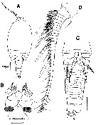 issued from : G.A. Boxshall in Bull. Br. Mus. nat. Hist. (Zool.), 1983, 44 (2). [p.104, Fig.1]. Female (from 34°57'N, 32°55'W): A, habitus (dorsal); B, rostrum and labrum (ventral); C, urosome (ventral); D, A1 (dorsal). Scale bars 0.100 mm unless otherwide indicated. Nota: Prosome apparently 4-segmented but with 1st free thoracic somite entirely concealed beneath a carapace-like extension from the posterior margin of the maxilliped)bearing somite. Nauplius eye absent. Prominent rostrum visible from dorsal aspect, not fused to labrum. Cone organs not observed but large mass of glandular tissue present on sides of cephalosome beneath usual location of cone organs (see structure and function in Benthomisophria palliata). Urosome 6-segmented. Surface of prosome and urosome somites 1 to 5 ornamented with a reticulum of epicuticular lamellae. Urosome somite 6 without reticulate markings. Caudal rami longer than wide, armed with 2 long distal margin setae, 2 medium-length distal angle setae, a dorsal seta near the inner margin and a distally located lateral seta. Labrum small, with a posterior row of marginal denticles. A1 27-segmented. P5 uniramous, 3-segmented and positioned midventrally with inner margins almost touching at base; P6 reduced to a semicircular flap closing off the opening of the genital antrum, bearing an outer plumose seta and a short inner spine.
|
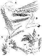 issued from : G.A. Boxshall in Bull. Br. Mus. nat. Hist. (Zool.), 1983, 44 (2). [p.106, Fig.2]; Female: A, A2 (anterior); B, Md (anterior); C, Mx1 (posterior); D, Mxp (posterior). Nota: Md with well developed gnathobase bearing distally 2 multicusped blades, 5 strong spines and an extensive fringe of pinnules; mandibular palp comprising basis, 2-segmented endopod and 4-segmented exopod. Mx1 gnathobase with 14 distal elzements; endite 1 with 1 spiniform and 3 setiform armature elements, endite 2 with 3 spiniform elements; outer lobe rudimentary represented by 6 plumose setae on outer surface of segment; maxillulary palp biramous with 2-segmented endopod and 1-segmented exopod. Mx2 6-segmented. Mxp 8-segmented
|
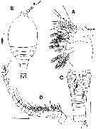 issued from : G.A. Boxshall in Bull. Br. Mus. nat. Hist. (Zool.), 1983, 44 (2). [p.107, Fig.3]. Female: A, Mx2; Male: B, habitus (dorsal); C, urosome (ventral); D, A1 (dorsal). Scale bars 0.100 mm unless otherwise indicated. Nota: A1 25-segmented, unigeniculate with the articulation between segments XIX and XX.
|
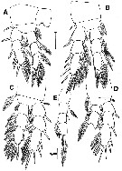 issued from : G.A. Boxshall in Bull. Br. Mus. nat. Hist. (Zool.), 1983, 44 (2). [p.109, Fig.4]. Male: A-D, P1 to P4 (anterior); E, P5 (anteroventral). Scale bars 0.100 mm unless otherwise indicated. Nota: swimming legs 1-4 incomplete in holotype and paratype females, assumed to be similar to those for a paratype male. P5 male uniramous, 4-segmented and with bases of legs almost touching at ventral midline as in female. P6 represented by a flattened plate bearing a long outer plumose seta and a short inner spine.
| | | | | NZ: | 1 | | |
|
Distribution map of Archimisophria discoveryi by geographical zones
|
| | | | Loc: | | | N Atlant. (SW off Azores) | | | | N: | 1 | | | | Lg.: | | | (606) F: 1,4-1,1; M: 1,3-1,1; {F: 1,10-1,40; M: 1,10-1,30} | | | | Rem.: | hyperbenthic (depth 23-25 m off the bottom of about 3000 m).
For Boxshall (1983, p.121) the 27-segmented A1 of female is of interest concerning the nature of this limb in the ancestral copepod. Giesbrecht (1892, 1899) analysed the segmentation and armature of the antennules of many copepods in an attempt to reduce the antennule of all copepod to a common type. Giesbrecht's basic copepod A1 was 25-segmented and by studying the arrangement of the armature elements he was able to determine which segments had fused in those forms with fewer segments. This basic limb closely resembles that of Calanus finmarchicus both in number of segments and in setation. The typical armature present on each antennulary segment is 2 setae and 1 aesthetasc, at least in te female (one or more of these elements is often lost, most commonly the aesthetasc). The arrangement of these 3 elements, which Giesbrecht called a 'trithek', follows a constant pattern. As Gurney (1931) noted, many calanoids possess 3 complete tritheks on the 2nd segment and a single proximal seta plus a distal trithek on the 1st segment. He interpreted this as evidence that the 2nd segment of calanoid antennules is derived from 3 fused segments and that the 1st segment may be derived from 2 fused segments. On the basis of this interpretation he postulated that the ancestral copepod A1 comprised 27 or possibly 28 segments. The discovery of Archimisophria discoveryi with its 27-segmented A1 provides a remarkable corroboration of Gurney's hypothesis. | | | Last update : 16/09/2021 | |
|
|
 Any use of this site for a publication will be mentioned with the following reference : Any use of this site for a publication will be mentioned with the following reference :
Razouls C., Desreumaux N., Kouwenberg J. and de Bovée F., 2005-2026. - Biodiversity of Marine Planktonic Copepods (morphology, geographical distribution and biological data). Sorbonne University, CNRS. Available at http://copepodes.obs-banyuls.fr/en [Accessed March 22, 2026] © copyright 2005-2026 Sorbonne University, CNRS
|
|
|
|


;)
;)
;)
;)
