|
|
 |
|
Misophrioida ( Order ) |
|
|
|
Speleophriidae ( Family ) |
|
|
|
Boxshallia ( Genus ) |
|
|
| |
Boxshallia bulbantennula Huys, 1988 (F,M) | |
| | | | | | | Ref.: | | | Huys, 1988 a (p.140, figs.F,M); Huys & Boxshall, 1991 (p.93, 437, 461, figs.F,M); Boxshall & Halsey, 2004 (p.223: figs.); Vives & Shmeleva, 2010 (p.152, figs.F,M, Rem.) | 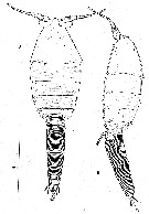 Issued from : R. Huys in Stygologia, 1988, 4 (2). [p.141, Fig.1] Female (from Lanzarote): A-B, habitus (dorsal and lateral, respectively). Nota : Prosome 5-segmented, 1st pedigerous somite free and not concealed beneath a carapace-like extension from the posterior margin of the Mxp-bearing somite. Nauplius eye not observed. Rostrum well developed, anteroventrally deflexed but still visible in dorsal aspect, not defines at base, not fused to labrum Ventrolateral margins of dorsal cephalic shield not indented, cone organs absent. 1st pedigerous somite without areas of folded, flexible integument. Dorsal and dorsolateral surfaces of prosomal somites (except cephalosome) ornamented with pattern of minute denticles. Posterolateral areas of last prosome somite furnished also with hair-like pinnules. Urosome 5-segmented, with genital and 1st abdominal somites fused (to form genital double-somite) ; all urosomites with squamose ornamentation. Caudal rami slightly longer than wide ; armed with 6 setae : anterolateral seta dorsally displaced and plumose, posterolateral seta tripinnate, outer and inner terlminal setae strongly debveloped, terminal accessory seta well developed and tripinnate, dorsal seta smooth and located near the midposterior margin, anterolateral accessory seta missing ; inner and posterior margins furnished with fine spinules.
|
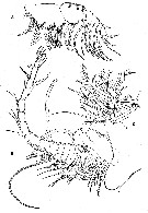 Issued from : R. Huys in Stygologia, 1988, 4 (2). [p.143, Fig.2] Female: A, Mxp; B, A1 (setation of segments 5 to 11 omitted); C, A1 segments 5 to 12. Nota : A1 27-segmented and borne on an expanded basal pedestal ; outer margin of proximal antennular segment produced into a conspicuous anteriorly directed bulb-shaped process bearing a ventral spinular row. Segments II to XI fused along the outer margin and the angle of fusion directs the distal segments more laterally. The aesthetascs on the anteroventral surfaces of segments XI and XVI are large, that on segment XXVII very short. The seta on segment XI looks aesthetasc-like and the inner one on segment XXVI is swollen. A2 : protopod comprising separate coxa and basis. Endopod indistinctly 3-segmented, segments 2 and 3 fused anteriorly. Exopod 7-segmented. Labrum small, not fused with rostrum, bilobed. Mxp : 8-segmented, praecoxa and coxa forming syncoxa. Basis with 2 inner margin setae and 3 fringes of fine spinules along outer margin. Endopd 6-segmented ; 1st segment fused with basis at posterior surface; segment 6 longest , bearing 2 plumose and 3 bare setae.
|
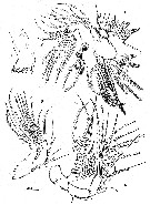 Issued from : R. Huys in Stygologia, 1988, 4 (2). [p.144, Fig.3] Female: A, A2; B, Md (mandibular palp); C, Md (mandibular gnathobase); D, Mx1. Nota : Md gnathobase well developed bearing several teeth along the distal margin. Palp biramous, comprising basis, 2-segmented endopod and indistinctly 3-segmented exopod. Mx1, setules along inner margin and 4 setae and 2 curved spines distally. :with praecoxa and coxa separated by well developed articulation. Praecoxal arthrite bearing 15 setae or spines around the distal margin. Coxal exite (epipodite) represented by 7 unequal plumose setae located on the outer margin, the 2 proximal-most setae being spatulate at tip ; coxal endite well developed, with 1 patch of spinules on the anterior surface, setules along inner margin and 4 setae and 2 curved spines distally. Basis elogated, with 2 endites ; proximal endite produced medially, bearing 4 plumose setae distally, and being spinulose along inner margin ; distal endite vestigial, represented by 4 plumose setae. Endopd 1-segmented, bilobed ; proximal inner lobe (equivalent of 1st endopod segment)with 3 apical plumose setae ; distal lobe forming apical process bearing 8 plumose setae. Exopod forming a flattened, 1-segmented plate, narrowing proximally and armed with 8 plumose setae around distal margin and with proximal fringe of long setules on inner margin.
|
 Issued from : R. Huys in Stygologia, 1988, 4 (2). [p.146, 147, Figs.4, 5] Female: left side A, P1; B, P2; right side A, P3; B, P4.
|
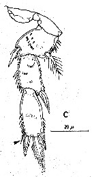 Issued from : R. Huys in Stygologia, 1988, 4 (2). [p.148, Fig.6, C] Female: C, P5 and intercoxal sclerite (arrow indicating minute seta). Nota: P5 biramous, comprising 2-segmented protopod, 2-segmented exopod and vestigial endopod represented by a bipinnate spine. Coxa unarmed ; basis spinulose along outer and distal margins, bearing plumose outer seta and some patches of spinules on surface. Exopod segment 1 with unipinnate spine at outer distal corner ; segment 2 with 4 distal margin elements, 2 short pinnate spines, 1 median longer spine and 1 inner minute seta. Members of P5 pair joined by well developed intercoxal sclerite as in legs P1 to P4.
|
 Issued from : R. Huys in Stygologia, 1988, 4 (2). [p.145] Setal and spine formula of swimming legs P1 to P4.
|
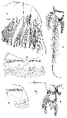 Issued from : R. Huys in Stygologia, 1988, 4 (2). [p.149, Fig.7, A, D-G] Female: A, Mx2; D, gonopores and P6 (ventral view); E, same (lateral view); F, caudal ramus (dorsal); G, same (ventral). Nota : Mx2 with praecoxa and coxa separated. Praecoxa bearing 2 short distal endites, closely set to each other, the 1st bearing 5 plumose setae, the 2nd 3 setae ; outer margin with several spinular rows. Coxa with 2 long cylindrical endites, not closely set to each other and furnished each with 3 plumose setae. Basis with single endite with fringe of long spinules along inner margin and produced into strong claw, being spinulose along inner distal half bearing 2 setae on both anterior and posterior surfave and 1 seta at basis on the claw near joint with endopod. Endopod 3-segmented ; segment 1 and 3 each with 2 inner margin setae ; segment 3 elongated and bearing 8 setae.
|
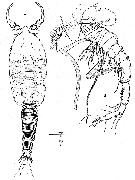 Issued from : R. Huys in Stygologia, 1988, 4 (2). [p.148, Fig.6, A-B] Male: A, habitus (dorsal); B, A1. Nota : Genital somite as long as wide, containing 2 oval spermatophores visible through integument on either side of midline. On either side of cephalosome and genital somite a testis is visible, apical part recurvede dorsally and posteriorly at level of maxillipedal somite. A1 23-segmented, unigeniculate on both sides with geniculation located between segmentsXIX and XX and distal part medially directed. Outer margin of segment 1 produced into conspicuous anteriorly directed bulb-shaped process. Segments IX to XI fused along outer margin. Segment XV fused ventrally with segment XVI ; both segments free and fully exposed dorsally. Segments XIX and XXII each represent two (XIX-XX and XXV-XXVI, respectively) fused segments ; segment XX represents three (XXI-XXIII) segments of the female. The aesthetascs on the anteroventral surfaces of segments XI and XVI are large, that on segment XXIII very short ; the seta on segment XI looks aesthetasc-like.
|
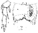 Issued from : R. Huys in Stygologia, 1988, 4 (2). [p.149, Fig.7, B, C] Male: B, genital somite and P6 (arrows indicating location of P5); C, P5. Nota : P5 biramous comprising unisegmented protopod, 2-segmented exopod and vestigial endopod represented by 1 bipinnate spine. Armament of exopod basically as in female. P6 represented by flattened plate bearing 3 unequal armature elements on lateral portion of posterior margin ; inner and distal part covered with numerous hair-like pinnules. Members of 6th pair of legs meeting in ventral midline but not fused ; articulation with somitic wall not well developed.
| | | | | NZ: | 1 | | |
|
Distribution map of Boxshallia bulbantennula by geographical zones
|
| | | | Loc: | | | Canary Is. (Lanzarote: anchialine lava pool) | | | | N: | 1 | | | | Lg.: | | | (605) F: 0,683; M: 0,577; {F: 0,68; M: 0,58} | | | | Rem.: | Anchialine cave | | | Last update : 15/02/2015 | |
|
|
 Any use of this site for a publication will be mentioned with the following reference : Any use of this site for a publication will be mentioned with the following reference :
Razouls C., Desreumaux N., Kouwenberg J. and de Bovée F., 2005-2025. - Biodiversity of Marine Planktonic Copepods (morphology, geographical distribution and biological data). Sorbonne University, CNRS. Available at http://copepodes.obs-banyuls.fr/en [Accessed May 21, 2025] © copyright 2005-2025 Sorbonne University, CNRS
|
|
 |
 |


